Bone Tumours
Dr. Wahby GhalibFJMC, CABO, MRCS
Classification
BenignMalignant : primary
secondaryBenign bone tumors
Bone : osteoid osteoma & osteoblastomaCartilage : enchondroma, chondroblastoma &
osteochondromaBlood vessels : haemangioma
Others : giant cell tumour
Benign tumour - like lesions
Bone cysts : simple & aneurysmalFibrous cortical defect
Primary malignant bone tumoursBone : osteosarcoma
Cartilage : chondrosarcomaBone marrow : Ewing sarcoma & myeloma
Connective tissue : fibrosarcomaOthers : chordoma & adamantinoma
Secondary malignant bone tumours
Prostate
BreastLung
ColonKidney
ThyroidStaging of malignant tumours (Enneking)
I : low gradeII : high grade
III : sarcoma with metastasisA : intra- compartmental
B : extra-copartmental
Surgery for malignant tumours
Wide excision : safe marginsThis includes : amputation
limb – salvage surgeryChemotherapy
Preoperative : (neoadjuvant) 8-12 wAfter tumour resection : check tumour necrosis
Postoperative : 6-12 mRadiotherapy
Residual tumourInaccessible tumour
Painful metastasis
Benign bone tumoursFibrous cortical defect
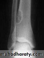
= non-ossifying fibromaVery common
ChildAccidentally on XR
Pain or pathologic fractureNo malignant potential
Rx : curettage + bone graft
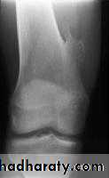
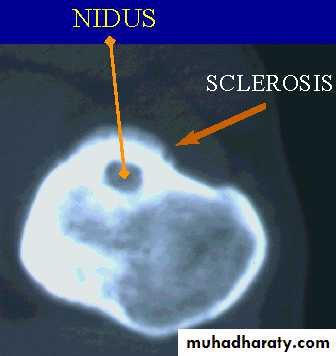
Osteoid osteoma
Patient < 30 yrPain > at night relieved by aspirin
In spine painful scoliosis
No malignant potential
XR : radioluscent nidus surrounded bysclerosis
Any bone except the skull
Rx : removal of nidus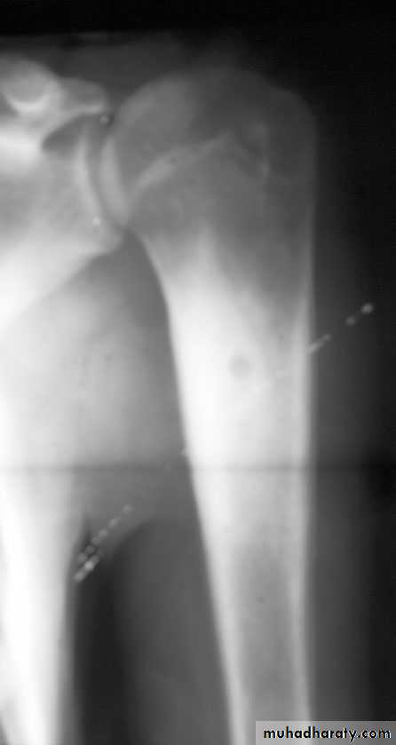
osteoblastoma
= O.O. but nidus > 1.5 cmCompact (ivory) osteoma
RareYoung adult
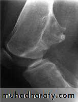
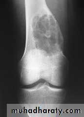
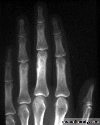
Painless lump on outer or inner table of skullEnchondroma
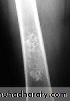
More in tubular bones of handAccidentally or pain / pathologic #
XR : lytic lesion + flecks of calcification
Malignant risk : 2%
Rx : curettage + bone graft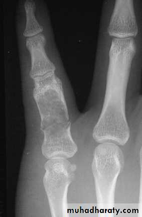
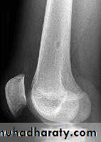
Picture 4. Frontal radiograph of the right thigh demonstrates coarse calcifications in the distal femur.
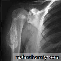
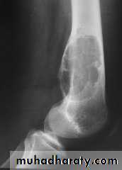
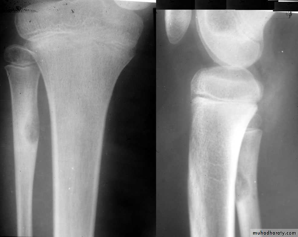
Osteochondroma
= exostosisCommonest benign bone tumour
It is bone outgrowth covered by cartilageHereditary multiple exostosis : AD
Malignant risk : 1% solitary
6% multipleIf continues to grow > 18 yr suspect malignancy
Rx : excision