
Urinary system imaging
lecture (1)
5
TH
stage
By
Dr. Firas Abdullah
Thiqar college of medicine

Aims of our lecture:
To know the different radiological techniques used in
urinary tract
To know different renal pathologies.
Urinary bladder diseases
Prostate and urethra disease
Scrotal and testicular disorders
Female genital organs imaging

I) Radiological techniques used in urinary
tract imaging:
Ultrasonography
Urography
CT scan
MRI
Radionuclide scanning
Special techniques:
o
Retrograde and antegrade pyelography
o
Voiding cystourethrogram (micturating cystogram) and videourodynamics
o
Urethrography
o
Renal arteriography.

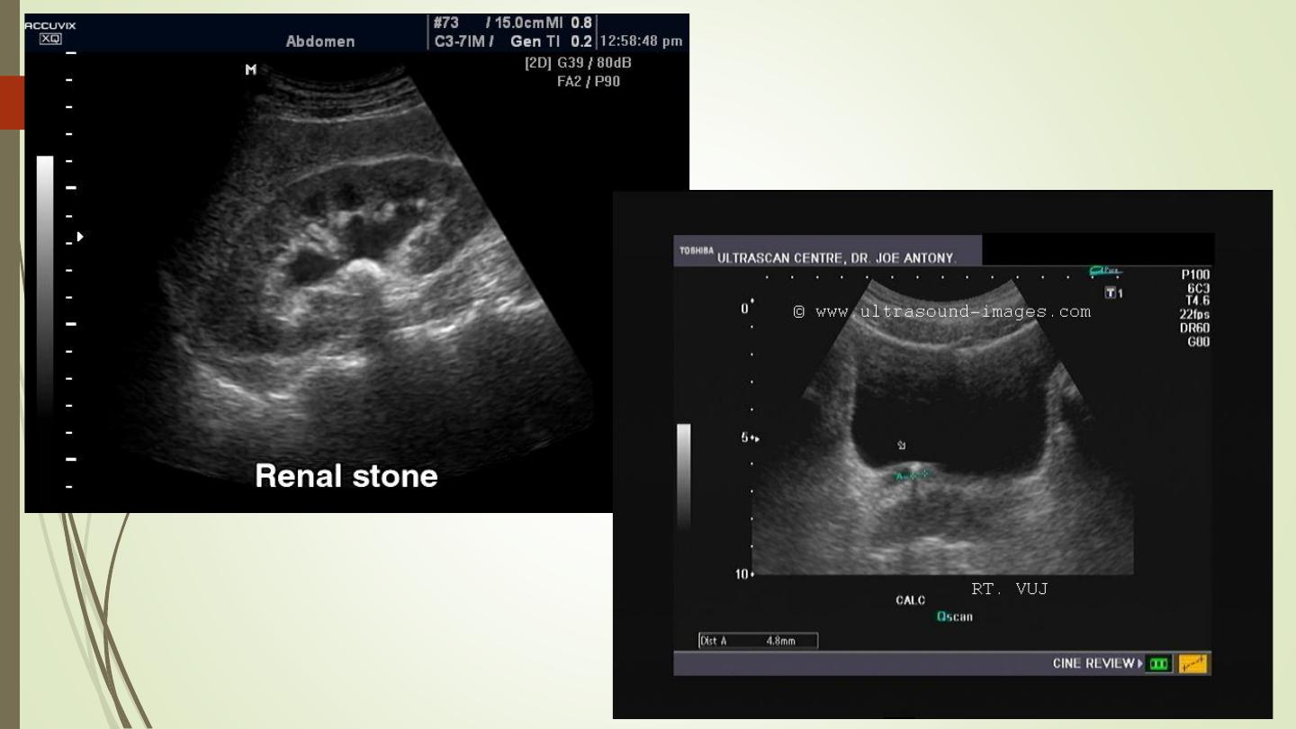
Ultrasonography:
Investigate patients with symptoms thought to arise
from the urinary tract.
Demonstrate the size of the kidneys and exclude
hydronephrosis in patients with renal failure.
Diagnose hydronephrosis, renal tumours, abscesses
and cysts including polycystic disease.
Assess and follow-up renal size and scarring in
children with urinary tract infections.
Assess the bladder and prostate.
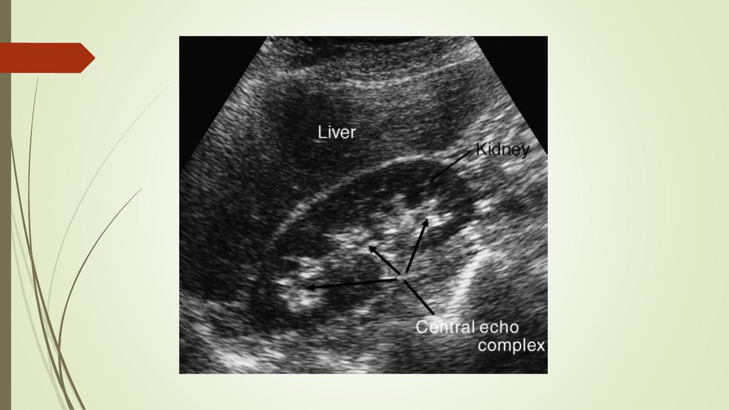

❑The normal adult renal length, measured by
ultrasound, is 9–12 cm.
❑Renal length varies with age, being maximal in
the young adult.
❑There may be a difference between the two
kidneys, normally less than 1.5 cm.
❑A kidney with a bifid collecting system is usually
1–2 cm larger than a kidney with a single
pelvicaliceal system.
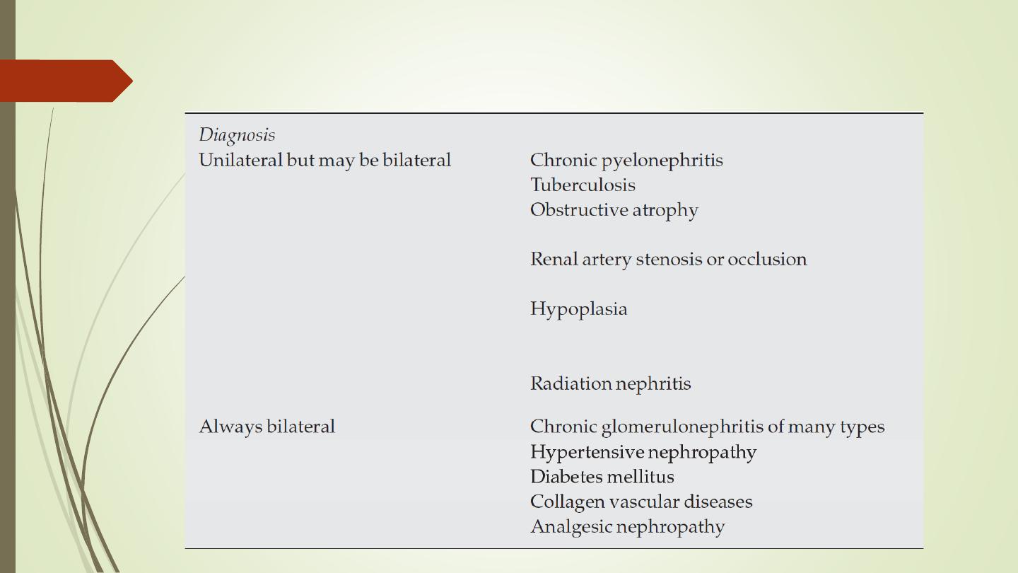
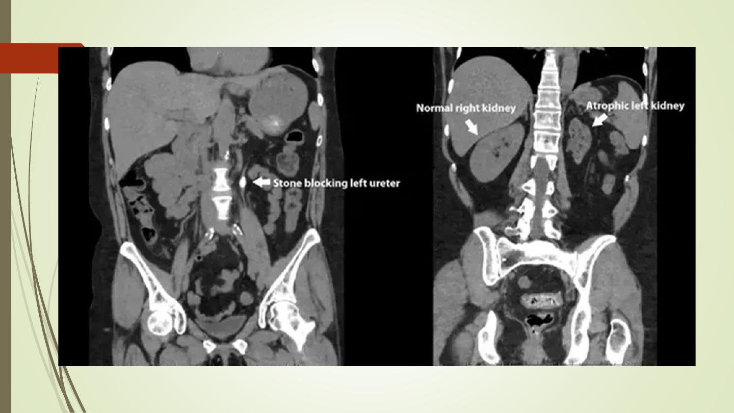
Causes of small kidney
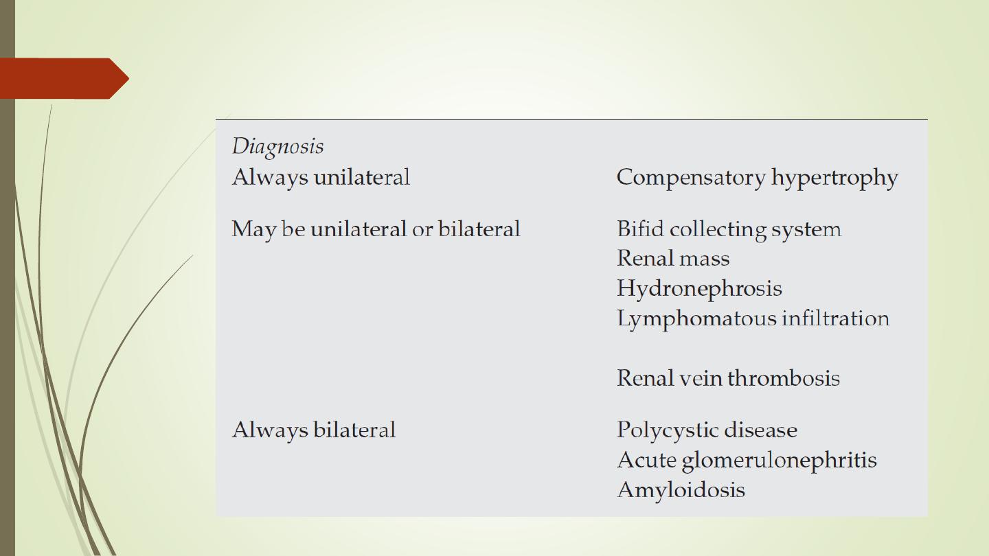
Causes of large kidneys

Urography:
Indications:
When detailed demonstration of the
pelvicaliceal system and ureters are required
Suspected ureteric injury, e.g. following pelvic
surgery or trauma
Assessment of acute ureteric colic

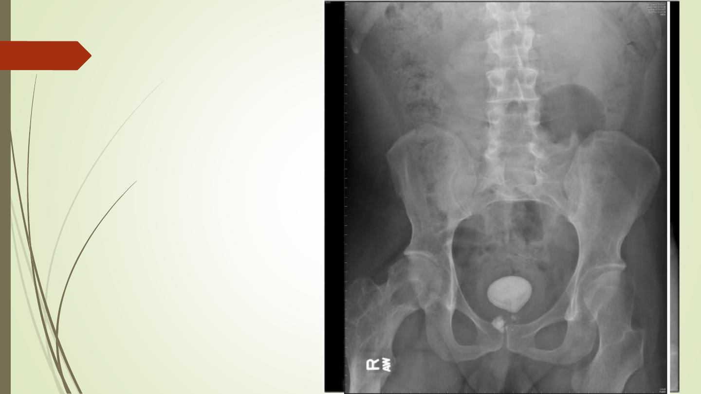
Intravenous (Excretory) Urography
Preliminary
Immediate
Compression
5 minutes
Release
Post -micturation
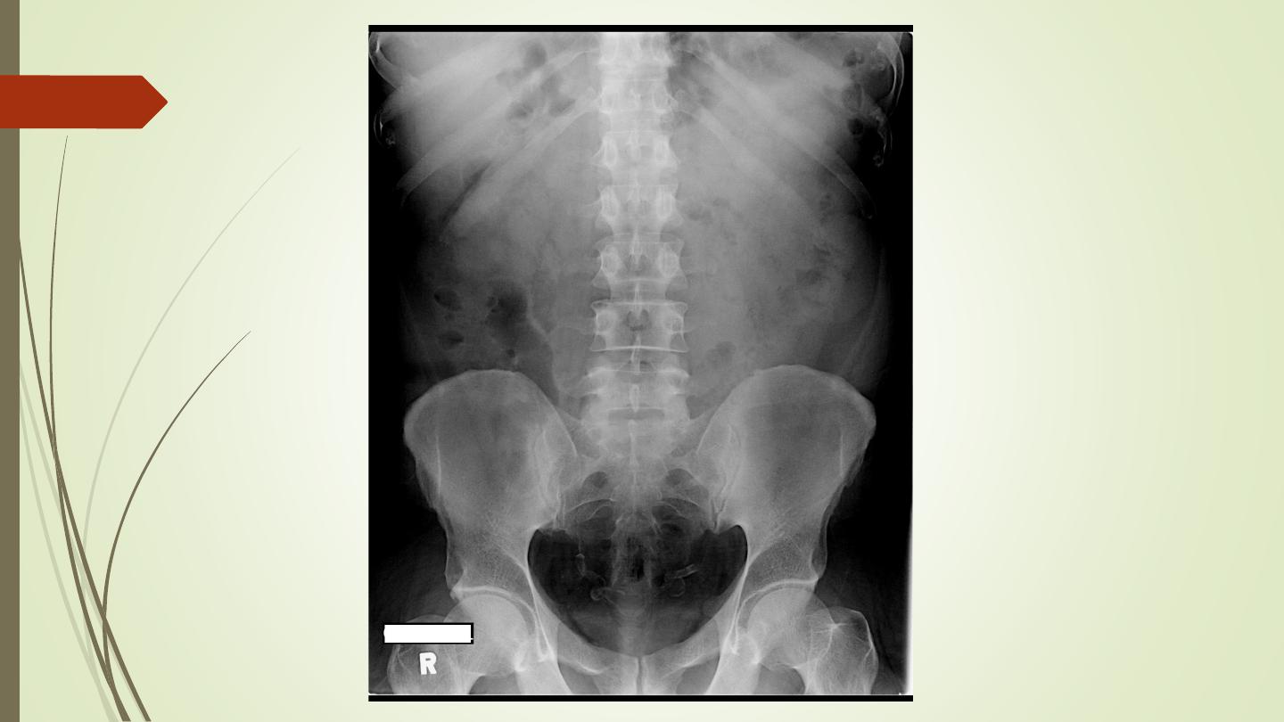
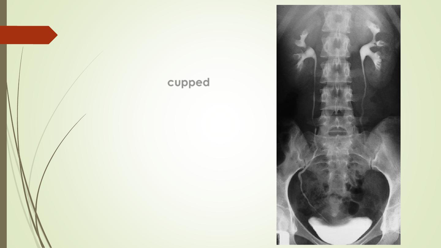
1. Check the Kidneys: outline, size, site
2. Check the calyces: cupped
3. Check renal pelvis and ureter
4. Check the bladder
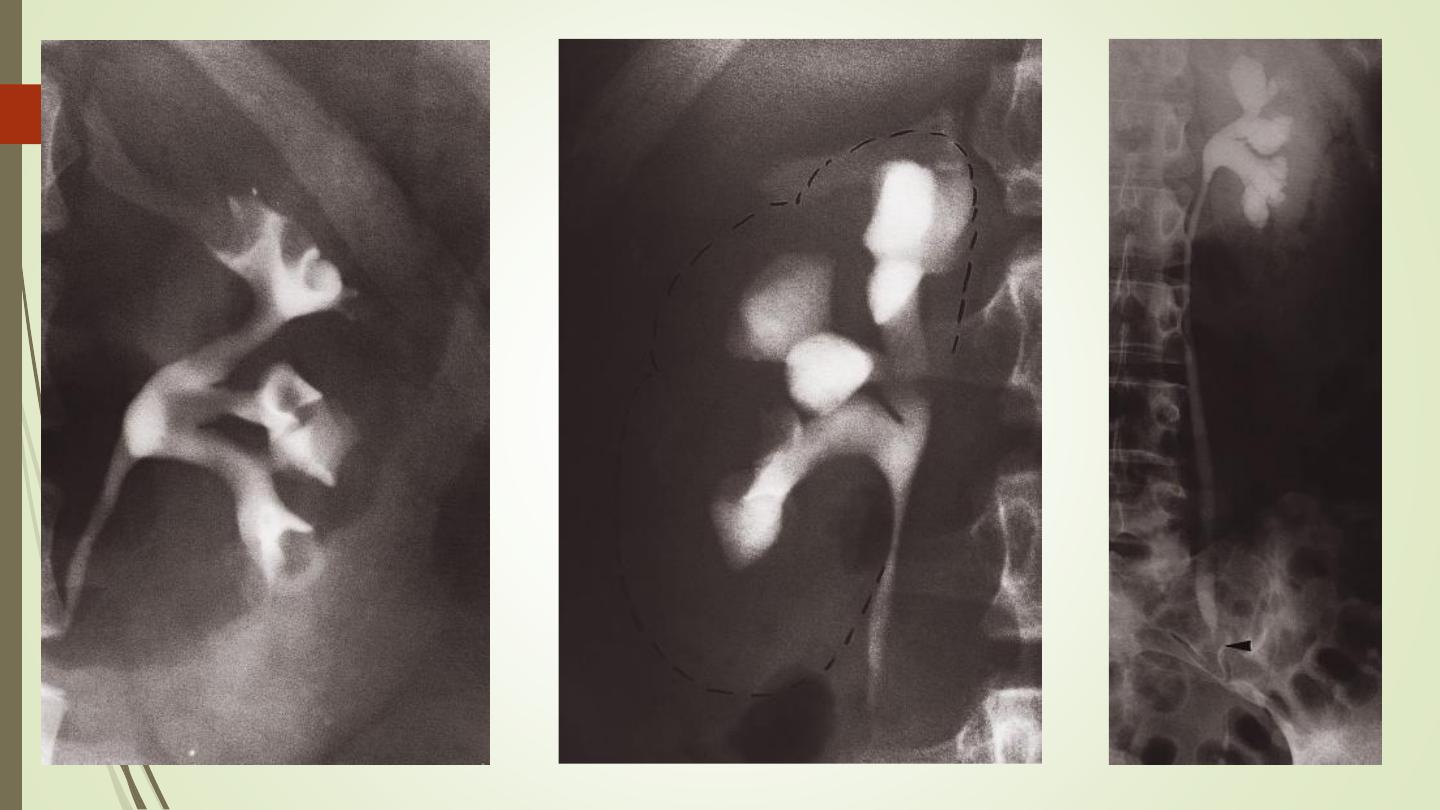

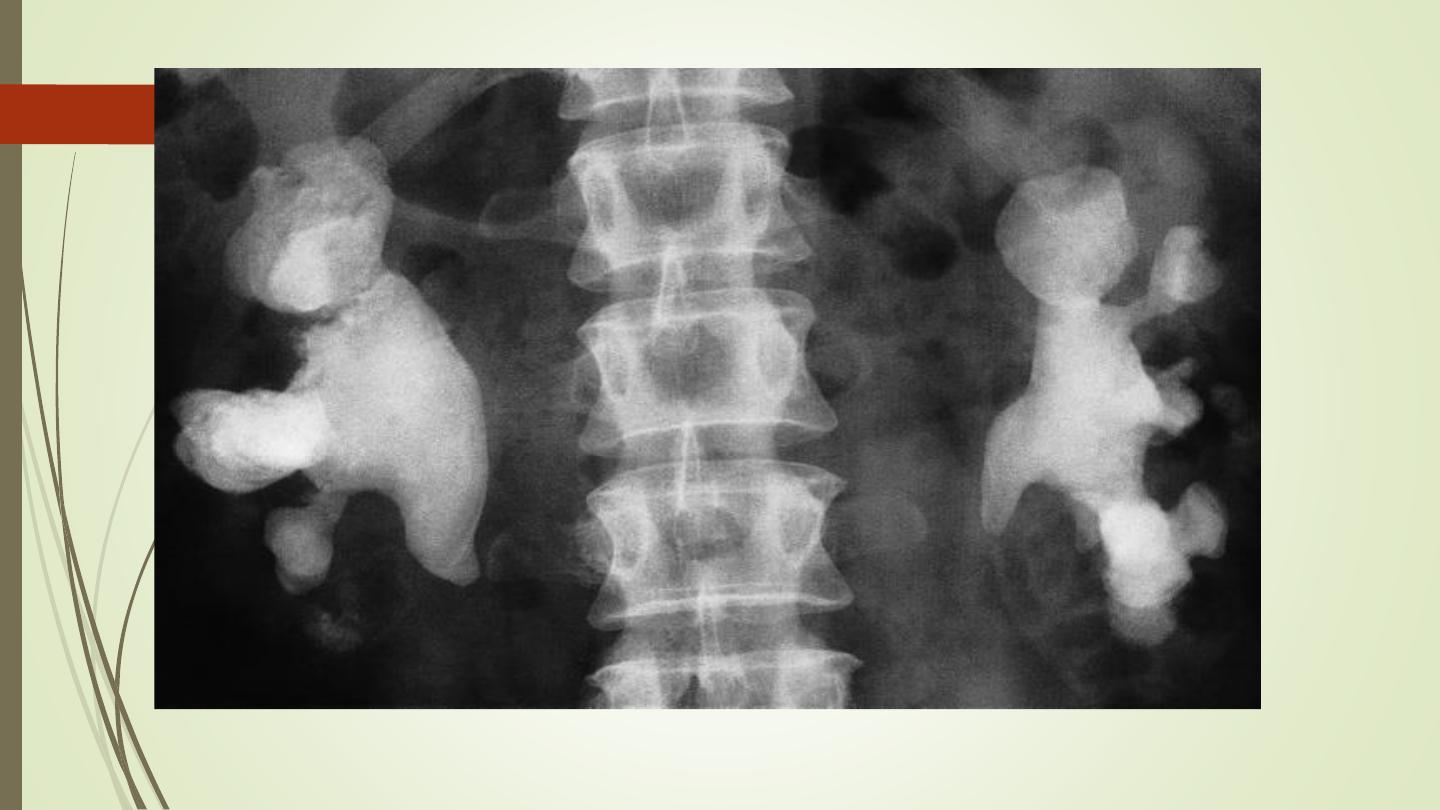
Causes of calyceal dilatation:
A: Due to obstruction
1.
Within the lumen:
I.
calculus
II.
blood clot
III.
sloughed papilla
2.
Within the wall of the collecting system
I.
intrinsic pelviureteric junction obstruction
II.
transitional cell tumour
III.
infective stricture (e.g. tuberculosis or schistosomiasis)
3.
Extrinsic compression
I.
retroperitoneal fibrosis
II.
pelvic tumour, e.g. cervical, ovarian or rectal carcinoma
III.
aberrant renal artery or retrocaval ureter
B) Due
to pap
i
llary atrophy or destruction:
1.
Reflux nephropathy
2.
Papillary necrosis
3.
Tuberculosis

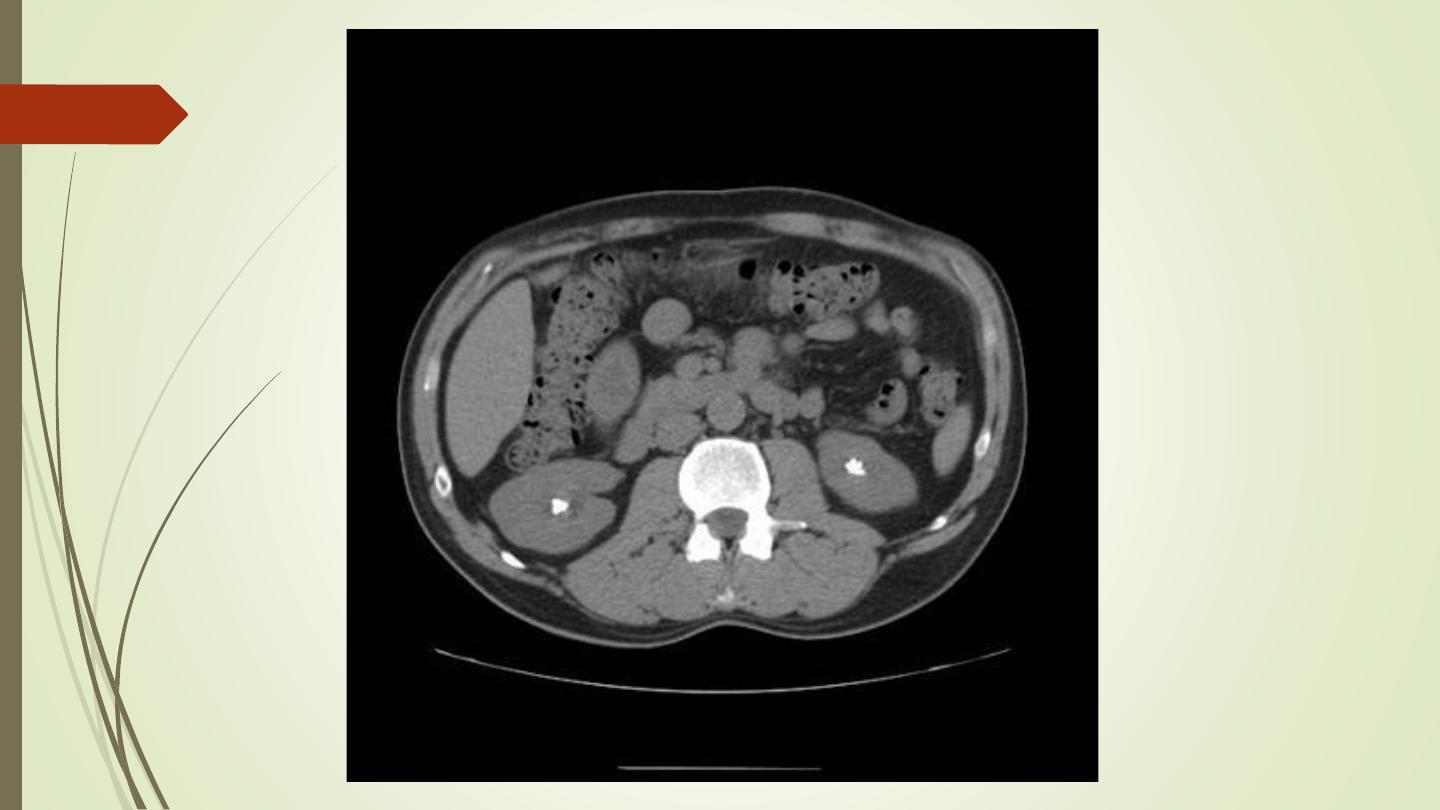
Indication of CT urography
Investigation of renal calculi
Investigation of haematuria
Characterization of a renal mass
Staging and follow-up of renal carcinoma
To delineate renal vascular anatomy (e.g.
suspected renal artery stenosis or prior to live
related kidney donation)
To diagnose or exclude renal trauma
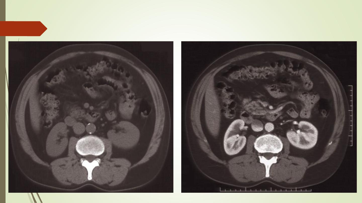
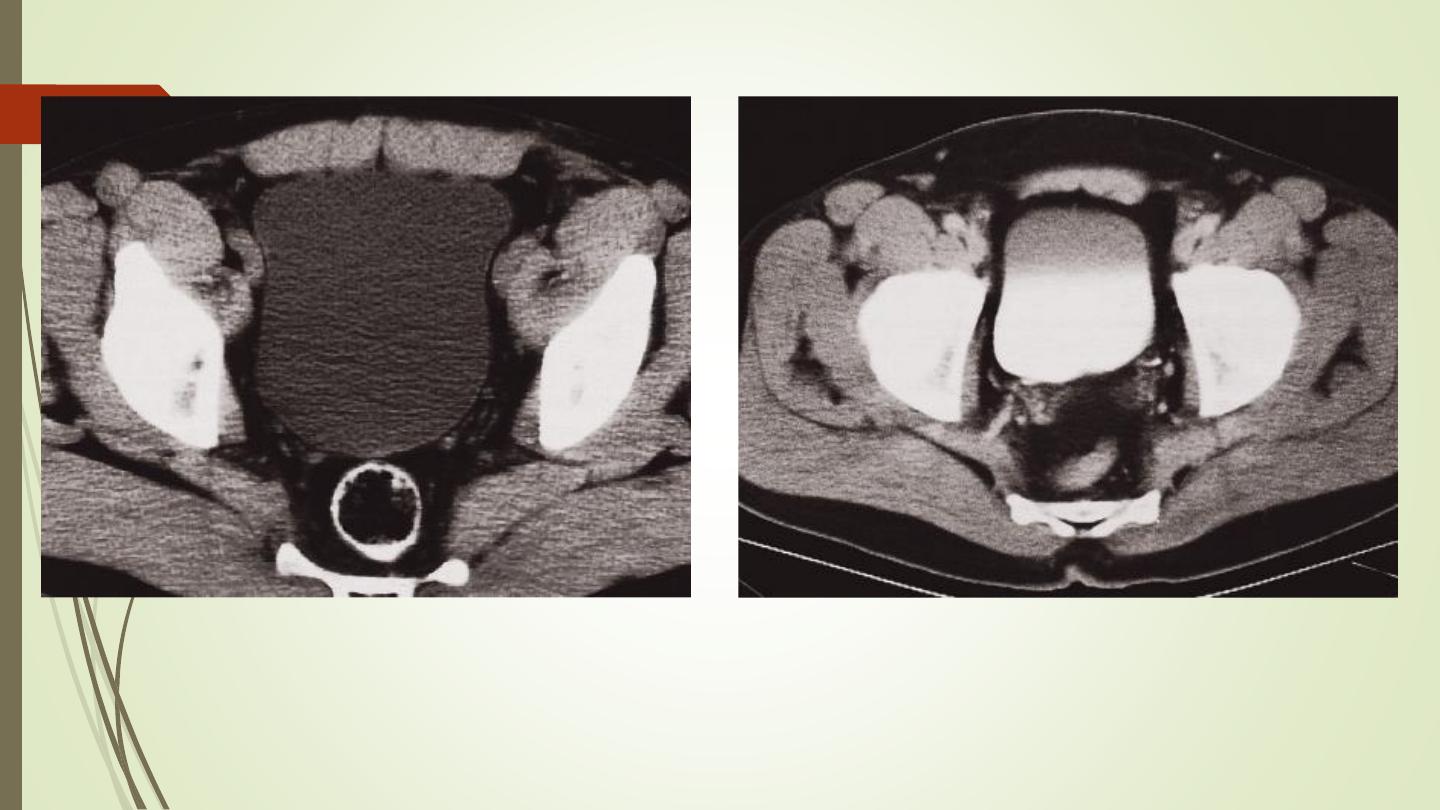
CT urography
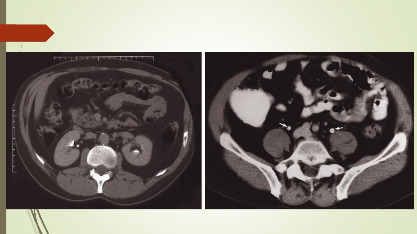
CT urography
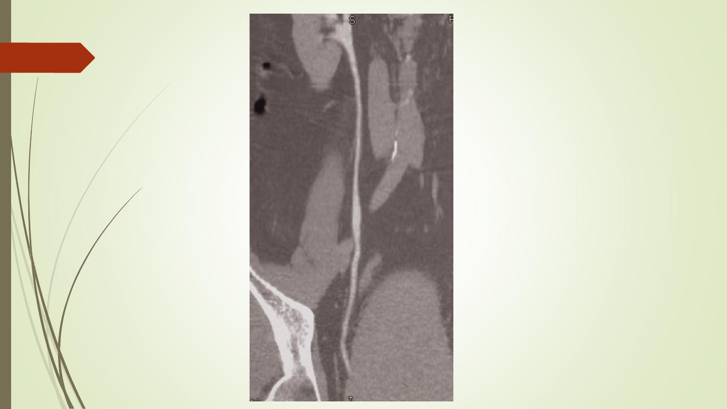
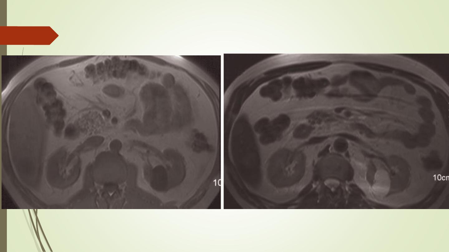
MRI
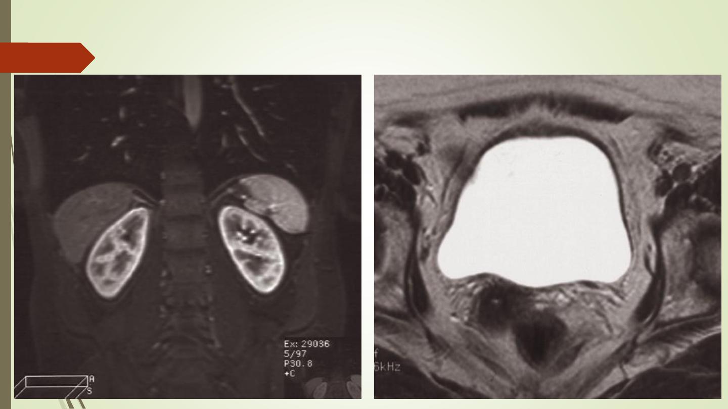
MRI
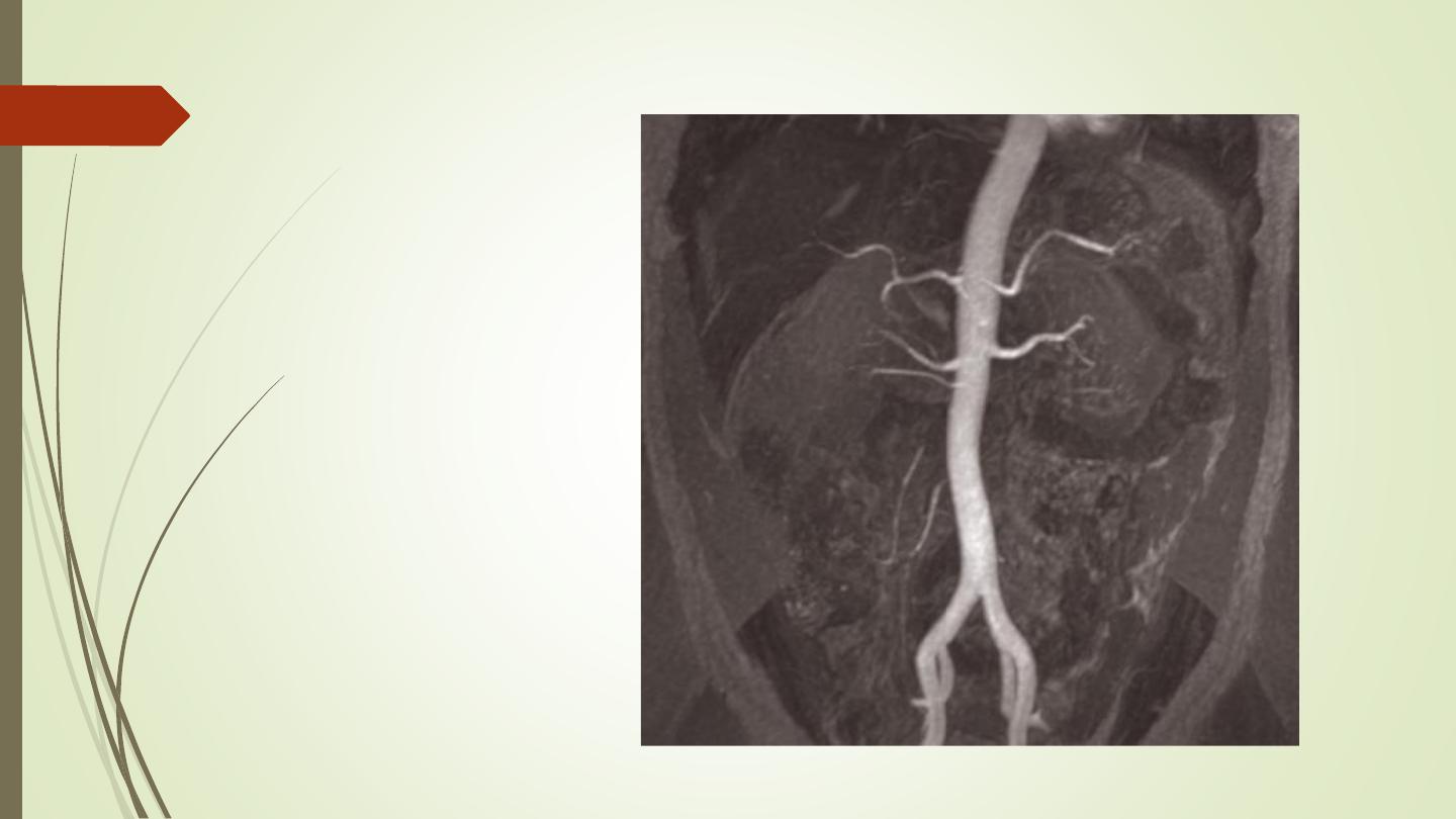
MRA
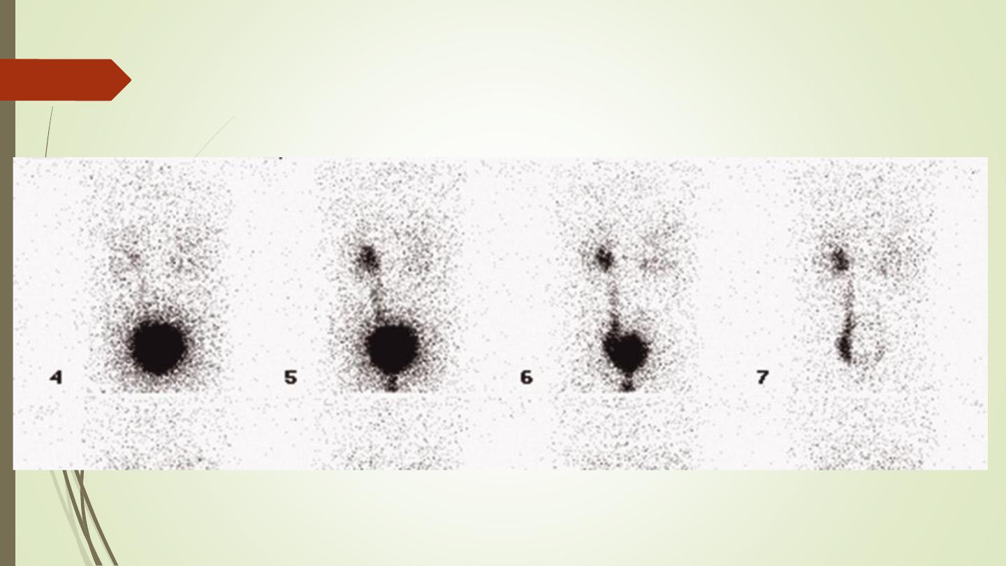
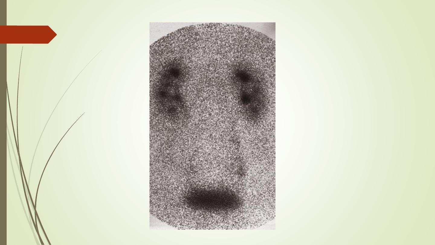
Radionuclide examination
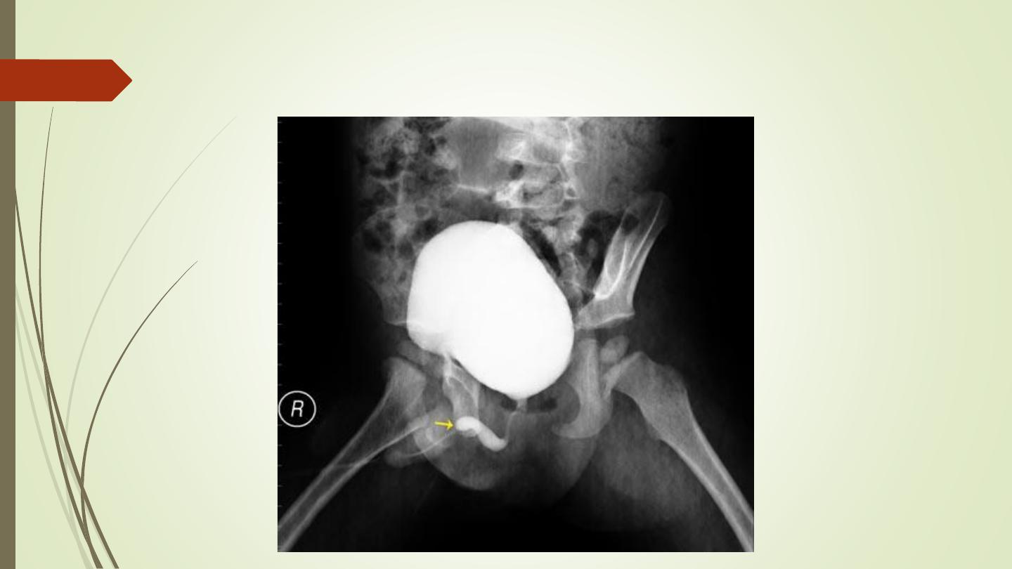
Voiding cystourethrogram (micturating cystogram)
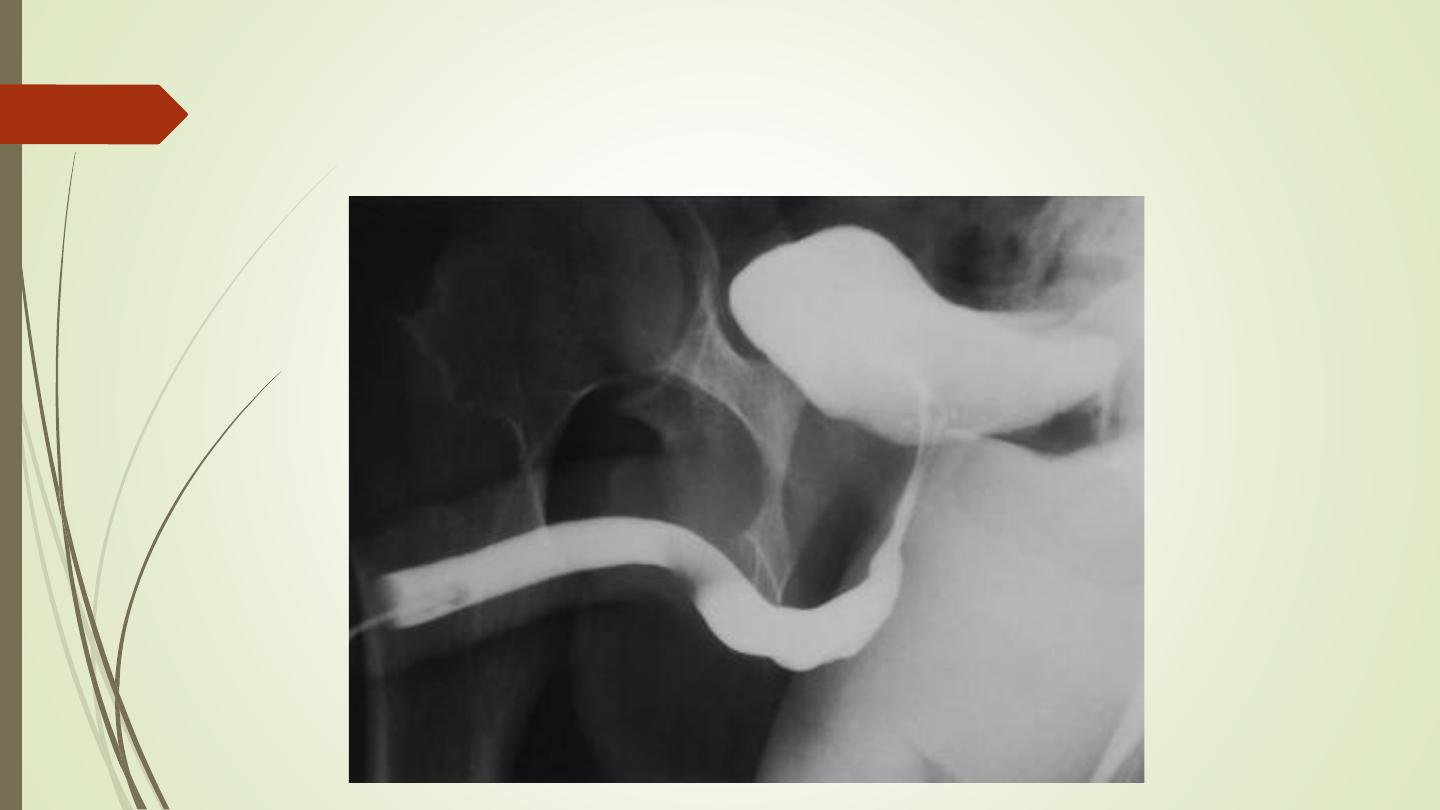
Retrograde urethrogram
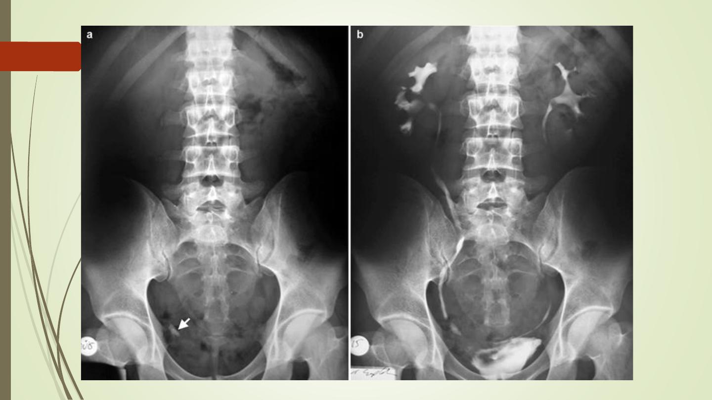
Urinary calculi

Nephrocalcinosis
Deposition of calcium salts in the
medulla or cortex of the kidney.

Medullary Nephrocalcinosis

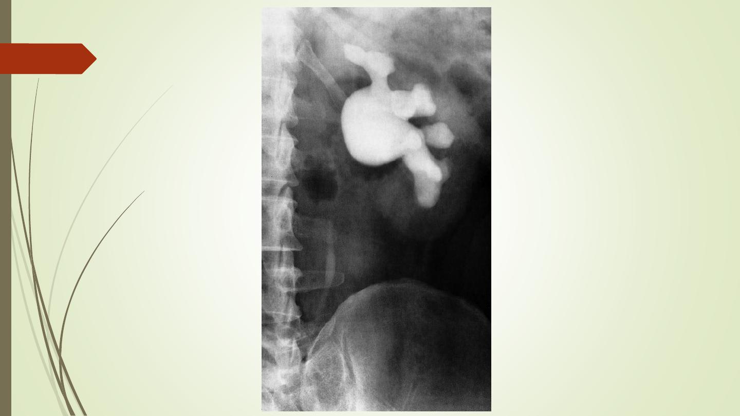
Congenital intrinsic pelviureteric junction (PUJ) obstruction
In this disorder, peristalsis is not transmitted across the
pelviureteric junction.
Childern and young adult
Dilatation of the pelvis and calices, with an abrupt
change in caliber at the pelviureteric junction
the ureter is either narrow or normal in size.

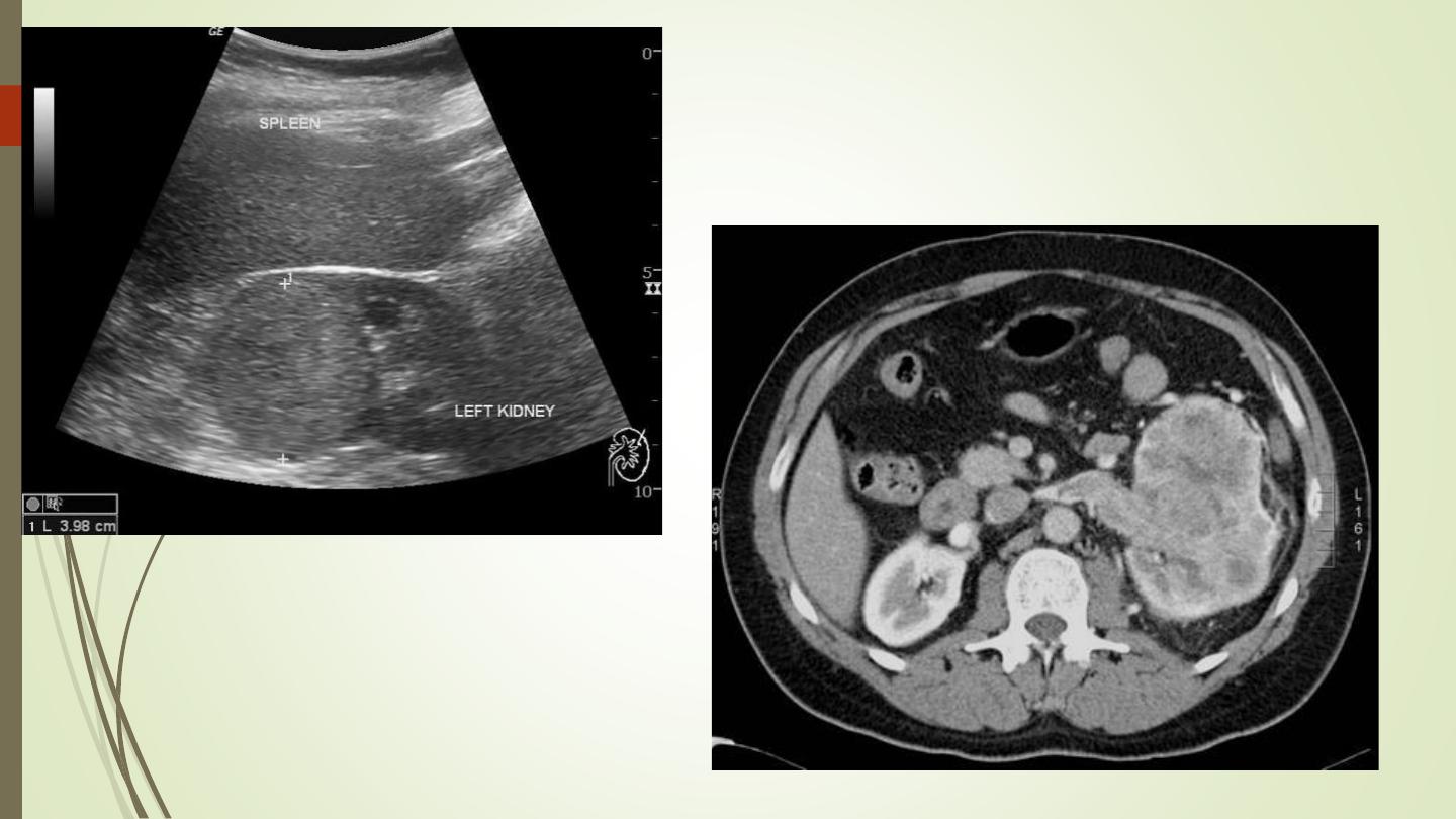
Renal parenchymal masses
In adults, the most common malignant tumour is
renal cell carcinoma, whereas in young children
the common neoplasm is Wilms’ tumour.
Other masses: renal abscess, benign tumour
(oncocytoma or angiomyolipoma), hydatid cyst,
and metastasis.
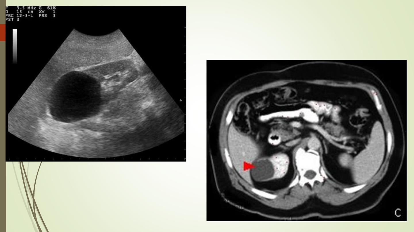
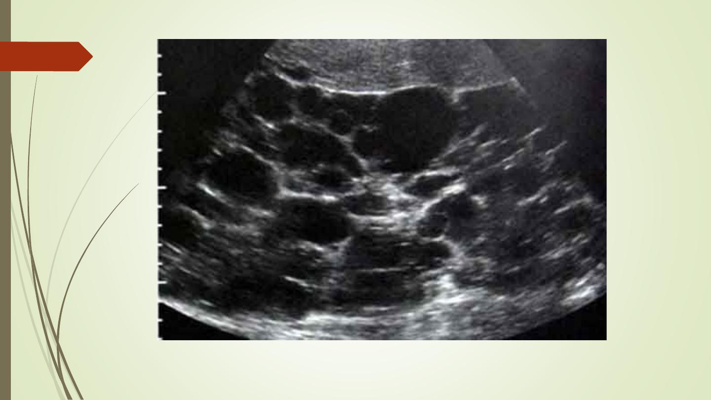
Renal cysts
‘renal pseudotumour’ or column of Bertin

Multiple renal masses include:
• Multiple simple cysts
• Polycystic disease
• Malignant lymphoma
• Metastases
• Inflammatory masses.

Thank you