
• Oral Histology
Your mouth under microscopeLecture one
Preparation of histological specimen
Assist. Lecturer
Anes A. AlshamaaOral histology
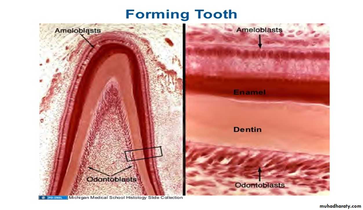
Branch of Human histology that describes the histological components of the teeth, periodontium, and the surrounding oral mucosa within the cellular and histological level (Under microscope).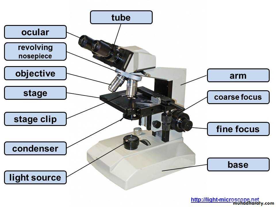
1. Light microscope
(compound microscope)Light microscope: a type of microscope uses visible light and magnification power of lens to observe specimens.
حضور
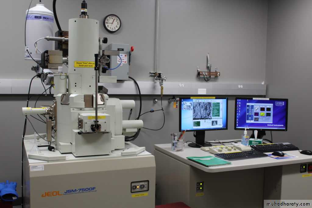
2. Electronic microscope
a type of microscope produces a beam of electrons that scan the surface of a specimen then electrons emitted from the specimen producing a 3D image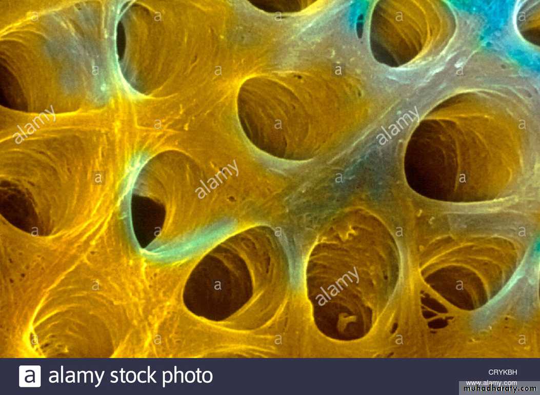
So we need to prepare histological slides to examine tissue under microscope
• How to Prepare a Slide
• In order to view the cellular components of a tissue, it has to be processed in a step• wise manner to produce a “slide” which can be viewed under a microscope.
• Observation
• Staining
• Mounting
• Sectioning
• Embedding
• Dehydration
• Fixation
Processing
Clearing
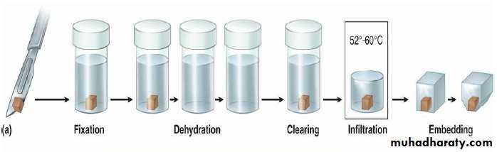
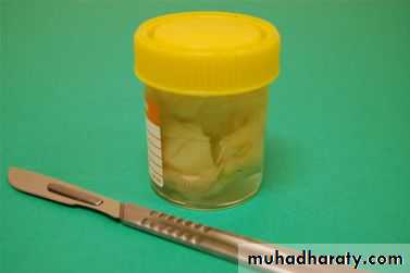
• (1) Fixation:
• •• Purpose: Maintenance of tissue structure
• proteins and inhibiting autolysis.
• cross
• linking
• Procedure:
• Takes 24 hours
• For L/M it is dipped in formalin
• E/M: is dipped in Glutraldehyde
• It is cut to smaller pieces
• A large specimen is received
• *Factors affect fixation:
• a-PH: The pH should be kept in the physiological range, between pH 4-9. If formalin is allowed to fall to a lower pH this can produce formalin pigments• b-Temperature: Affects the morphology of the tissue. It will increase the rate of penetration.
• c-Penetration of fixative: Fixatives penetrate the tissue at different rates. • The tissue is fixed starting at the periphery of the tissue and working inward toward the center of the tissue
• d-Volume of tissue: Ideal size of the tissue should be 3mm. Volume of fixative is at least 15 to 20 times greater than volume of tissue
• According to previous factors we can determine the concentration of fixative and fixation time. Prolonged fixation in aldehydes can cause shrinkage
• Types of fixative:
• Simple fixation: Formaldehyde (formalin), Glutaraldehyde,• .
• Compound fixation: Cold acetone, Ethanol, Zenker’s fluid.
• (2) Dehydration & Clearing:
• •• Purpose:
• To
• remove
• all
• the
• water
• because
• the
• Paraffin
• (embedding medium) is immiscible in water.
• •
• Dehydration: Solution is placed in increasing concentrations
• of alcohol beginning with 50% to 100%. Each step takes about
• 2 to 3 hours.
• •
• Clearing: This involves removing the alcohol and replacing it
• with a chemical that is miscible in both alcohol and paraffin.
• The chemical is Xylene solution which will now infiltrate the
• tissues.
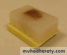
• (3) Embedding:
• •• Tissues are then placed in an oven containing
• that “Infiltrates” it.
• The high temperatures of 52-60⁰C evaporates
• A block of Paraffin obtained.
• L/M: uses Paraffin and plastic resins.
• E/M : uses resins as embedding medium.
• liquid Paraffin
• •
• •
• •
• •
• the Xylene.
• •
• Advantages of Paraffin: it stains reliably and is
• with
• easy to work
• •
• Disadvantages: slices cannot be cut very thinly.
• The properties of paraffin wax are improved for histological purposes by the inclusion of substances added alone or in combination to the wax:
• improve ribboning.
• increase hardness.
• decrease melting point.
• improve adhesion between specimen and wax.
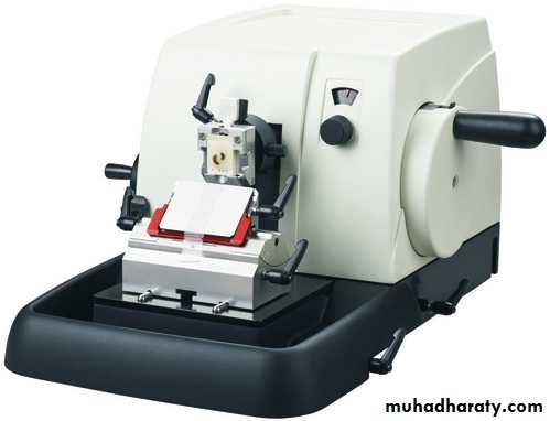
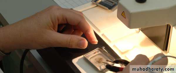
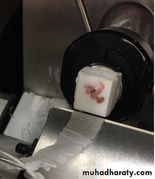
Microtome
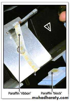
• (4) Sectioning:
• “Sectioning”• involves using a machine called a microtome that cuts sections very thinly in the form of ribbons . Sections are cut from these ribbons and mounted on to the glass slide.
• L/M: the sections are usually 10μm thick
• E/M: 0.02-0.1μm thick.
• The disadvantage of thick sections: Overlapping and therefore
• decreased resolution.
Types of sections for studying oral tissues
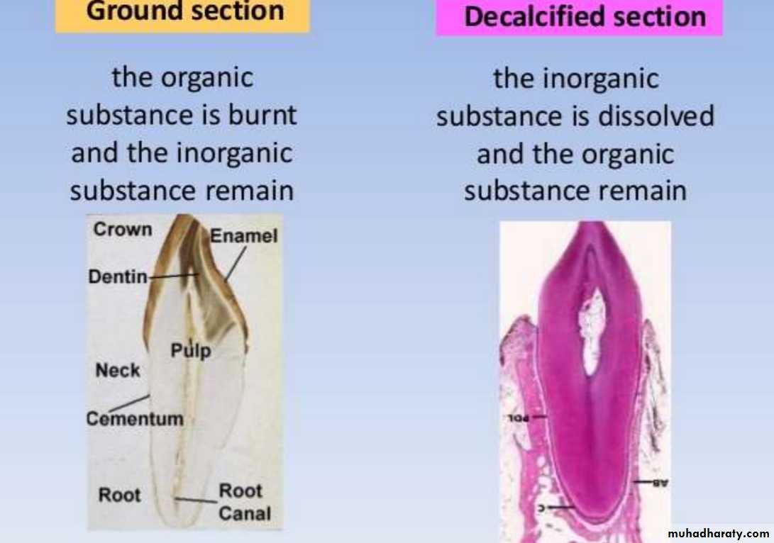
• Ground section: A section of bone or tooth prepared for histological study by polishing until thin enough for microscope viewing.
• Decalcified section: Decalcification softens the hard structure of tooth by removing the hard substance calcium. Also this technique is useful in analysing the soft tissue component of teeth, such as the pulp and periodontal ligament.
• Frozen section: is a pathological laboratory procedure to perform rapid microscopic analysis of a specimen. It is used most often in oncological surgery. The technical name for this procedure is cryoprotection.
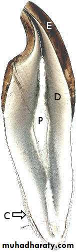
Ground section
PDL
CEMENTAlveolar bone
Frozen suction
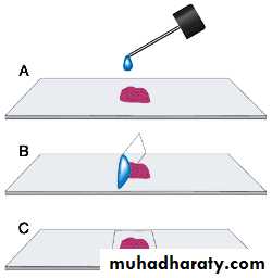
Mounting
• Mounting: the thin sections obtained from the microtome are mounted upon glass slides.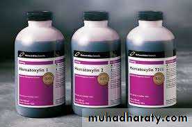
• (5) Staining:
• •• •
• Staining:
• The
• most
• common stains are Hematoxylin and Eosin.
• Hematoxylin:
• • Is a BASIC dye.
• •It colors the “Acid ic Components” of cells
• giving them a bluish tint.
• •Egs. Of structures stained by it are usually
• protein rich areas like Nuclei, RER. And also
• Extracellular matrix like the collagen matrix.
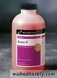
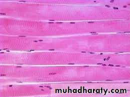
• Eosin:
• • Is an Acidic dye.
• •It colors the “Basic Comp on ents” of cells
• giving them a pinkish tint.
• •Egs. cytoplasm, Collagen fibres,
• Mitochondria, lysosomes, muscle, connective
• tissue, colloid, red blood cells.
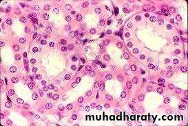
• Nuclei are
• stained with• hematoxylin
• Remaining
• cytoplasm stains
• with Eosin
• Nuclei are
• stained with
• hematoxylin
• Muscle fibers stained with Eosin
Observation