Lecture one: Introduction to General Histology
Histology is the microscopic study of various tissues of the body. A tissue is made
up of groups of cells performing the same function.
Body components
1- Cell: smallest living unit of organization in the body, since each cell is capable
of performing necessary functions by its living components:
Epithelial cell, neuron, myofibril, chondrocyte, osteocyte, fibroblast, erythrocyte,
macrophage.
2- Tissue: collection of similarly (form and function) specialized cells are grouped
together:
Epithelium, nervous tissue, muscle, cartilage, bone, connective tissue, blood.
3- Organ: various tissue types are bounded together to form independent body part
that performs a specific function.
Skin, brain, heart, liver.
4- System: many organs are functioning together.
CNS, RS, IS, CVS.?
Cells:
Each cell has a cell membrane, cytoplasm, organelles, and inclusions. Cell
membrane surrounds the cell and acts as a barrier between the tissue fluid
(extracellular fluid) and the cytoplasm (intracellular fluid and organelles).
Cytoplasm consists of a cytoplasmic matrix with organelles and inclusions
suspended in it. Organelles are living units while the inclusions are non-living
entities. The major cytoplasmic organelles are the endoplasmic reticulum, Golgi
apparatus, mitochondria, lysosomes, ribosomes and cytoskeleton.
Inclusions: lipids, glycogen (energy sources) and melanin (pigment source). Also
Many of inclusions include bodies, which are spent lysosomes and their digested
material.
Intercellular junctions
Mechanical attachments formed between cells, and also between cells and adjacent
noncellular surfaces. All intercellular junctions involve attachment device, which
includes an attachment plaques and tonofilaments.
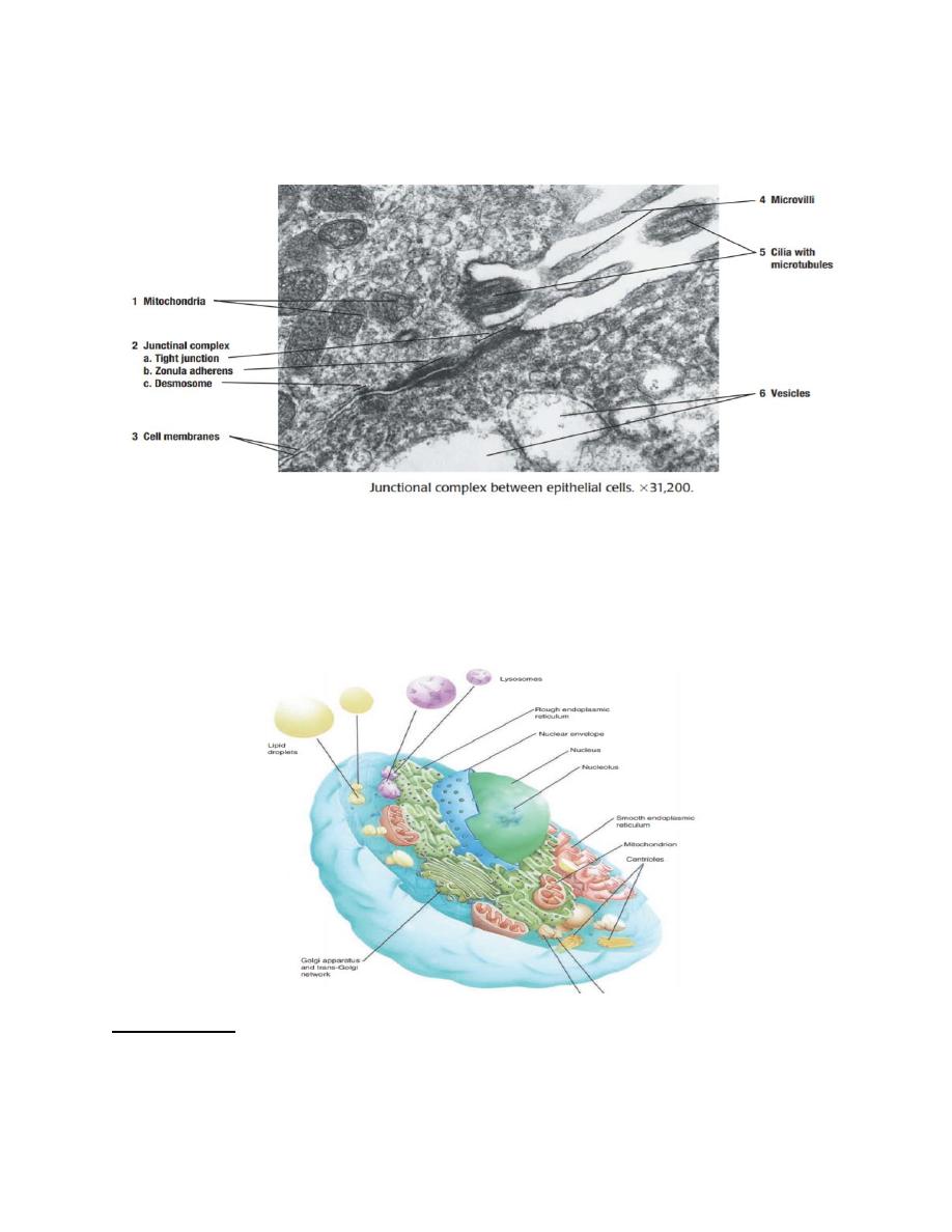
An intercellular junction between cells is formed by desmosome, while attachment
of a cell to an adjacent noncellular surface is formed by hemidesmosome.
These are characterised by local thickening of the adjacent cell membrane of the two
cells. Desmosomes are bands present below and parallel to the tight junctions. These
strongly anchor one cell to the next one. Hemidesmosomes are present at the basal
border of the cells. These help to attach the cells firmly to the underlying basement
membrane.
Basic Tissues
Tissue types are categorized according to four basic histological types includes
epithelial, connective, muscle, and nerve tissue.
Tissue
Types
Subcategories
Epithelium
Simple
Squamous, cuboidal, columnar,
pseudostratified
Stratified
Squamous (keratinized, nonkeratinized)
Cuboidal, columnar, transitional.
Connective tissue
Solid soft
Connective tissue proper, specialized
Adipose, fibrous, elastic, reticular
Solid firm
Cartilage
Sold rigid
Bone
Fluid
Blood, lymph
Muscle
Involuntary Smooth , cardiac
Voluntary
Skeletal
Nerve
Afferent
Sensory
Efferent
Motor
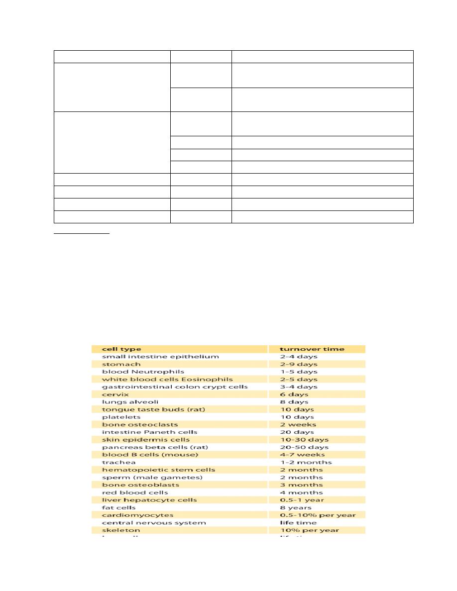
Regeneration refers both to the regular and repeated renewal of a particular structure
or tissue throughout the life of an organism, that is, the cellular renewal that occurs
during normal aging (also called tissue homeostasis or physiological regeneration),
as well as to restoration of injured tissue or lost body. Epithelial proliferation is the
term given to the ability of any epithelium to be able to renew itself after cells die,
that is, the ability to turnover cells. This occurs in the stratum basale, which is
situated on deepest layer of cells in thin epithelia (e.g. the bottom of the mouth), and
in the lower two to three cells layers of thicker epithelia (e.g. buccal mucosa or inside
of the cheek).

Lecture two: Epithelium
Epithelium (plural, epithelia) consists of sheets of cells that cover the external
surfaces of the body, line the internal cavities, form various organs and glands, and
line their ducts. Epithelial cells are in contact with each other, either in a single layer
or multiple layers. The structure of lining epithelium, however, differs from organ
to organ, depending on its location and function. For example, epithelium that covers
the outer surfaces of the body and serves as a protective layer differs from the
epithelium that lines the internal organs.
Major Features
• Classification based on number of cell layers and cell morphology
• Basement membrane separates epithelium from connective tissue
• All epithelia are nonvascular; delivery of nutrients to cells and removal of
metabolic waste occurs via diffusion
• Surface modifications include motile cilia, microvillli, and stereocilia
FUNCTIONS OF EPITHELIAL TISSUE/EPITHELIUM
1-Protective: The stratified squamous keratinized epithelium of skin offers
mechanical protection including conservation of moisture.
2-Secretory: The glands which are derivatives of the epithelium secrete
useful chemical substances.
3-Absorptive: Epithelia of small intestine and of proximal convoluted
tubules of kidney are modified to specialize in absorptive functions.
4-Excretory: Epithelium of distal convoluted tubules and collecting ducts
of kidney function as excretory organs.
5-Sensory: The rods and cones of retina and hair cells of olfactory mucous
membrane are specialized sensory cells.
Importantly, for dental professionals, both the epithelium of the skin and oral mucosa
are of similar ectodermal origin. In comparison, those lining the respiratory and
digestive tract are of endodermal origin, and those lining the urinary tract are derived
from mesoderm.

Types of Epithelia
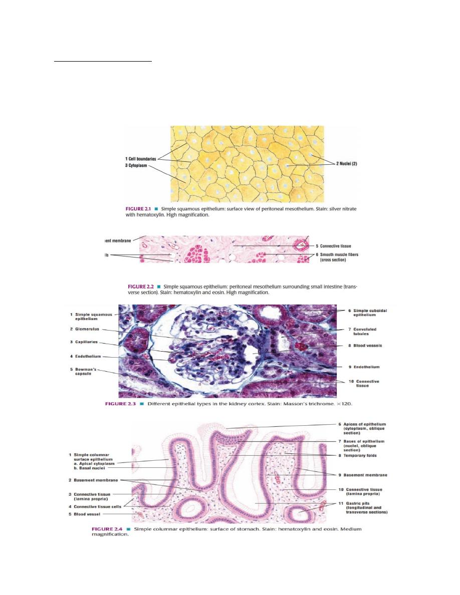
Simple Epithelium
Simple squamous epithelium that covers the external surfaces of the digestive
organs, lungs, and heart is called mesothelium. Simple squamous epithelium that
covers the lumina of the heart chambers, blood vessles, and lymphatic vessels is
called endothelium.
Simple cuboidal epithelium lines small excretory ducts in different organs. In the
proximal convoluted tubules of the kidney, the apical surfaces of the simple cuboidal
epithelium are lined with a brush border consisting of microvilli.
Simple columnar epithelium covers the digestive organs (stomach, small and large
intestines, and gallbladder). In the small intestine, simple columnar absorptive cells
that cover the villi also exhibit microvilli. Villi are fingerlike structures that project
into the lumen of the small intestine. In the female reproductive tract, the simple
columnar epithelium is lined with motile cilia.
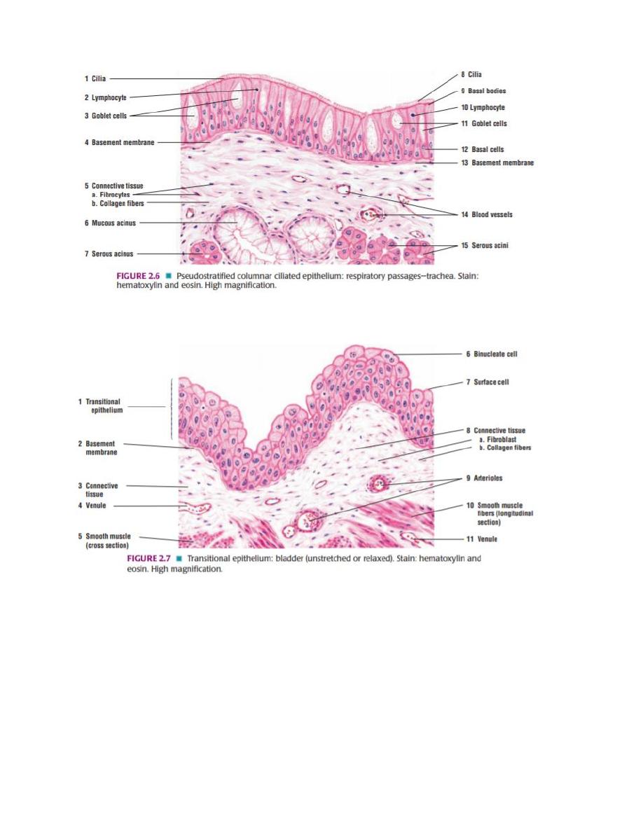
Pseudostratified Columnar Epithelium
Pseudostratified columnar epithelium lines the respiratory passages and lumina of
the epididymis and vas deferens. In trachea, bronchi, and larger brochioles, the
surface cells exhibit motile cilia; in the epididymis and vas deferens, the surface cells
exhibit nonmotile stereocilia, which are branched or modified microvilli.
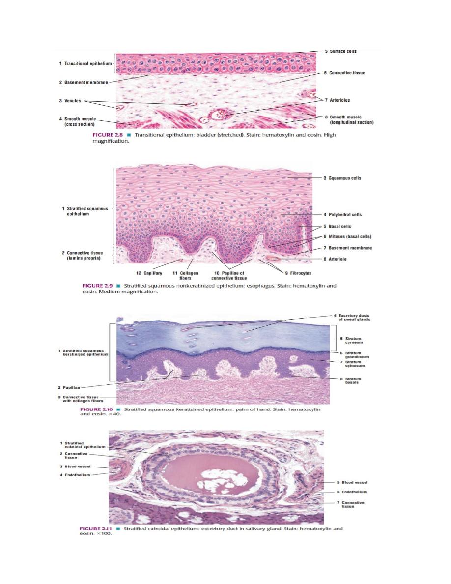
Stratified Epithelium
Stratified squamous epithelium contains multiple cell layers. The basal cells are
cuboidal to columnar; these cells give rise to cells that migrate toward the surface
and become squamous. There are two types of stratified squamous epithelia:
nonkeratinized and keratinized.
Nonkeratinized epithelium exhibits live surface cells and covers moist cavities such
as the mouth, pharynx, esophagus, vagina, and anal canal. Keratinized epithelium
lines the external surfaces of the body. The surface layers contain nonliving,
keratinized cells that are filled with the protein keratin. The exposed epithelium that
covers the palms and soles exhibits especially thick layers of keratinized cells.
Stratified cuboidal epithelium and stratified columnar epithelium have a limited
distribution in the body. Both types of epithelia line the larger excretory ducts of the
pancreas, salivary glands, and sweat glands. In these ducts, the epithelium exhibits
two or more layers of cells.
Transitional epithelium lines the minor and major calyxes, pelvis, ureter, and bladder
of the urinary system. This type of epithelium changes shape and can resemble either
stratified squamous or stratified cuboidal epithelia, depending on whether it is
stretched or contracted. When transitional epithelium is contracted, the surface cells
appear dome-shaped; when stretched, the epithelium appears squamous.