Lecture 1: Introduction to General Histology Anes Adnan
Histology is a microscopic study of various tissues of the body.
Body components
1- Cell: smallest living unit of organization in the body, since each cell is capable of performing necessary functions by its living components: Epithelial cell, neuron, myofibril, chondrocyte, osteocyte, fibroblast, erythrocyte, macrophage.
2- Tissue: collection of similarly (form and function) specialized cells are grouped together: Epithelium, nervous tissue, muscle, cartilage, bone, connective tissue, blood.
3- Organ: various tissue types are bounded together to form independent body part that performs a specific function. Skin, brain, heart, liver.
4- System: many organs are functioning together. CNS, RS, IS, CVS.
Tissues are composed of: Cells and Extracellular matrix
Organs are composed of: Parenchyma (the cells that perform the main function of organ) and Stroma (supporting tissue)
Basic Tissues
Tissue types are categorized according to four basic histological types includes epithelial, connective, muscle, and nerve tissue.
Tissue
Types
Subcategories
Epithelium
Simple
Squamous, cuboidal, columnar, pseudostratified
Stratified
Squamous (keratinized, nonkeratinized)Cuboidal, columnar, transitional.
Connective tissue
Solid soft
Connective tissue proper, specialized
Adipose, fibrous, elastic, reticular
Solid firm
Cartilage
Sold rigid
BoneFluid
Blood, lymphMuscle
Involuntary
Smooth , cardiac
Voluntary
SkeletalNerve
Afferent
Sensory
Efferent
MotorLecture 2: Respiratory system Anes Alshamaa
The complex of organs and tissue which are necessary to exchange blood carbon dioxide (CO2) with air oxygen (O2).
Organs of respiration are nose, nasal cavity, posterior nares including the paranasal air sinuses. Nasopharynx Larynx Trachea Pleura, lungs including the bronchial tree.

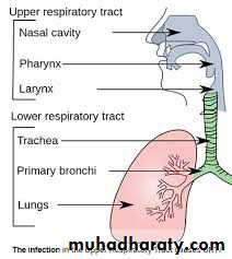
The respiratory system is divided into two parts:Upper respiratory tract: nose, mouth, and the beginning of the trachea.
Lower respiratory tract: trachea, the bronchi, broncheoli and the lungs.
The trachea: It connecting the throat to the bronchi.
The bronchi: It divides into two bronchi (tubes).
The broncheoli: the bronchi branch off into smaller tubes called broncheoli which end in the pulmonary alveolus.
The Lungs: The structure of the lungs includes the bronchial tree – air tubes branching off from the bronchi into smaller and smaller air tubes, each one ending in a pulmonary alveolus.
Functionally the respiratory system is divided into:
Conducting part which comprises nose, nasopharynx, larynx, trachea and bronchial tree till the level of terminal bronchioles.
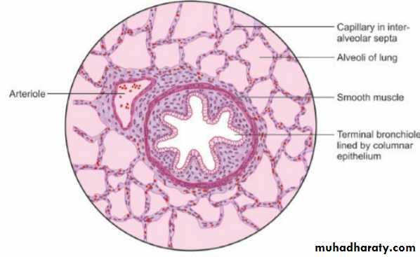
Respiratory part comprising of respiratory bronchioles, alveolar duct, atria alveolar sac and alveoli. These are present in the spongy part of lung for exchange of gases.
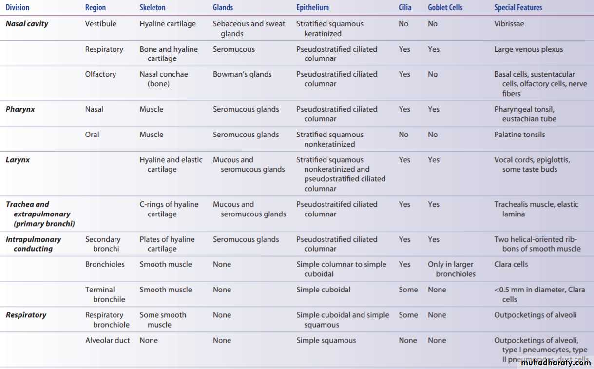
Histology of respiratory system
NOSE
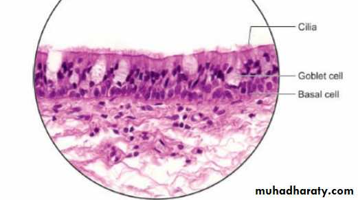
The nasal cavities are lined by a ciliated pseudostratified columnar epithelium containing the cell bodies of bipolar nerve (olfactory) cells. of these olfactory cells contain proteins that act as odorant receptors.
Functions
The cilia of the columnar cells carry foreign particles towards oropharynx to be swallowed. Goblet cells produce mucus to trap foreign particles. The cells in lamina propria provide necessary immunity.
Clinical considerations
The nose, nasopharynx get attacked by various bacteria pollutants and allergans causing sinusitis, and tonsillitis. The venous plexus may rupture due to picking/heat, leading to epistaxis (bleeding from nose).
Nasopharynx
It is situated between the posterior nares of nose and nasopharynx. It is lined by pseudo-stratified ciliated columnar epithelium with goblet cells.
Oropharynx
It contains mucous glands and it is lined by non-keratinized stratified squamous epithelium, that is continuous with this type of epithelium at the proximal end of the larynx.
Larynx
It is made up of number of cartilages. Most of the larynx is lined by pseudostratified ciliated columnar epithelium interspersed with goblet cells.
Trachea
The trachea is lined by pseudostratified ciliated columnar epithelium with interspersed goblet cells resting on a basement membrane. The lamina propria consists of elastic fibres, lymphocytes and short ducts of the glands.
Clinical considerations
Tracheitis is the viral or bacterial infection of trachea. This is characterized by acute inflammation of mucous membrane causing tissue congestion and profuse secretion of watery fluid.
BRONCHIAL TUBES
Intrapulmonary bronchus is lined by pseudostratified ciliated columnar epithelium with goblet cells resting on a thin basement membrane. Cilia prevent the accumulation of mucus in the bronchial tree. The lamina propria consists of reticular and elastic fibres. The submucous coat contains both mucous and serous acini. Outermost is the hyaline cartilage which is visible as small cartilaginous plates of varying sizes and shapes with tunica adventitia.
TERMINAL BRONCHIOLE
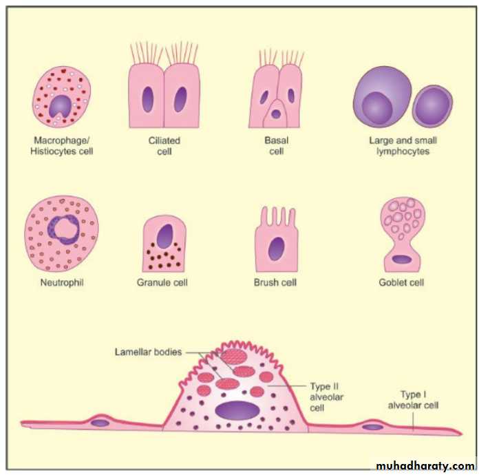
There are following distinct epithelial cell types in the conducting airways. These are:
Ciliated columnar cells: These cells form the force of mucociliary current in the bronchial tree. The 300 cilia project from the apical part of the cell.
Goblet cells: These cells are present from trachea to the smallest bronchi, excluding the bronchioles. In the smokers, the goblet cells increase in number and extend into the bronchioles as well.???? Clinical consedration!!
Clara cells: These are non-ciliated cuboidal cells, bulging into the lumen. These contain secretory granules and lysosomes. These cells may regulate in transport.
Basal cells: These cells are seen in the airways lined by pseudostratified epithelium. These are mitotic stem cells.
Brush cells: These are delicate non-ciliated cells with long apical microvilli, which are stiff in nature. These have sensory receptor function.
Neuroendocrine cells: These are rounded cells and form part of neuroendocrine system of amine precursor uptake and decaboxylation (APUD) cells. These cells are maximum in fetal lungs and their number decreases after birth.
Lymphocytes: Lymphocytes derived from thymus (T lymphocytes) are present. These are concerned with immune mechanism of the respiratory system.
Mast cells: These are present in the basal region of the epithelium. Their secretory granules release histamine in response to irritants.
Columnar cells: These line the terminal bronchiole.
RESPIRATORY PART RESPIRATORY BRONCHIOLE
The pseudostratified ciliated columnar epithelium of the trachea and bronchi gives way to a simple columnar ciliated epithelium in the bronchioles and then to the simple squamous epithelium of the alveolar ducts and alveoli.
The ciliated cells undergo a gradual reduction in height from trachea to terminal and respiratory bronchiole.
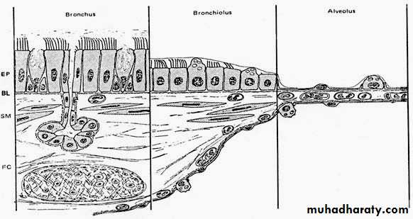
Change of airway wall structure at three principal levels in the lung. The epithelium (EP) gradually reduces from pseudostratified to cuboidal and then to squamous, but retains its organization as a mosaic of lining and secretory cells. The smooth-muscle layer (SM) disappears in the alveoli.
Histology of the Alveolar Region (Five major cells)
1. Alveolar Type I Cell (Squamous alveolar epithelial cell)These elongated thin cells line the alveoli and cover a large surface area (approximately 95% of the alveolar surface)
2.Alveolar Type II Cell (Great alveolar cell, granular pneumocyte): These cells form tight junctions with Type 1, and positioned at alveolar septal junctions.
3. Capillary Endothelial cell: The pulmonary capillary bed is the largest vascular bed in the body. It receives the entire cardiac output.
4. Alveolar macrophages
These large are located in the aqueous hypophase of the surfactant layer, they move over the alveolar surface ingesting microorganisms and inhaled particulate matter.5. Interstitial cells a progenitor cell: Interstitial cells of the alveolar region are fibroblasts with ramified cytoplasmic extensions.
Clinical consideration
Asthma: Mucous membrane and muscle layers of bronchi are thickened and the mucous glands enlarged causing reduced airflow in the lower respiratory tract.
Emphysema Decreased elasticity of the lungs due to the disruption of elastic tissue. large air sacs will be formed due to breakdown of the walls between the alveoli.
Pneumonia: The disease of lungs caused by inhaled or blood borne microbes. watery inflammatory exudates accumulate and fill up many alveoli in a lobule.
Reflexes associated with Respiratory System
Cough reflex: Swallowing reflex: Sneezing refleX
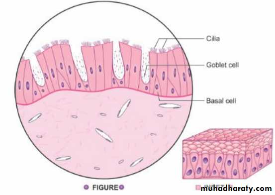
Pseudostratified columnar ciliated epithelium: Trachea. Stain: Haematoxylin-eosin, 400X
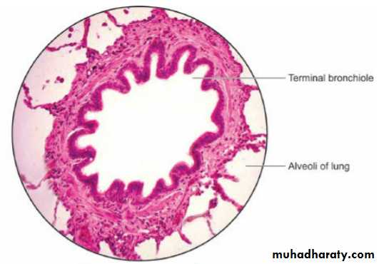
Structure of terminal bronchiole. Stain: Haematoxylin-eosin, 400X
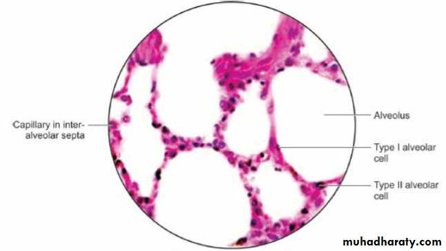
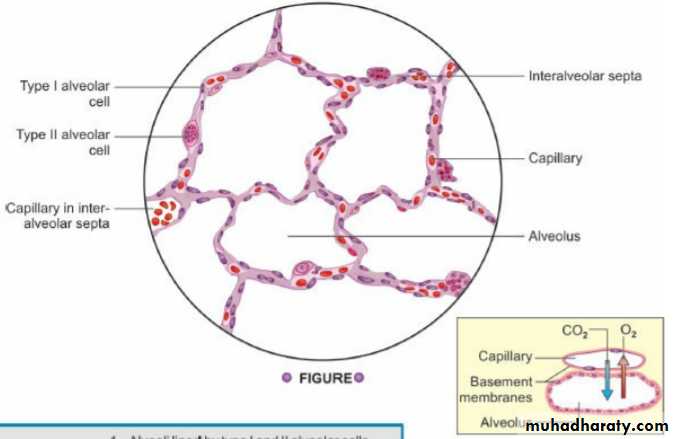
Alveoli and interalveolar septa. Haematoxylin-eosin, 400X
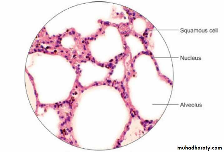
Squamous epithelium: Lung parenchyma. Stain: Haematoxylin-eosin, 400X