1
Morphology of skin lesions (language of dermatology)
Ass. Prof.
Dr. Zena S. Al-Fadhily /
Department of Medicine
Primary lesions: are basic lesions that are produced by the initial disease
pathology. Most skin diseases begin with a primary lesions.
Secondary lesions: are either evolve from primary lesions or develop as a
consequence of the patient's activities, or as complication of the disease.
Primary lesions:
Macule: Flat, circumscribed, non-palpable skin lesion less than 1 cm in diameter. Often hypo
or hyperpigmented but could be of any color (pink, red, violet, ..). It can be round, oval, or
irregular in shape • May be sharply marginated or blend into the surrounding skin.
Ex: freckles, vitiligo, nevi, m elasm a… …
Patch: sim ilar to m acule, but m ore than 1 cm in diam eter.
Ex: vitiligo, post inflam m atory hypo and hyper pigm entations, ecchym osis, ..
Papule: An elevated solid lesion less than 1 cm in diameter; color and shape varies. It could
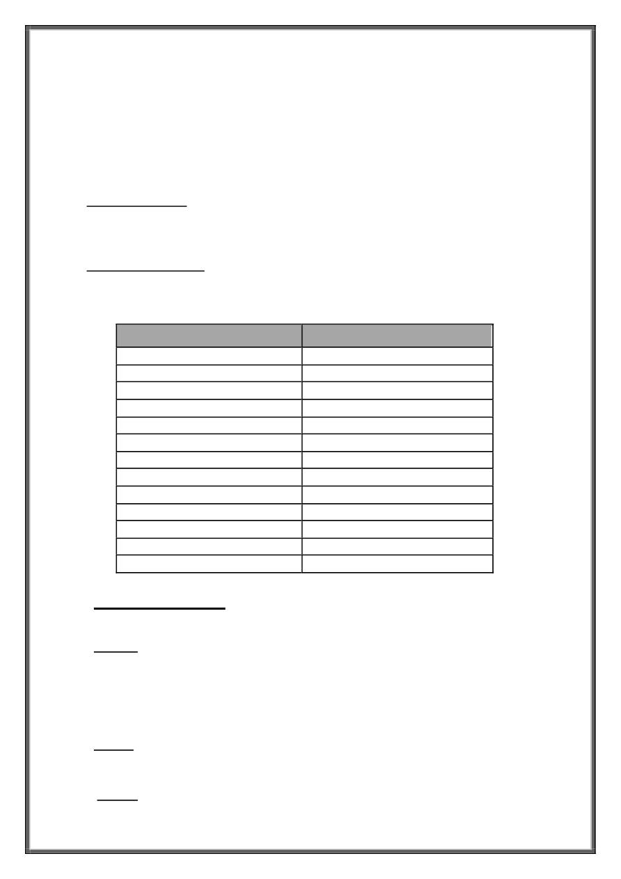
Primary lesions
Secondary lesions
Macule
scale
patch
crust
papule
erosion
plague
ulcer
nodule
fissure
cyst
atrophy
wheal
scar
vesicle
excoriation
bullae
lichenification
pustule
comedones
burrow
telangiectasia

2
be plain, verrucous, pedunculated, Dom-shaped, umbilicatd, fili-form, tinny, excoriated.
Ex: acne, wart, m olluscum , nevi, … .
Plague: sim ilar to papule, but m ore than 1 cm in diam eter.
Ex: psoriasis, derm atitis, tinea,..
Nodule: solid Elevated circumscribed lesion larger and deeper than papule Involves the dermis
and may extend to the subcutis. It is Infiltrated and deep on palpation.
Ex: skin tumors, nodular acne ….
Cyst: A circumscribed solid lesion with a wall and a lumen; the lumen may
contain fluid or solid matter.
Ex: epidermal cyst, nodulo-cystic acne
Vesicle: A circumscribed collection of free fluid inside the skin less than 1 cm
in diameter.
Ex: herpes labialis, chicken pox,…
Bullae: similar to vesicle but more than 1 cm in diameter.
Ex: pemphigus vulgaris, burn, …
Pustule: A circumscribed collection of pus inside the skin that varies in size.
Ex: folliculitis, acne, infected eczema,…
Wheal (hive): A firm edematous of the skin resulting from infiltration of the
dermis with fluid. Wheals are transient and may last only a few hours. They
blanch or disappear on pressure.
Ex: urticaria, insect bite
Comedone: A plug of sebaceous and keratinous material lodged in the
opening of a hair follicle; the follicular orifice may be dilated (blackhead,
opened) or narrowed (whitehead or closed).
Ex: acne, nevus comedonicus,..
Burrow: A narrow, elevated, tortuous channel produced by a parasite. It is
pathognomonic for scabies.
Telangiectasia: Dilated superficial blood vessels.
Ex: spider telangiectasia, steroid atrophy,..

3
Secondary lesions:
Scales: Excess dead epidermal cells that are produced by abnormal keratinization and
shedding. They could be fine or thick, white or brown, loose or adherent.
Ex: psoriasis, pityriasis rosea, dermatitis, icthyosis, …
Crust: A collection of dried serum, pus or debris covering skin surface. Color varies
yellowish to brown according to the dried material.
Ex: impetigo, wounds, burns,..
Lichenification: Combination of thickening of the skin with
hyperpigmentation, and accentuation of natural skin lines. It could occur in
any disease with chronic itching.
Ex: dermatitis, scabies, …
Erosion: A focal loss of epidermis. Erosions do not penetrate below the
dermo-epidermal junction and therefore heal without scarring.
Ex: impetigo, dermatitis, ….
Ulcer: A focal loss of epidermis and dermis; ulcers may heal with scarring.
Ex: bed sore, diabetic foot ulcer,…
Excoriation: removal of the top of the lesions by exogenous injury like
excessive itching. It could be linear or punctate.
Ex: excoriated acne, dermatitis, prurigo simplex, …
Fissure: A linear cleft of epidermis and dermis with sharply defined nearly
vertical walls. It is often paiful.
Ex: fissures heel, dermatitis,
Atrophy: A depression in the skin resulting from thinning of the epidermis or
dermis.
Ex: morphea, steroid atrophy, striae…
Scar: An abnormal formation of connective tissue implying dermal damage
after injury or surgery. Scars are initially thick and pink but with time become
white and atrophic. Scars could be atrophic, hypertrophic.
Ex: burn, surgical wounds, trauma, acne….

4
Important histopathological terms:
Parakeratosis: incom plete keratinization characterized by retention of
nuclei in the horny layer and associated with a marked underdevelopment of
or absence of the granular layer. as in psoriasis and porokeratosis. It
represents a physiologic event in mucous membrane.
Acanthosis: increase in thickness of the prickle cell layer. as in chronic eczema.
H ypergranulosis: increase in thickness of granular cell layer. as in chronic
eczem a and lichen planus.
Acantholysis: loss of cohesion between the epiderm al cells .It is either
prim ary which occurs am ong unaltered cells as a result of dissolution of the
intercellular substance as in pem phigus vulgaris, or secondary which occur
am ong altered or dam aged cells as in im petigo and herpes viral vesicles.
Spongiosis: intercellular edema between the squamous cells of the epidermis
causes an increase in the width of the spaces between the cells .It occurs
frequently in the inflammatory processes of the skin as in acute and subacute
eczema.
Papillomatosis: upward proliferation of sub-epidermal papillae causing the
surface of the epidermis to show irregular undulation .It seen in linear epidermal
nevus, solar keratosis, seborrheic keratosis, verruca vulgaris (wart), Acanthosis
nigricans and nevus sebaceous.