Bones of the Leg
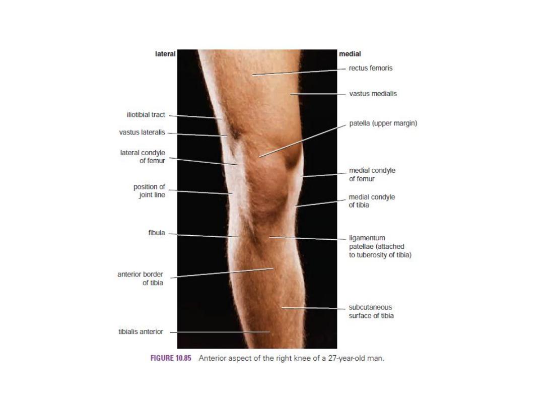
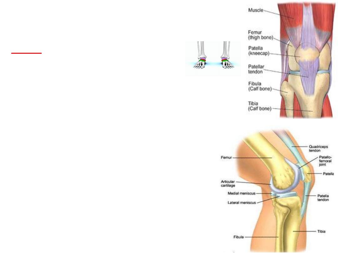
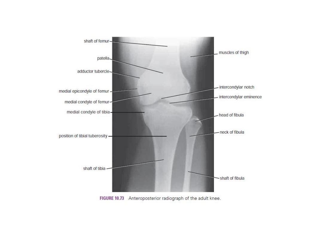
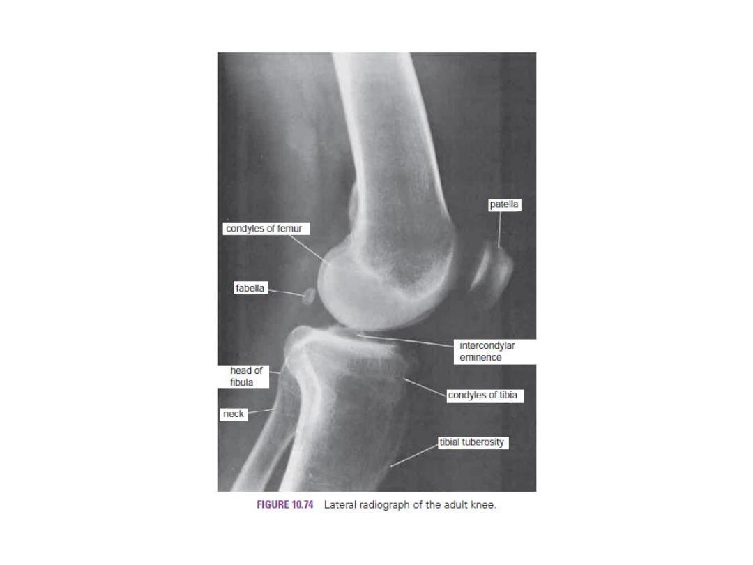
Patella
The largest sesamoid bone.
Within the tendon of the quadriceps femoris muscle.
Triangular, its apex lies inferiorly; and connected to the tuberosity
of the tibia by the ligamentum patellae.
The posterior surface articulates with the condyles femur.
It is in front of the knee and can easily be palpated.
Separated from the skin by subcutaneous bursa.
The upper, lateral, and medial margins give attachment to
the quadriceps femoris .
It is prevented from displaced laterally by horizontal fibers
of the vastus medialis and by the large size of the lateral
Condyle of the femur.
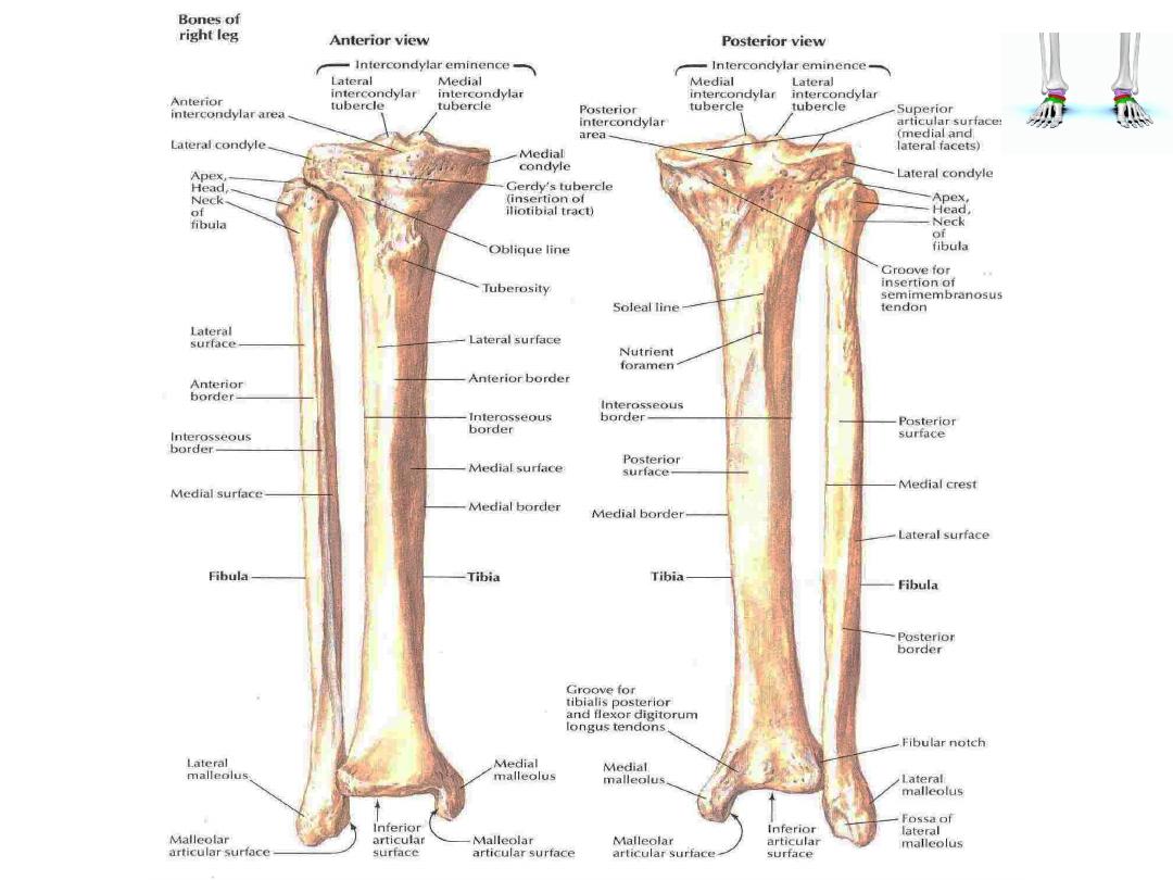
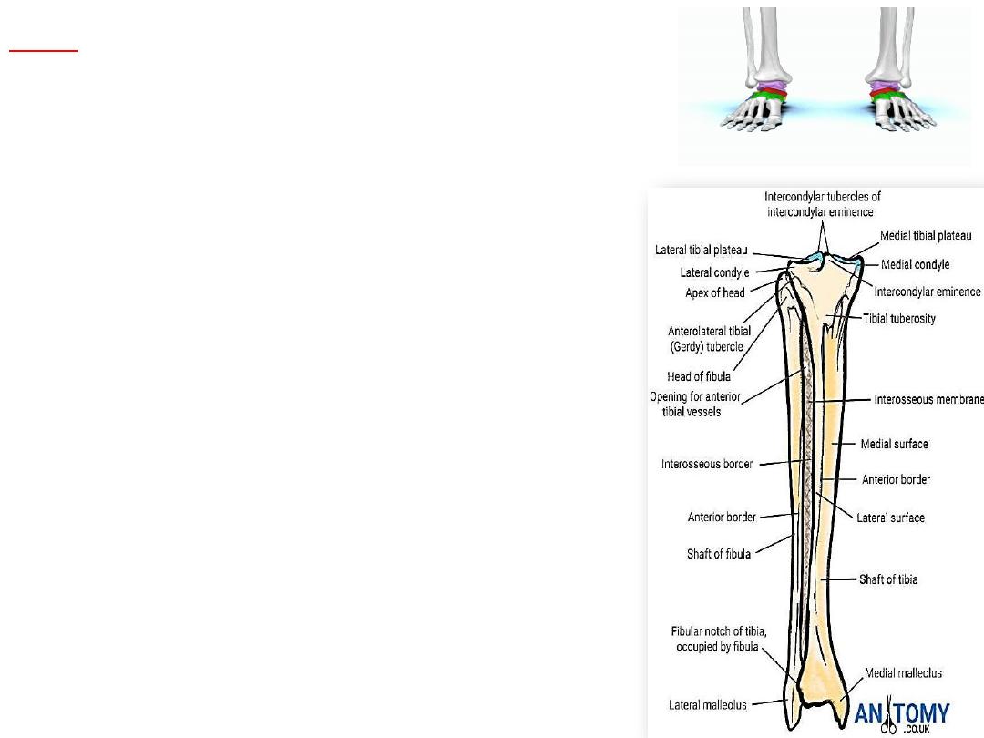
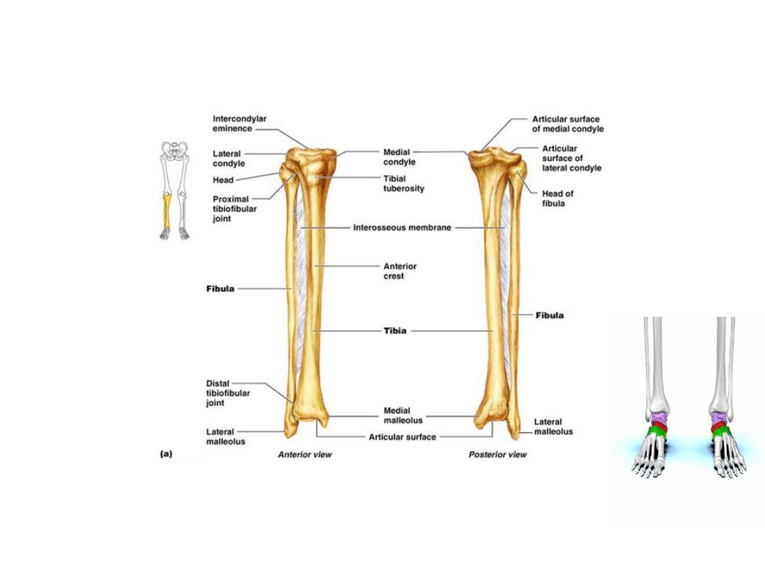
Tibia
The large weight-bearing medial bone of the leg.
Articulates with the condyles of the femur and
the head of the fibula above and with the talus and
the distal end of the fibula below.
At the upper end are the lateral and medial condyles
(sometimes called lateral and medial tibial plateaus),
which articulate with the lateral and medial condyles of
the femur and the lateral and medial menisci intervening.
Separating the upper articular surfaces of the tibial condyles
are anterior and posterior intercondylar areas; lying
between these areas is the intercondylar eminence.
The shaft of the tibia is triangular in cross section,
presenting three borders and three surfaces.
Its anterior and medial borders, with the medial surface
between them, are subcutaneous.
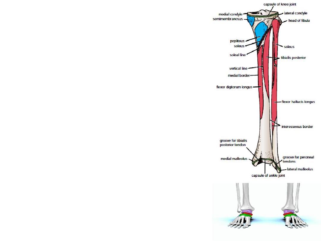
The anterior border is prominent and forms the shin.
At the junction of the anterior border with the upper
end of the tibia is the tuberosity, which receives the
attachment of the ligamentum patellae.
The anterior border becomes rounded below, where
it becomes continuous with the medial malleolus.
The lateral or interosseous border gives attachment
to the interosseous membrane.
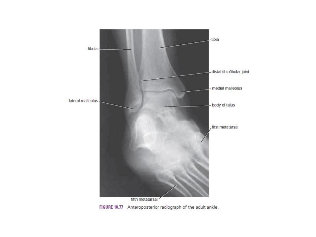
The lower end of the tibia is slightly expanded and
on its inferior aspect shows a saddle-shaped articular
surface for the talus.
The lower end is prolonged downward medially to form
the medial malleolus.
The lateral surface of the medial malleolus articulates
with the talus. The lower end of the tibia shows a wide,
rough depression on its lateral surface for articulation
with the fibula.
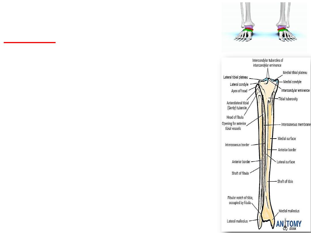
Fibula
The fibula is the slender lateral bone of the leg.
It takes no part in the articulation at the knee joint, but
below it forms the lateral malleolus of the ankle joint.
It takes no part in the transmission of body weight, but
it provides attachment for muscles.
The upper end, or head, possesses an articular surface
for articulation with the lateral condyle of the tibia.
The shaft of the fibula is long and slender, typically, it has
four borders and four surfaces.
The medial or interosseous border gives attachment to the
interosseous membrane.
The lower end of the fibula forms the triangular lateral
malleolus, which is subcutaneous. On the medial surface
of the lateral malleolus is a triangular articular facet for
articulation with the lateral aspect of the talus.
Below and behind the articular facet is a depression called
the malleolar fossa.
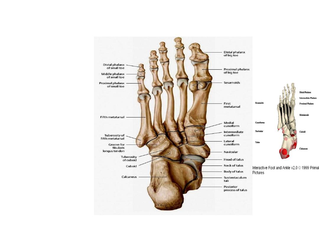
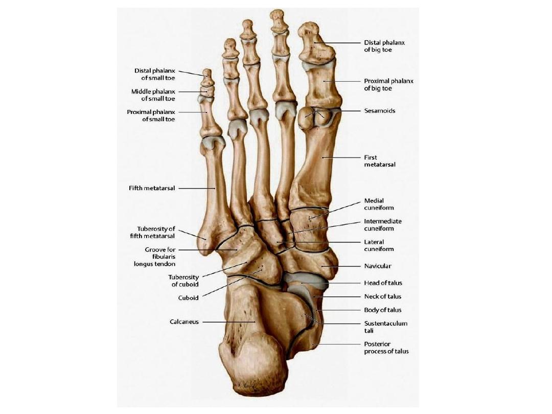
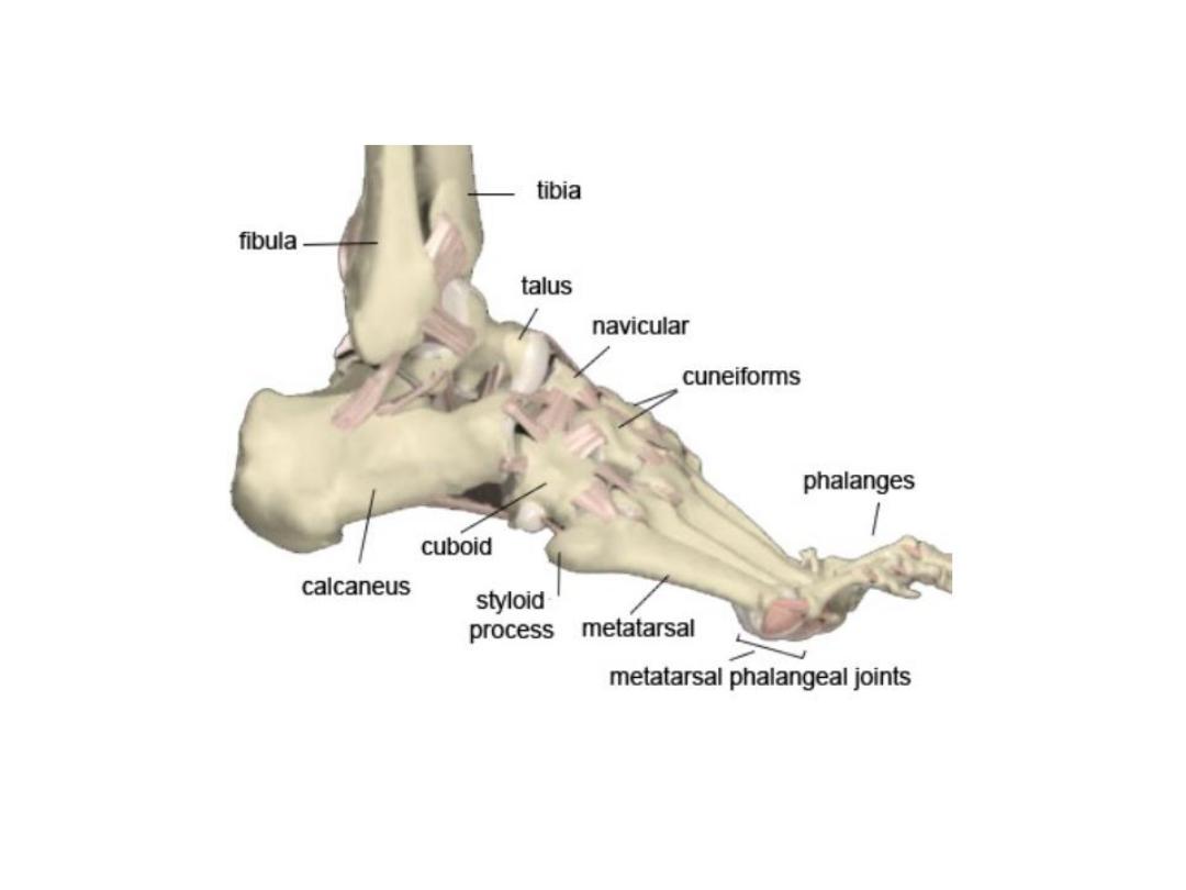
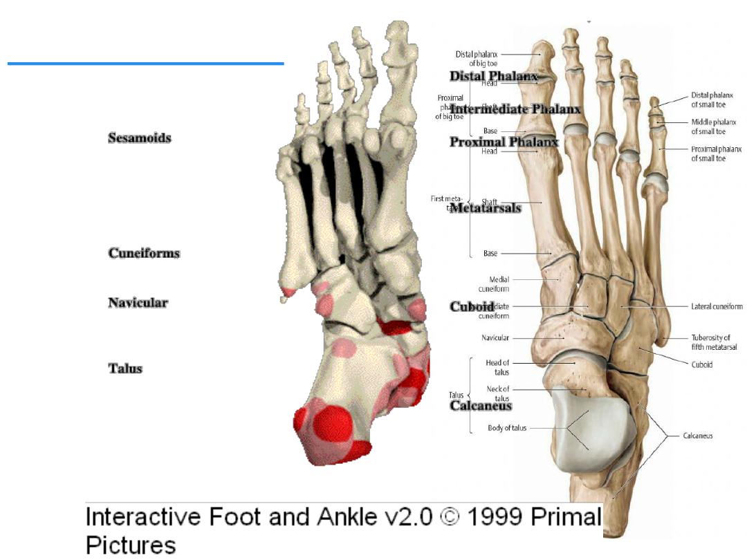
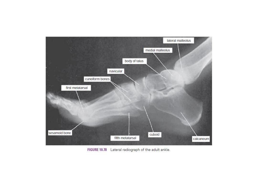
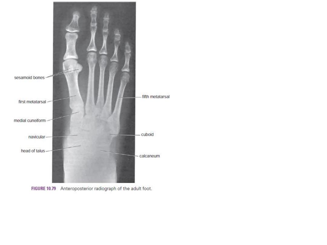
Bones of the Foot
tarsal bones
Metatarsals
phalanges.
Tarsal Bones
Calcaneum
Talus,
Navicular
Cuboid
Three Cuneiform.
Only the talus articulates with the tibia and the fibula at the ankle joint.
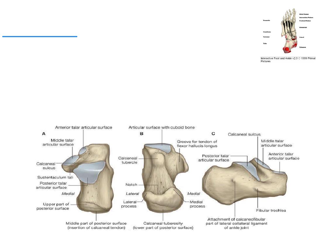
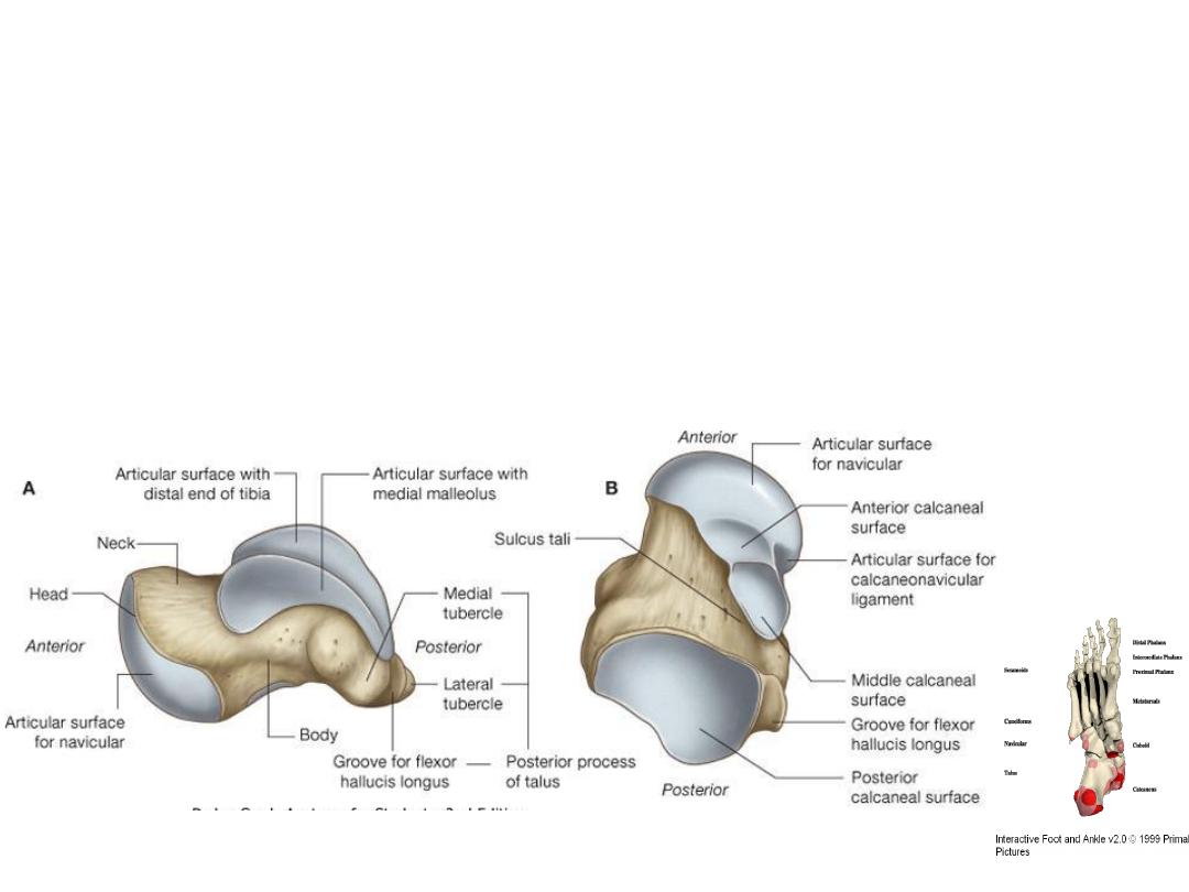
Calcaneum
It is the largest bone of the foot and forms the prominence of the heel.
It articulates above with the talus and in front with the cuboid.
It has six surfaces.
1-
The anterior surface is small and forms the articular facet that articulates with the
cuboid bone.
2 -
The posterior surface forms the prominence of the heel and gives attachment to the
tendo calcaneus (Achilles tendon)

3 -
The superior surface is dominated by two articular facets for the talus,
separated by roughened groove, the sulcus calcanei.
4 -
The inferior surface has an anterior tubercle in the midline and a large medial
and smaller lateral tubercle .
5 -
The medial surface possesses a large, process, termed the sustentaculum
tali, which assists in the support of the talus.
6 -
The lateral surface is almost flat. On its anterior part is a small elevation called
the
peroneal tubercle.
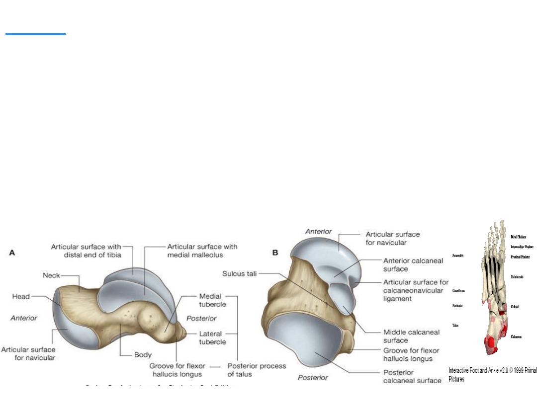
Talus
It articulates above at the ankle with the tibia and fibula, below with the calcaneum,
and in front with the navicular bone.
It possesses a head, a neck, and a body:-
o The head of the talus is directed distally and has an oval convex surface for
articulation with the navicular bone.
o The neck of the talus lies posterior to the head and is slightly narrowed. Its upper
surface is roughened and gives attachment to ligaments, and its lower surface shows
a deep groove, the sulcus tali. The sulcus tali and the sulcus calcanei in the
articulated foot form a tunnel, the sinus tarsi, which is occupied by the strong
interosseous talocalcaneal ligament.

o The body of the talus is cuboidal. Its superior surface articulates with the distal end
of the tibia; it is convex from before backward and slightly concave from side to side.
Its lateral surface presents a triangular articular facet for articulation with the lateral
malleolus of the fibula. Its medial surface has a small, comma-shaped articular facet
for articulation with the medial malleolus of the tibia. The posterior surface is marked
by two small tubercles, separated by a groove for the flexor hallucis longus tendon.
Numerous important ligaments are attached to the talus, but no muscles are
attached to this bone.
Cuneiform Bones
The three small, wedge-shaped cuneiform
bones articulate proximally with the navicular
bone and distally with the first three metatarsal
bones. Their wedge shape contributes greatly
to the formation and maintenance of the
transverse arch of the foot.
The tarsal bones, unlike those of the carpus,
start to ossify before birth.
Centers of ossification for the calcaneum
and the talus, and often for the cuboid, are
present at birth.
By the fifth year, ossification is taking place
in all the tarsal bones.
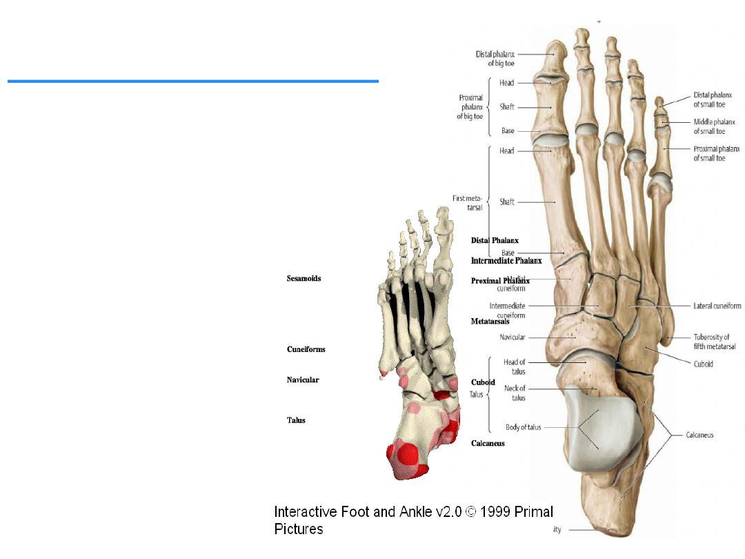
Metatarsal & Phalanges
The metatarsal bones and phalanges
resemble the metacarpals and phalanges
of the hand, and each possesses a head
distally, a shaft, and a base proximally.
The five metatarsals are numbered from
the medial to the lateral side.
The first metatarsal bone is large and
strong and plays an important role in
supporting the weight of the body.
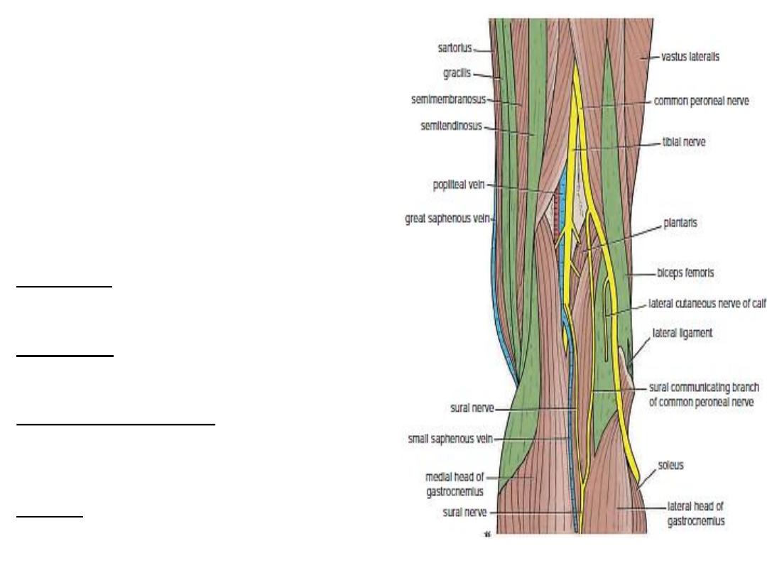
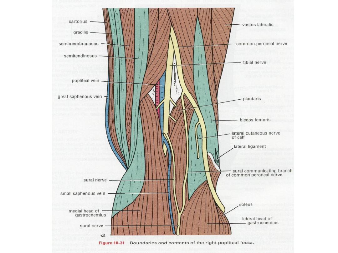
Popliteal Fossa
It is a diamond-shaped intermuscular space at
the back of the knee .
It is most prominent when the knee joint is
flexed.
It contains the popliteal vessels, the small
saphenous vein, the common peroneal and
tibial nerves, the posterior cutaneous nerve of
the thigh, the genicular branch of the obturator
nerve, connective tissue, and lymph nodes.
Boundaries
■■ Laterally: The biceps femoris above and the
lateral head of the gastrocnemius and plantaris
below.
■■ Medially: The semimembranosus and
semitendinosus above and the medial head of
gastrocnemius below.
The anterior wall or floor of the fossa is formed by
the popliteal surface of the femur, the posterior
ligament of the knee joint, and the popliteus
muscle.
The roof is formed by skin, superficial fascia, and
the deep fascia of the thigh.
Boundaries and
contents
of the right popliteal fossa.
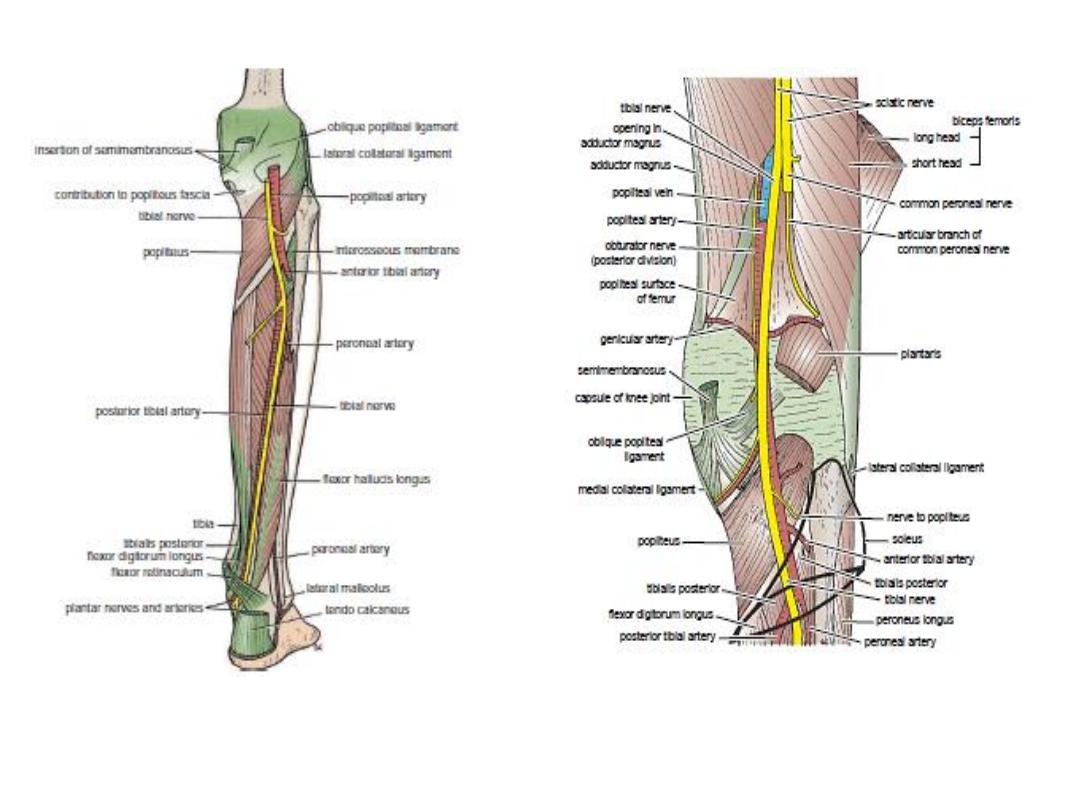
Deep structures in the posterior aspect
of the right leg.
Deep structures in the right popliteal
fossa. The proximal end of the soleus
muscle is shown in outline only.

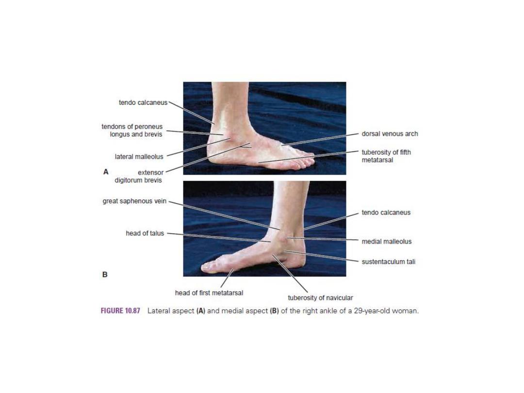
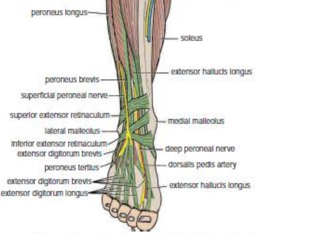
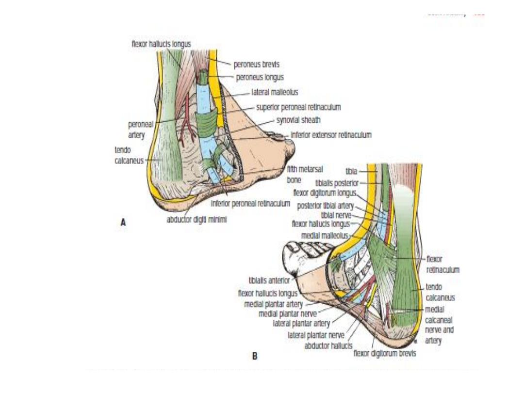
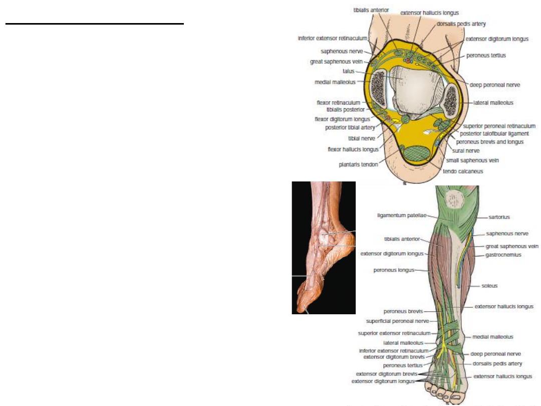
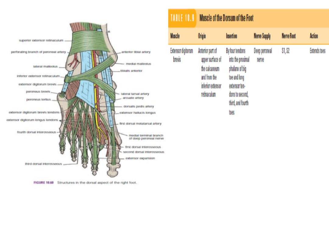
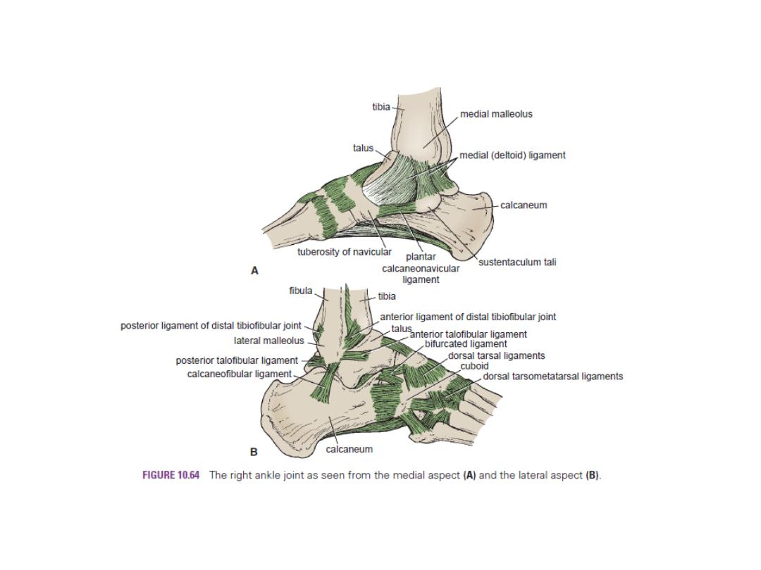
Retinacula of the Ankle
The retinacula are thickenings of the deep fascia that keep
the long tendons around the ankle joint in position and act as pulleys.
o
Superior Extensor Retinaculum
The superior extensor
retinaculum is attached to the distal ends of the anterior
borders of the fibula and the tibia.
o
Inferior Extensor Retinaculum
It is a Y-shaped fibrous
band located in front of the ankle joint separate the tendons
into compartments each of which is lined by a synovial sheath.
o
Flexor Retinaculum
It extends from the medial malleolus downward
and backward to the medial surface of the calcaneum .
o
Superior Peroneal Retinaculum
The superior peroneal retinaculum
connects the lateral malleolus to the lateral surface of the calcaneum.
o
Inferior Peroneal Retinaculum
It binds the tendons of the peroneus
longus and brevis muscles to the lateral side of the calcaneum .
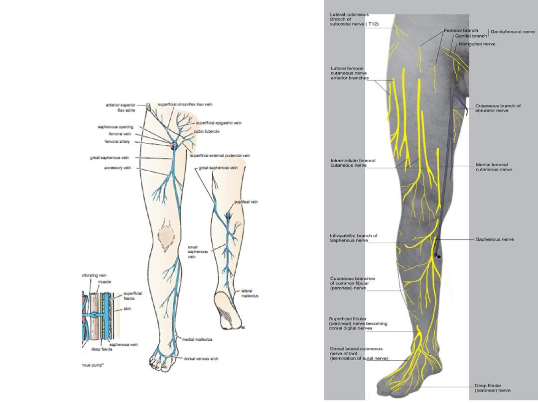
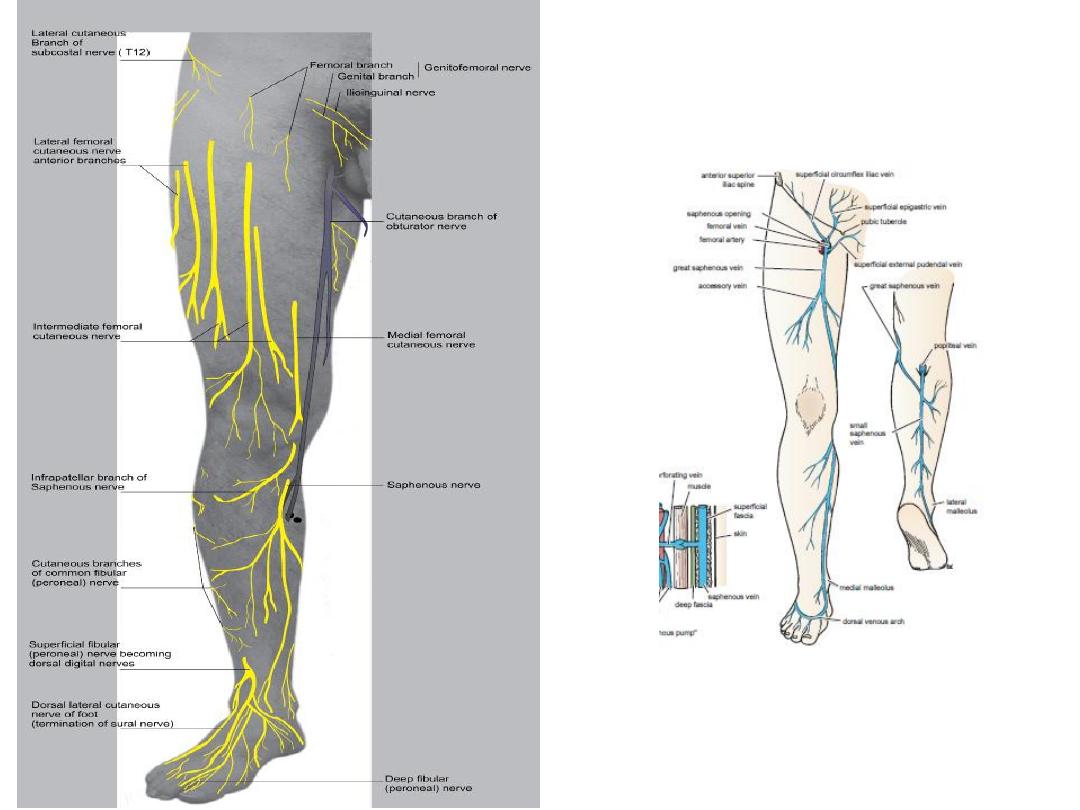
The Front of the Leg
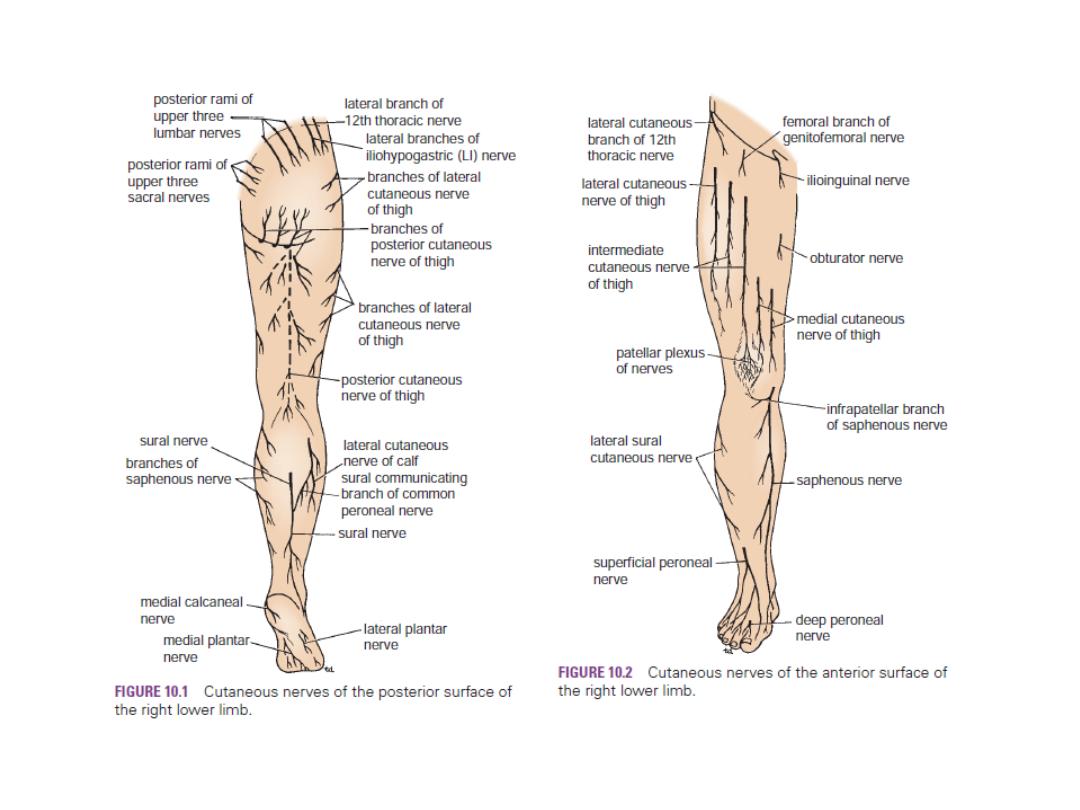
Skin
o Cutaneous Nerves a branch of the common peroneal
supplies skin on upper part of the lateral surface of leg.
o The superficial peroneal nerve, a branch of the
common peroneal supplies skin of lower part
of the anterolateral surface of the leg.
o saphenous nerve, a branch of femoral
supplies skin on the anteromedial surface of leg.
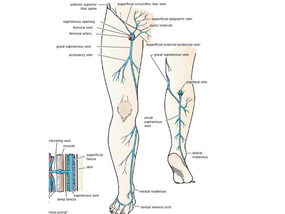
Superficial Veins
Numerous small veins curve around the medial
aspect of the leg and ultimately drain into
great. saphenous vein.
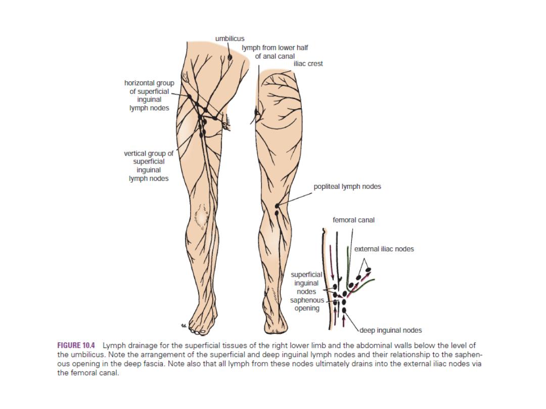
Lymph Vessels
The greater part of the lymph from skin and
superficial fascia on the front of leg drains
upward and medially in vessels that follow
great saphenous vein, to end in the vertical group
of superficial inguinal lymph nodes .A small amount
of lymph from upper lateral part of the front of
leg may pass via vessels that accompany the small
saphenous vein and drain into the popliteal nodes.
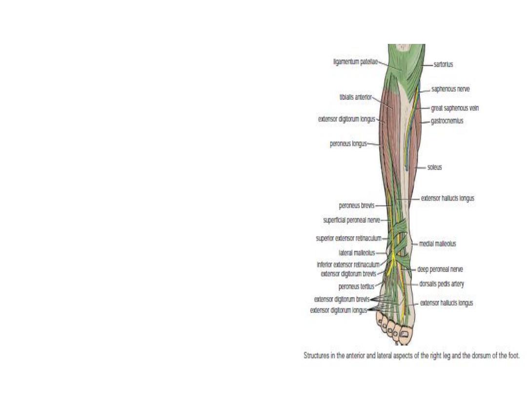
Contents of the
Anterior Fascial Compartment of the Leg
■■ Muscles:
The tibialis anterior, extensor digitorum longus,
peroneus tertius, and extensor hallucis longus
■■ Blood supply:
Anterior tibial artery
■■ Nerve supply:
Deep peroneal nerve
Contents of the
Lateral Fascial Compartment the Leg
■■ Muscles: Peroneus longus and peroneus brevis
■■ Blood supply: Branches from the peroneal artery
■■ Nerve supply: Superficial peroneal nerve
Contents of the
Posterior Fascial Compartment
The deep transverse fascia of the leg is a septum that divides the
muscles of the posterior compartment into superficial and deep
groups.
■■ Superficial group of muscles: Gastrocnemius, plantaris, and soleus
■■ Deep group of muscles: Popliteus, flexor digitorum longus, flexor
hallucis longus, and tibialis posterior
■■ Blood supply: Posterior tibial artery
■■ Nerve supply: Tibial nerve
The Region of the Ankle
Anterior Aspect of the Ankle
Structures Pass Ant.to the Ext.Ret from M to L.
■■ Saph. N and great saph V(in front of the m.m)
■■ Superficial per. N (medial and lateral branches)
Structures Pass Beneath or Through Ext.Ret
from M to L
■■ Tibialis anterior tendon
■■ Extensor hallucis longus tendon
■■ Anterior tibial artery with venae comitantes
■■ Deep peroneal nerve
■■ Extensor digitorum longus tendons
■■ Peroneus tertius
As each of above tendons passes beneath or
through the extensor retinacula, it is surrounded
by a synovial sheath.
The tendons of extensor digitorum longus and
peroneus tertius share a common synovial she.
Structures That Pass in Front of the Medial Malleolus
■■ Great saphenous vein
■■ Saphenous nerve
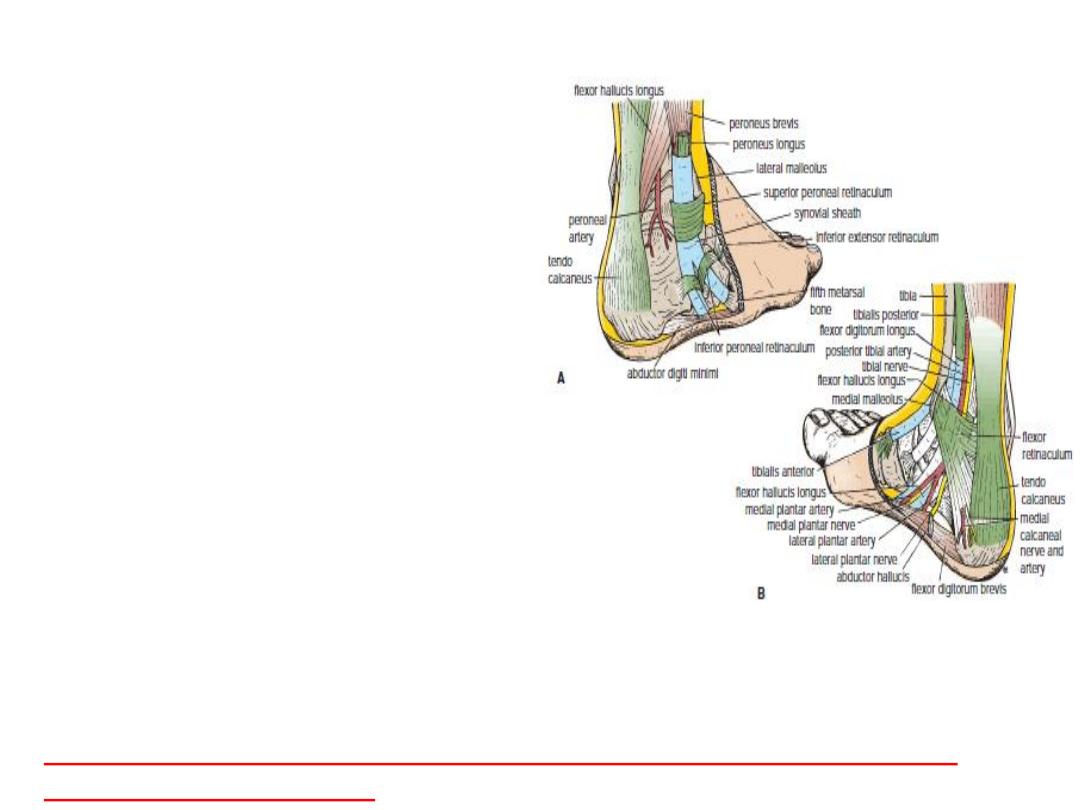
Posterior Aspect of the Ankle
Structures Pass behind Medial Malleolus
beneath Flex.Retin.From M to L
■■ Tibialis posterior tendon
■■ Flexor digitorum longus
■■ Posterior tibial artery with venae comit.
■■ Tibial nerve
■■ Flexor hallucis longus
As each of these tendons passes beneath
flexor retin,it is surrounded by a synov.sheath.
Structures Pass behind Lat. Mal.Superficial
to the Sup. Per. Ret
■■ The sural nerve
■■ Small saphenous vein
Structures Pass behind Lat. Mal. beneath
Superior Peroneal Retinaculum
The peroneus longus and brevis tendons share
a common synovial sheath. Lower down, beneath
the inferior peroneal retinaculum, they have separate
sheaths.
Structures Lie Directly behind the Ankle
The fat and the large tendo calcaneus lie behind the ankle.
Tenosynovitis and Dislocation of the Peroneus Longus and Brevis Tendons
Ruptured Tendo Calcaneus
The Foot
The foot supports the body weight and provides
leverage for walking and running.
It is constructed in the form of arches.
It also serves as to absorb shocks, in jumping.
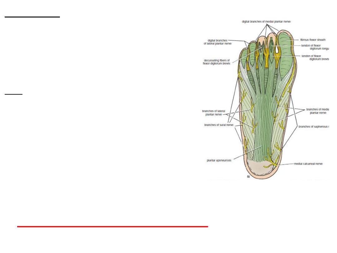
The Sole of the Foot
Skin
The skin is thick and hairless with a large
numbers of sweat glands.
It is firmly bound down to the underlying
deep fascia by numerous fibrous bands.
Deep Fascia
The plantar aponeurosis is a triangular
thickening of the deep fascia that protects
the underlying nerves, blood vessels, and muscles.
Its apex is attached to the medial and lateral tubercles of the calcaneum.
The base of the aponeurosis divides into five slips that pass into the toes.
Plantar Fasciitis & calcaneal spur
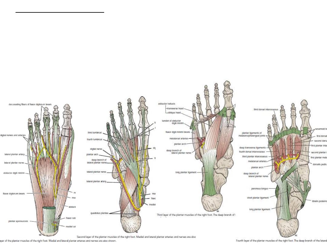
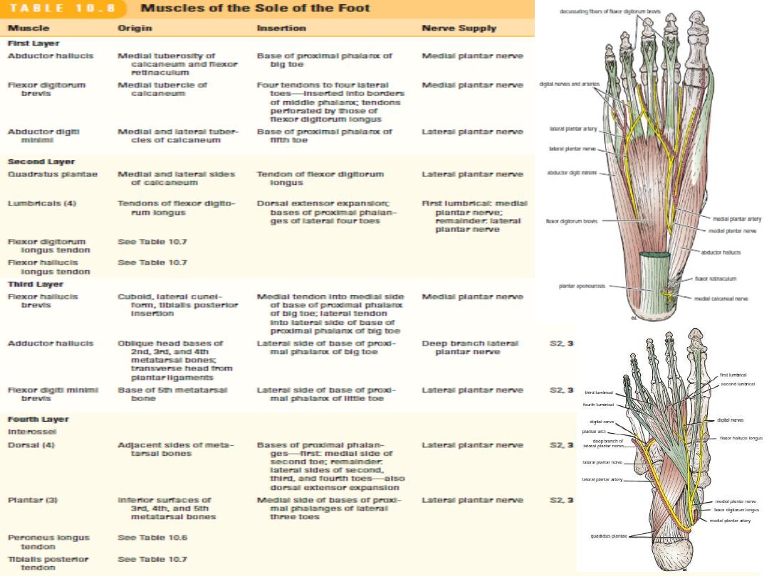
The muscles of the sole are in four layers from the inferior layer superiorly.
■■
First layer: Abductor hallucis, flexor digitorum brevis, abductor digiti minimi
■■
Second layer: Quadratus plantae, lumbricals, flexor digitorum ongus tendon, flexor
hallucis longus tendon
■■
Third layer: Flexor hallucis brevis, adductor hallucis, flexor digiti minimi brevis
■■
Fourth layer: Interossei, peroneus longus tendon, tibialis posterior tendon
Unlike the small muscles of the hand, the sole muscles have few delicate functions
and are chiefly concerned with supporting the arches of the foot.
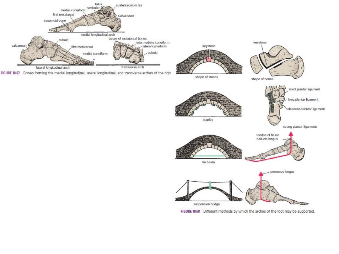
The Arches of the Foot
A segmented structure can hold up weight
only if it is built in the form of an arch.
The foot has three such arches, which are
present at birth: -
Medial longitudinal arch: This consists of the
calcaneum, the talus, the navicular bone, the
three cuneiform bones, and the first three
metatarsal bones.
Lateral longitudinal arch: This consists of the
calcaneum, the cuboid, and the 4th and 5th
metatarsal.
Transverse arch: This consists of the bases of
the metatarsal bones and the cuboid and the 3
cuneiform.
Pes planus & Pes cavus
DVT
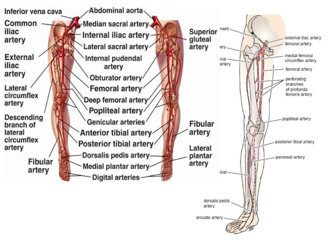
Injury to post.tibial & ant tibial artery
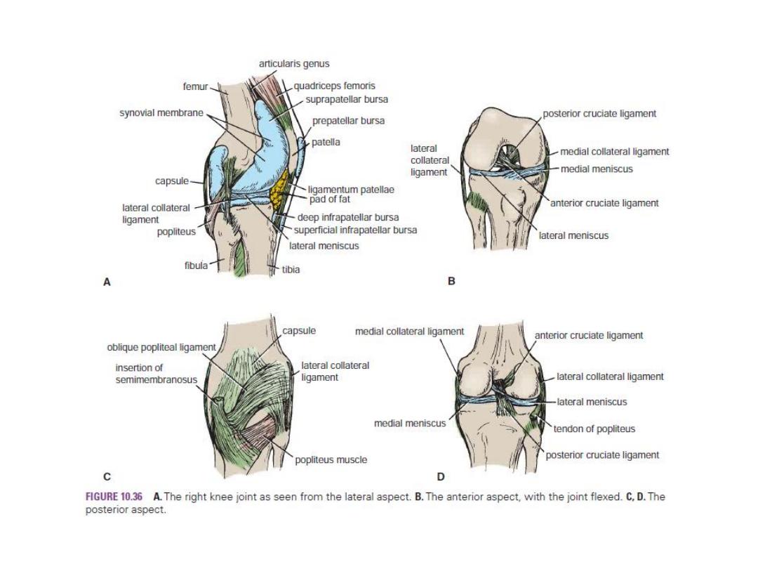
Bursae & ligaments Related to the Knee Joint
Bursitis
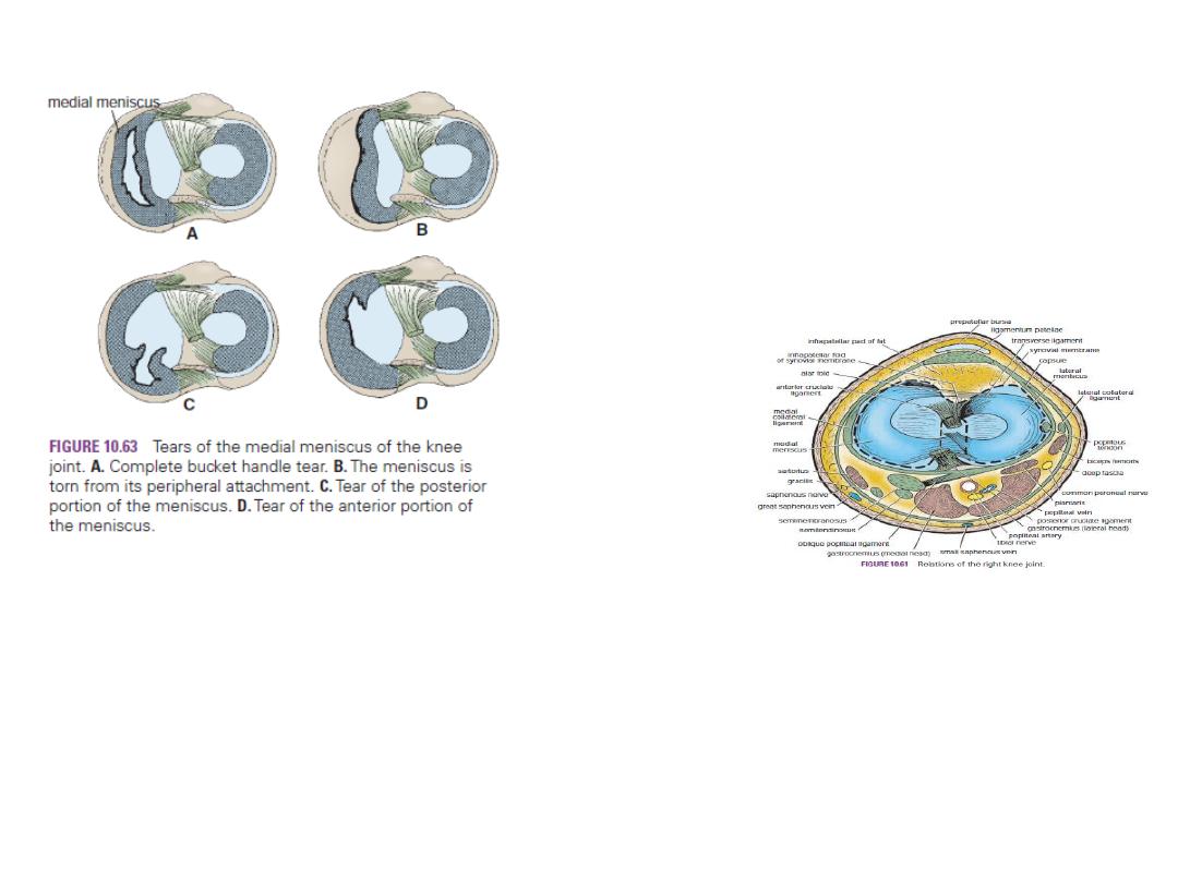
Meniscus
Strength of the Knee Joint
Ligamentous Injury of the Knee Joint
Meniscal Injury
Arthroscopy
Ligaments
Ankle Joint Stability, sprain, fractures arround.
Metatarsophalangeal Joint of the Big Toe (Hallux valgus &hallux rigidus).