Disorders of the salivary glands Dr. Ali Jaffer Alghazzawi
THE PAROTID GLAND- AnatomyBounded by : the ramus of the mandible
the base of the skull
the mastoid process
Posterior relation : the carotid sheath
CNs XI&XII
Structures run through the gland
Facial nerve that divides into five branches
The terminal branch of the external carotid artery (maxillary & superficial temporal a.)
Retromandibular vein
Intraparotid LN
The facial nerve divides the gland into two lobes:
superficial 80%
deep 20%
accessory (ant. To the superficial lobe)
THE PAROTID GLAND- inflammatory disorders
Viral infectionsMumps is the most common cause of acute painful swelling
Predominantly affects children
Spread: airborne infected saliva
Prodromal period of 1-2 days (fever&headache)
Clinically: pain exacerbated by eating and drinking , swelling
Provides lifelong immunity
Rx: paracetamol& adequate oral fluid
Cx: orchitis, oophoritis, pancreatitis, sensorineural deafness & meningoencephalitis
Recurrent parotitis of childhood
It may be caused by an incompetent punctum that leads to soiling of the parotid ducts with contaminated oral fluids.
Children 3-6 years ,rapid swelling of one or both glands,
fever& malaise last 3-7 days. The symptoms worsen
by chewing and eating.
Sialography: punctate sialectasis (snowstorm)
Rx: endoscopic washouts& antibiotics
Bacterial infections ( acute ascending bacterial siladentis)
Previously described in dehydrated elderly patients following major surgery.
Reduced salivary flow secondary to dehydration results in ascending infection via the parotid duct into the parotid paranchyma.
Currently , it is commonly associated with a salivary calculus.
Tender, painful swelling, generalised malaise, pyrexia and cervical lymphadenopathy
Symptoms exacerbated by eating or drinking
Intraoral exam; pus exuding form the parotid gland papilla
Staphylococcus aureus or streptococcous virdans
Rx; appropriate antibiotics
absecss… aspiration with large pore needle or formal drainage under anaesthesia (the skin incision should made low to avoid damage to the lower branch of the facial nerve)
Sialography is contraindicated during acute infection.
HIV- associated sialadenitis
May present as chronic parotitis in children.
May present as classical Sjogren syndrome in adults clinically and histologically but there is lack autoantibody.
May present with multiple parotid cysts which cause gross parotid swelling and facial disfigurement.
CT&MRI; characteristic Swiss cheese appearance of multiple large cystic lesions.
The swollen glands usually painless and may regress on the institution of antiviral therapy.
Cysts may be aspirated
THE PAROTID GLAND- obstructive parotitis
Stone formation (sialolithiasis) and stricturesParotid sialolithiasis 20%
The stones are usually radiolucent.
Locations; confluence of the collecting ducts, at the point the courses over the masseter muscle or in the distal aspect of the parotid duct adjacent to the parotid papilla.
Presentation: intermittent swelling particularly in the mealtimes.
Ix : US
Rx: Small stones (less 4mm) retrieved by basket
up to 8mm broken with lithotripsy
larger than 8 mm removed by endoscopic assisted surgery
Strictures of the parotid duct 20%
Stricture lead to stagnation and mucus plug obstriction
Clinically; meal time syndrome starting at the breakfast as swelling which persists. Massage release the plug with a gush of salty saliva.
Rx dilatation and endoscopic washouts with steroid solutions
Papillary obstruction
trauma to the parotid results in inflammatory oedema and obstruction of salivary flow.
May result dilation of the duct (mega-duct) which visible coursing the patient cheek
Rx progressive dilation of the punctum& stent insertion.
Papillotomy should be avoided as it cause stricture.
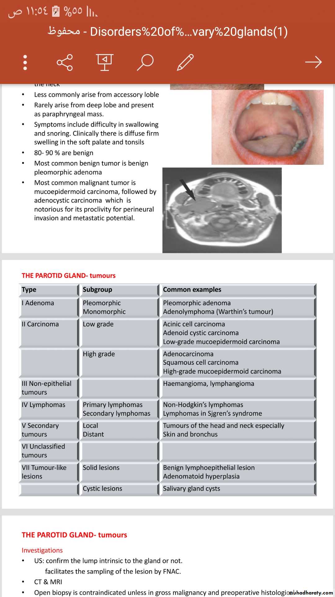
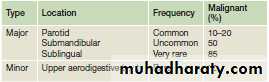
THE PAROTID GLAND- tumours
The most common site of salivary tumours
Most tumors arise from the superficial lobe.
Slow-growing , painless swelling below ,in front of the ear or in the upper aspect of the neck
Less commonly arise from accessory loble
Rarely arise from deep lobe and present as paraphryngeal mass.
Symptoms include difficulty in swallowing and snoring. Clinically there is diffuse firm swelling in the soft palate and tonsils
80- 90 % are benign
Most common benign tumor is benign pleomorphic adenoma
Most common malignant tumor is mucoepidermoid carcinoma, followed by adenocystic carcinoma which is notorious for its proclivity for perineural invasion and metastatic potential.
Investigations
US: confirm the lump intrinsic to the gland or not.facilitates the sampling of the lesion by FNAC.
CT & MRI
Open biopsy is contraindicated unless in gross malignancy and preoperative histological diagnosis is required as a preclude to radical parotidectomy
Parotidectomy
Superficial parotidectomy: the part of the gland superficial to the facial nerve is removed
- Benign, low grade and low stage malignant tumours
- Partial superficial parotidectomy for small tumours
- Extracapsular dissection: benign parotid gland tumours
B. Radical parotidectomy: is performed when there is clear histological evidence of high grade malignant tumors eg squamous cell carcinoma with invasion of the facial nerve.
it involves removal of all parotid tissue and division of the facial trunk through the main trunk with removal of the ipsilateral masseter muscle and may require neck dissection particularly when there is clinical, radioliogical and cytological evidence o f lymph nodes metastasis.
COMPLICATIONS OF PAROTID GLAND SURGERY
haematoma formation;
infection;
deformity: unsightly scar and retromandibular hollowing;
temporary facial nerve weakness;
transection of the facial nerve and permanent facial weakness;
sialocele;
facial numbness;
permanent numbness of the ear lobe associated with great auricular nerve transection;
Frey’s syndrome.
Frey’s syndrome
(gustatory sweating) is considered an inevitable consequence of parotidectomy unless preventive measures are taken .
It results from damage to the autonomic innervation of the salivary gland with inappropriate regeneration of the postganglionic parasympathetic nerve fibres of the auriculotemporal nerve that aberrantly stimulate the sweat glands of the overlying skin.
Clinically; sweating and erythema (flushing) over the region of surgical excision of the parotid gland
Diagnosis ;starch iodine test. This involves painting the affected area with iodine, which is allowed to dry before applying dry starch, which turns blue on exposure to iodine in the presence of sweat.
Sweating is stimulated by salivary stimulation.
PREVENTION
● sternomastoid muscle flap;
● temporalis fascial flap;
● insertion of artificial membranes between the skin and theparotid bed.
All these methods replace the barrier between the skin and the parotid bed to minimise inappropriate regeneration of autonomic nerve fibres.
MANAGEMENT OF ESTABLISHED FREY’S SYNDROME
● antiperspirants, containing aluminium chloride;
● denervation by tympanic neurectomy;
● the injection of botulinum toxin into the affected skin. ( most effective and can be performed as an out-patient).
THE PAROTID GLAND- tumour- like lesions
Sialasenosis (sialoisis)Non inflammatory swelling
Associated with conditions eg DM, alcoholism, pregnancy, bulimia and idiopathic.
Prolong malnutrition produces sialosis by process of hypertrophy to compensate for swings in acid balance.
Drug induced ; commonly sympathomimetic.
Age; 40-70, soft and often symmetrical swelling, hamster-like appearance
In DM and drug induce silaosis, may which may associated with neuropathy which interferes with salivary gland function and subsequent acinar cell atrophy.
Rx; no effective treatment ; treat the underlying cause.
THE PAROTID GLAND- degenerative conditions
Sjogren’s syndrome
autoimmune condition causing progressive destruction of the salivary and lacrimal gland s.
Primary Sjogren’s syndrome occurs without an associated connective tissue diseases, the symptoms are more sever than the secondary one and it has higher lymphomatous transformation.
Incidence 0.5%-2%, F:M 10:1, enlarged salivary gland (parotid more than submadiblular), pain, xerostomia.
Pathology: progressive lymphocytic infiltration, acinar cell destruction and proliferation of duct epithelium in all salivary and lacrimal gland tissue.
Dx; depends on history.
Mx; symptomatic, ophthalmological assessment and artificial tear for keratoconjunctivitis sica , artificial salivary substitute for dry mouth.
Cx; non Hodgkin’s B-cell lymphpma 4.3%
Xerostomia
salivary flow decreases with age
Causes.
Chronic anxiety states and depression.
Dehydration.
Anticholinergic drugs especially antidepressants
Salivary gland disorders eg Sjogren’s syndrome
Radiotherapy to the head an neck.
Sialorrhea
Increase salivary flow
Caused by certain drugs and oral infection.
Mx
antisialogogues.
Intraparanchymal botulinum toxin injection.
Uncotrollable drooling managr by surgery:
Blilateral submandibular duct repositioning and simultaneous sublingual gland excision.
Bilateral submanidibular gland excision.
Transposition of parotid ducts and simultaneous submandibular gland excision
* Restiing salivary flow arise from the submandibular gland
THE SUBMANDIBULAR GLANDS-anatomy
Paired glands lie below the mandible on either side.
There is a larger superficial lobe and a smaller deep lobe.
Important anatomical relationship
Lingual nerve
Hypoglossal nerve
Anterior facial vein
Facial artery
Marginal mandibular branch of the facial nerve
It drains by Wharton’s duct and opens in the anterior floor of the mouth at the sublingual papilla.
Ectopic / aberrant salivary gland tissue
Stafne bone cyst, the most common ectopic salivary tissue.
Asymptomatic clearly demarcated radiolucency of the angle of the mandible.
No treatment
THE SUBMANDIBULAR GLANDS-inflammatory disorders
Acute submandibular sialadenitis
viral. Paramyxovirus (mumps)
Bacterial. More common than viral sialadenitis and occurs secondary to stone obstruction.
Chronic submandibular sialadenitis.
THE SUBMANDIBULAR GLANDS-obstruction and trauma
sialolithiasis; the most common cause of obstruction 80%, because the submandibular secretions usually viscus.
80% radio-opaque and can identified by x-ray
The stones composed mainly of phosphate and oxalate salts.
Stricture is the 2nd most common cause of obstruction.
Floor of mouth pathology or external pressure accounts 5-10%
C/F: painful swelling, precipitated by eating , the swelling occurs rapidly and resolve spontaneously over 1-2 hrs.(meal time syndrome)
The most common sites of impaction are the of the gland and near the punctum.
Examination: enlarged firm tender swelling. Pus may be visible from sublingual papilla or expressed by bimanual palpation.
THE SUBMANDIBULAR GLANDS-obstruction and trauma
Mx
small less than 4 mm .. Retrieved by Dormia basket (min invasive procedure performed under local anaesthesia either endoscopically –sialendoscopy- or under US control.
Larger … extrscorporeal or intracorporial lithotripsy. Then retrieved as above.
Stone in submandibular duct in the floor of the mouth anterior to the point crossing the lingual nerve (second molar region)…. The stone removed under local anaesthetics.
Stone at the hilum of the gland…. Stone retrieval via intraoral approach under GA
Stone retrieval success rate 95%
Failure of stone retraction….. Submandiular gland excision
Indications of submandibular gland excision
Sialadentis when min invasive methods have failed.
Salivary tumours
Complications of submandibular gland excision
Haematoma
Wound infection
Marginal mandibular nerve injury
Lingual nerve injury
Hypoglossal nerve injury
Transection of the nerve to the mylohoid muscle producing submental skin anaesthesia.
THE SUBMANDIBULAR GLANDS-tumours
It presents as a slow-growing, painless swelling
on examination,it is difficult to differentiate from submandibular lymphadenopathy.
This can be resolved by US examination.
Most salivary neoplasms, even malignant tumours, are often slow-growing, painless swellings. The difficulty is to always distinguish between benign and malignant lesions prior to excision.
Pain is not a reliable indication of malignancy
rapid growth, facial nerve palsy, lymph node enlargement and skin tethering are signs of a high-grade malignant lesion.
The most common malignant tumour is an adenocystic carcinoma (40%),
Investigation
US with FNAC/True-Cut biopsy , the investigation of choice ( with carful history 95% of malignancy can be identified)CT&MRI scanning for preoperative planning.
Open surgical biopsy is contraindicated , it may seed the tumour into surrounding tissue making it impossible to eradicate microscopic deposit.
Management
Benign: surgical excision
Malignant depend on stage of the disease. Larger and more aggressive the lesion the more radical surgery required.