
43
College of Medicine/Babylon University
Medical Physics Module
Session 4:
Lecture 7:
Physics of Cardiovascular System
Objectives: after the end of this lecture, the student must know:
1- The principle laws that govern the heart structure and function
2- Blood pressure and its measurement
3- Types of blood flow (laminar and turbulent) and its application in diseases.
The blood is pumped by contraction of the heart muscle, from left ventricle at
pressure of 125 mmHg and finally into very fine meshwork or capillary bed for
few seconds the blood supplies O2 to cells and picks up CO2. Adult has about 4.5
liters of blood, each section of heart pumps 80 ml with each contraction.
The combination of RBC and plasma causes blood to have flow properties
different from those of fluid like water.
Starling law:
fluid movement through capillary wall= the hydrostatic pressure
(p) across the capillary wall+ osmotic pressure bringing fluid in.
Work Done By The Heart
Left side of the heart systole pressure= 120 mmHg
Right side of the heart diastolic pressure= 80 mmHg
Above caused by following:
1- The left side of the heart three times thicker than right side.
2- The circular shape of left ventricle producing pressure larger than elliptical
shape of right ventricle.
College of Medicine/Babylon University
Medical Physics Module
The work done= P∆V
Blood Pressure And Its Measurement
The instruments called Sphygmomanometer, the sound heard with
stethoscope called Kortokoff or K sounds.
The onset of K indicate systolic pressure precision ± 2 mmHg
The fade of k indicates diastolic pressure precision ± 5 mmHg
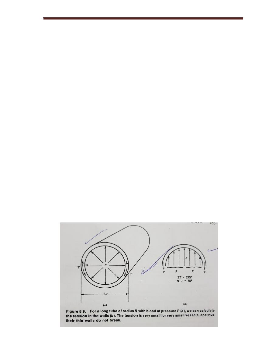
Pressure Across Blood Vessel Wall
The pressure drops in the capillaries which have thin walls (1 μm) permit easy
diffusion of O2 and CO2, the capillary do not burst according to Laplace law
Consider a tube of radius R
F = 2 R P
P blood pressure
T tension in the wall
2T = 2 R P
T = R P
College of Medicine/Babylon University
Medical Physics Module
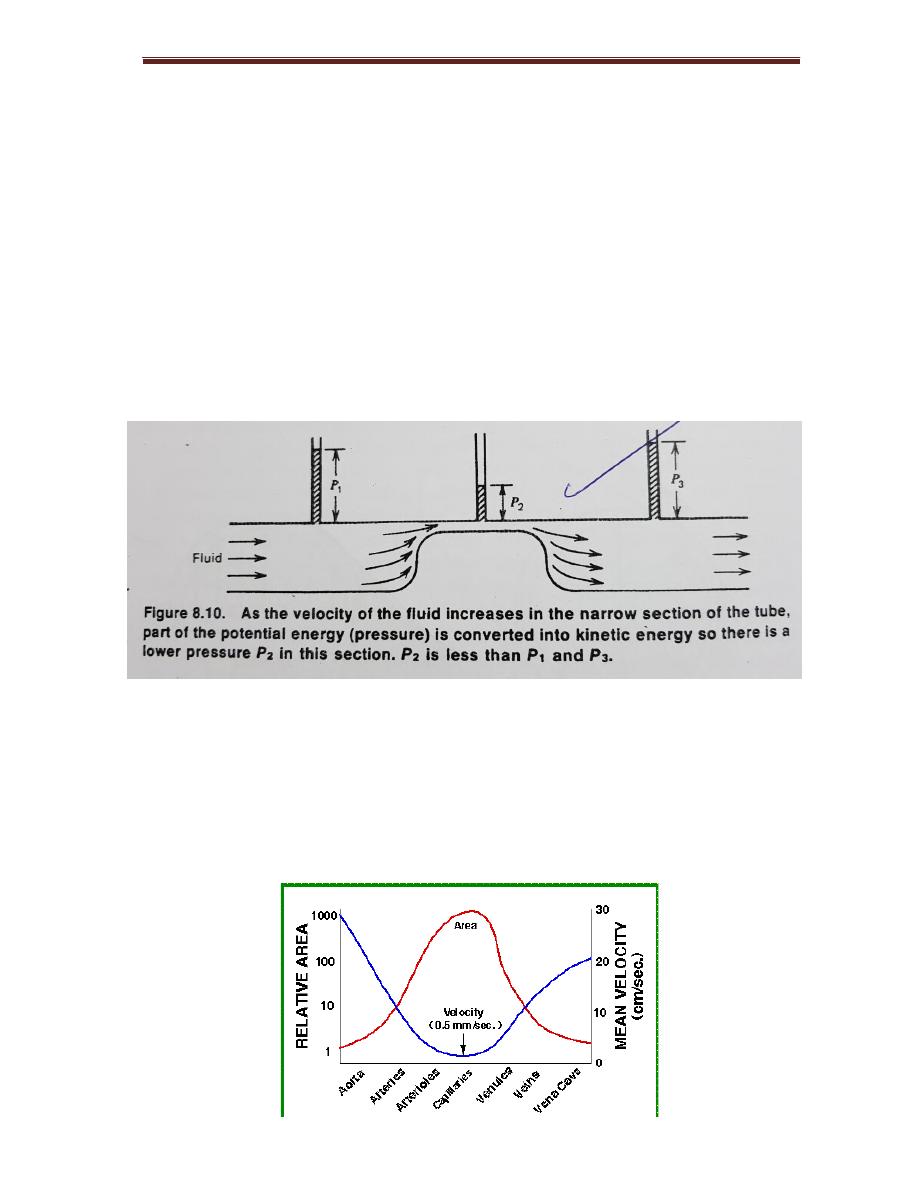
Bernoulli’s principle Applied To Cardiovascular System
Bernouli’s principle
is based on the law of conservation of energy
Increasing KE (kinetic energy) obtained by reduction of PE, the pressure in the
tube
Average K E of 1 gm ( 1 cm
3
) of blood as it leaves the heart
KE= 1/2 mv
2
v= 30cm/ sec
KE=1/2x1x(30)= 450 erg/ cm³
How Fast Does Your Blood Flow
Capillaries about 20 μm diameter, there total cross-sectional area 30 cm. The
blood velocity V inversely related to the total cross-section area of the vessels
carrying blood.
College of Medicine/Babylon University
Medical Physics Module
Figure 7-1:
relationship between cross sectional area of vessel and velocity of
blood flow.
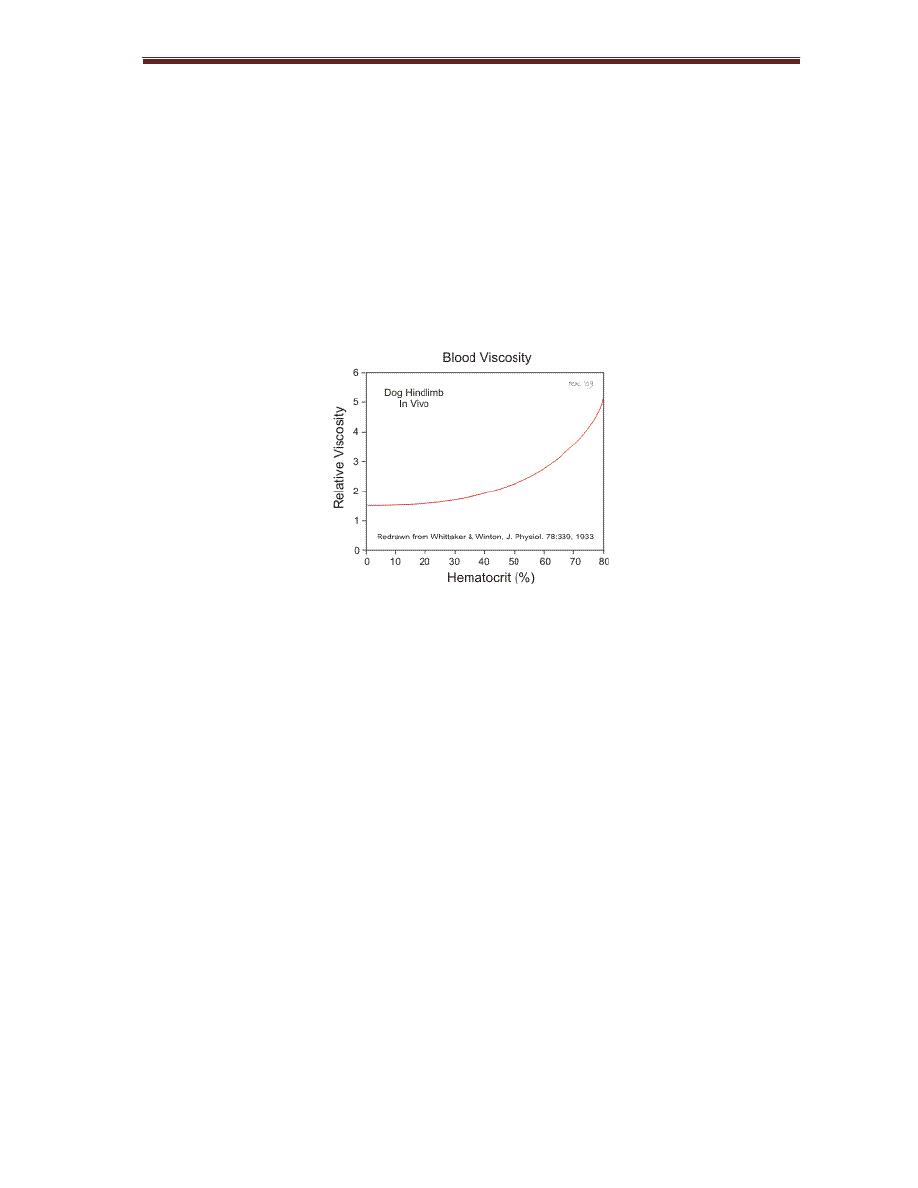
Viscosity of blood= 3x10
-3
to 4x10
-3
Pas. Depending on RBC in the blood or
Hematocrit.
Hematocrit α Viscosity
Viscosity α Temperature
Figure 7-2
: relationship between hematocrit and velocity of blood flow
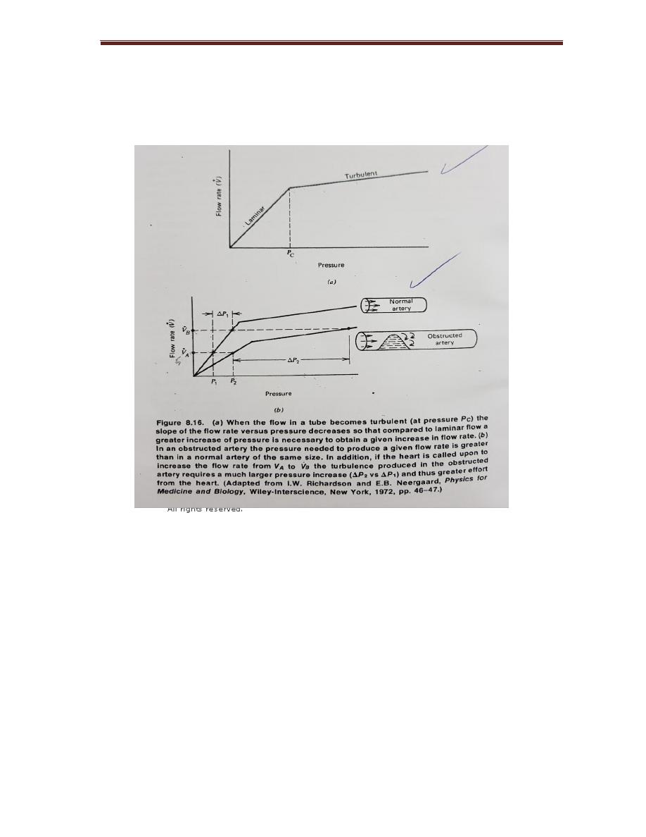
Blood Flow Laminar And Turbulent
Laminar (
silent) if all blood flow were laminar information could not be
obtained from the heart with stethoscope
If increase the velocity of the fluid in the tube by reduction the radius it will
reach the critical velocity Vc, when laminar flow change into turbulent flow. The
critical velocity will be lower if there is restriction or obstruction in the tube.
Osborne Rynold studied the property in 1883
Vc = k η / ρ R
R ; radius of the tube
K ; constant 1000 for many fluid
For aorta has radius = 1 cm in adults
College of Medicine/Babylon University
Medical Physics Module
Vc = ( 1000 ) ( 4 x 10
-3
pas ) / ( 10 ³ kg / cm³ ) ( 10̄
-2
m ) = 0.4 m /s
Figure 7-3:
effect of gradual tapering of tube on velocity
The Physics Of Some Cardiovascular Diseases
Work load of heart increases by ;
1- Hypertension
2- Tachycardia
Disease
1- Heart attack, caused by blockage one or more arteries of the heart muscle.
the blockage does not always immediately affect the electrical signal and a

48
College of Medicine/Babylon University
Medical Physics Module
person has heart attach may still have normal ECG.
2- Congestive heart failure, characterized by enlargement of the heart and
reduction in the ability of the heart to provide adequate circulation.
Medical treatment of congestive heart failure help to reduce the work load on
the heart.
3- Heart valve defects are of two types, the valve either does not open enough
(stenosis) or it does not close well enough (insufficiency)
The Physics OF Cardiovascular Diseases Involving In The Blood Vessels
1- A more common vessel problems is the formation sclerotic plaques on the
wall of the artery, increase the velocity in that region with a decrease in
wall pressure because of Bernoulli effect.
2- If the valve defective and let the blood run back down it will pool in the
vein, and the vein will become varicose.
The viscosity of blood depends on temperature change from (37-0 ) increases
the velocity of blood by a factor 2.5 in addition to viscosity ,
other factors affect the flow of blood in the vessel; the pressure difference
from one end to the other, the length of the vessel, and its radius.
Poiseulle’s law stats that the flow of a given tube depends on the pressure
difference from one end to the other P1-P2, the length of the tube L, the
radius R and viscosity of blood.
Flow rate = ( P1- P1 ) ( Π / 8 ) ( 1/ η ) ( R4 / L )
If radius is doubled the flow rate increases by 24 or by 16. This law applies
to the rigid tubes of constant radius, since the major arteries have elastic walls
and expand slightly at each heartbeat, so blood does not obey this law exactly

49
College of Medicine/Babylon University
Medical Physics Module
in addition the blood viscosity changes slightly with flow rate, however this
effect is negligible.
A disease clinically causes varicose vein, these enlarged surface veins in the
legs results from a failure of the one way valves in the veins.
The pressure in the leg vein is about 90 mmHg (115 cm of blood) due to a
column of blood above it.
The slandered treatment for varicose veins is surgical removal of offending
vessels. There are usually adequate parallel veins to carry the blood back to the
heart.