
Medical
biology
Histology & cell biology
and their methods of study
Dr. kaiser N. Madlum
Dep. of Anatomy &
Histology

•
All living things (or organisms) are built from
cells
•
Cells:
small
,
membrane enclosed
units filled
with a
concentrated aqueous solution of
chemicals
, and equipped with the
extraordinary
ability to create copies of themselves
by growing
and then dividing into two cells.
•
Higher organisms, including ourselves, are
communities of cells derived by growth and
division from a single founder cell.
Cell biology:
is the study of cells and their structure, function,
and behavior.
Histology
: is the study of the tissues of the body and how these
tissues are arranged to constitute organs.
Tissues are made of two interacting components: cells and
extracellular matrix. The main functions of extracellular matrix
are:
1- Provide a mechanical support for the cells,
2- Transport nutrients to the cells,
3- Carry away catabolites and secretory products.

❖ Cells produce the extracellular matrix, but it also influenced and
sometimes controlled by molecules of the matrix.
❖ Many components of the matrix recognized by and attaching to
receptors present on cell surfaces which are molecules that cross the
cell membranes and connect to structural components of the
intracellular cytoplasm.
❖ Each of the fundamental tissues (except the central nervous system)
is formed by several types of cells and typically by specific
associations of cells and extracellular matrix.

Tissues Preparation
It is the preparation of histological sections or tissue
slices that can be studied with the aid of the light
microscope.
They must be sectioned to obtain thin, transparent
sections and then attached to glass slides before they
can be examined.
The ideal microscopic tissue preparation should be
preserved so that the tissue on the slide has the same
structure and molecular composition as it had in the
body.

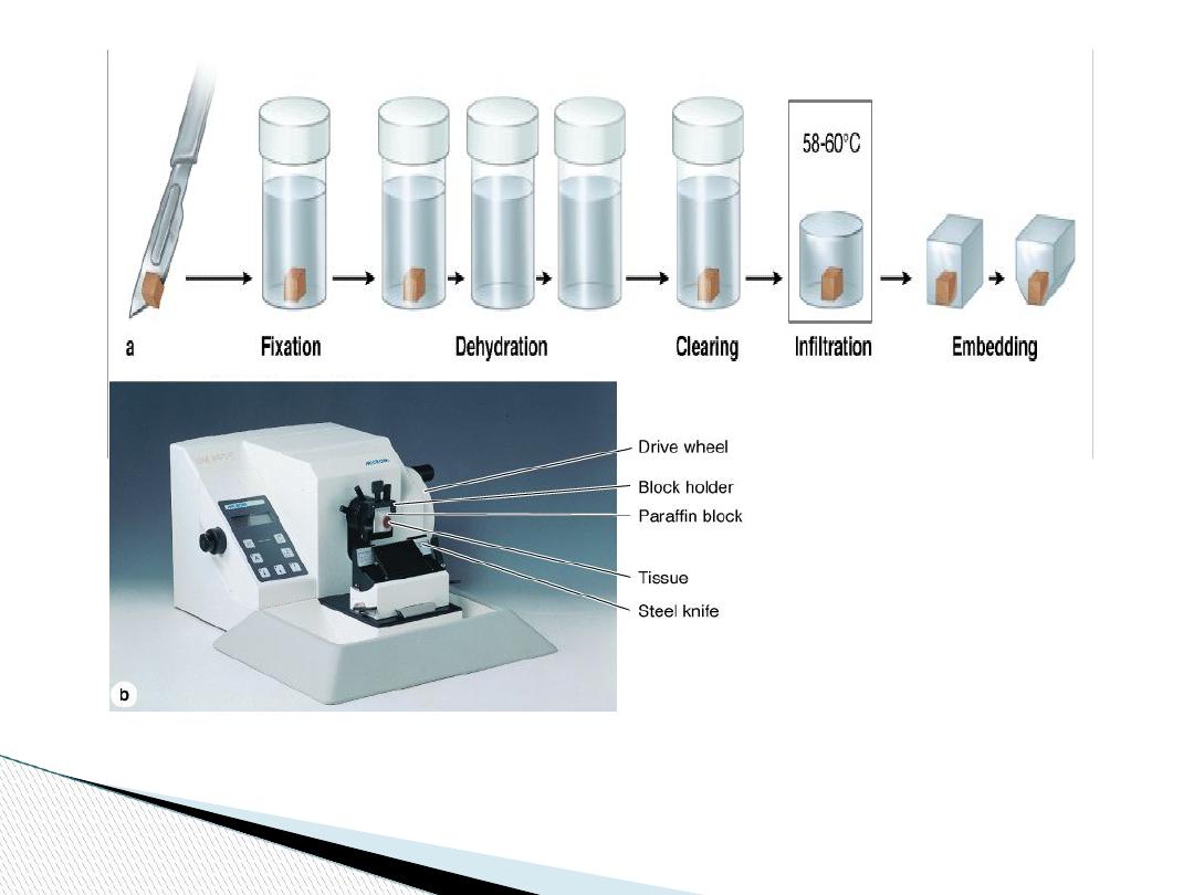
Preparation of Tissues for Study
1- Fixation
2- dehydration
3- clearing
4- Embedding
5- Sectioning
6- Staining
7- Mounting
To watch the processes online, please visit: https://youtu.be/TLm37BbR1mo
The basic steps used in tissue preparation for histology

The basic steps used in tissue preparation for histology are:
1-
Fixation
If a permanent section is desired, tissues must be fixed. Fixation is
used to:
1-
Terminate cell metabolism,
2- Prevent enzymatic degradation of cells and tissues by autolysis
(self-digestion).
3- Kill pathogenic microorganisms such as bacteria, fungi, and
viruses.
4- Harden the tissue as a result of either cross-linking or denaturing
protein molecules.

One of the best fixatives for routine light microscopy is
formalin
, a buffered isotonic solution of 37% formaldehyde.
Due to high resolution afforded by the electron microscope, a
double fixation procedure
, using a buffered
glutaraldehyde
solution followed by a second fixation in buffered
osmium
tetroxide
, is a standard procedure in preparations for fine
structural studies.
The effect of osmium tetroxide is to preserve and stain lipids and
proteins.

Fixation by freezing
It involves the submission of the tissues to rapid freezing.
A freezing microtome (
cryostat
) is then used to section the frozen
block with tissue. This method offers these advantages:
1- Allows the rapid preparation of sections
2- Effective in the histochemical study of very sensitive enzymes
or small molecules
3- Useful when structures containing lipids are to be studied (no
xylene).

2- Dehydration:
The water is first extracted by bathing tissue
successively in a graded series of mixtures of ethanol and water,
usually from 70% to 100% ethanol.
3- Clearing:
The ethanol is then replaced with other solvent xylene.
As the tissues are infiltrated with this solvent, they generally
become transparent.
4- Paraffin Embedding:
Tissues are usually embedded in a solid
medium to facilitate sectioning. Embedding substances gives a rigid
consistency to the tissue.
Embedding materials include paraffin and plastic resins.
Paraffin: for light microscopy
Resins: for both light and electron microscopy.

Once the tissue absorbs the solvent (xylene), it is placed in melted
paraffin in an oven, typically at 52–60°C. The heat causes the
solvent to evaporate, and the spaces within the tissues become filled
with paraffin.
The tissue together with its impregnating paraffin hardens after
removal from the oven.
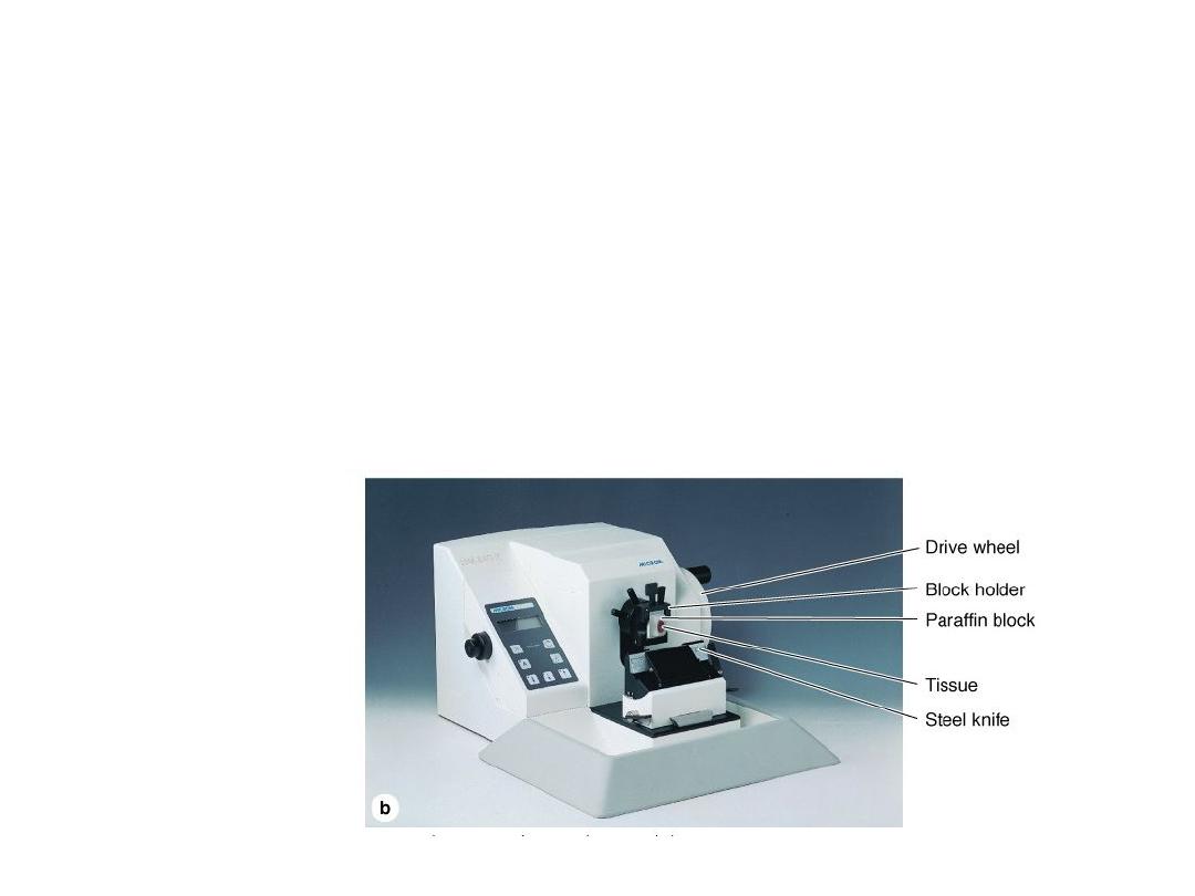
5- Sectioning
: The hard blocks containing the tissues are then
placed in the microtome and are sliced by the microtome's steel or
glass blade into sections 1 to 10 micrometers thick, (micrometer
(1um) = 1/1,000 of a millimeter (mm) = 10
–6
m). The sections are
floated on water and then transferred to glass slides to be stained.

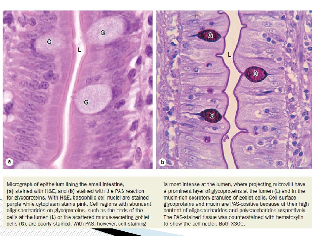
6- Staining
Sections must typically be stained or dyed because most tissues are
colorless
. Most of these dyes behave like
acidic
or
basic
compounds and have a tendency to form electrostatic (salt) linkages
with ionizable radicals of the tissues.
Tissue components with a net negative charge (
anionic
) stain more
readily with basic dyes and are termed basophilic;
cationic
components, such as proteins with many ionized amino groups,
have affinity for acidic dyes and are termed acidophilic.

Basic dyes
like toluidine blue, alcian blue, methylene blue and
Hematoxylin stain the nucleic acids, and acid glycoproteins.
Acid dyes
(eg, orange G, eosin, acid fuchsin) stain the
acidophilic components of tissues such as mitochondria,
secretory granules, and collagen
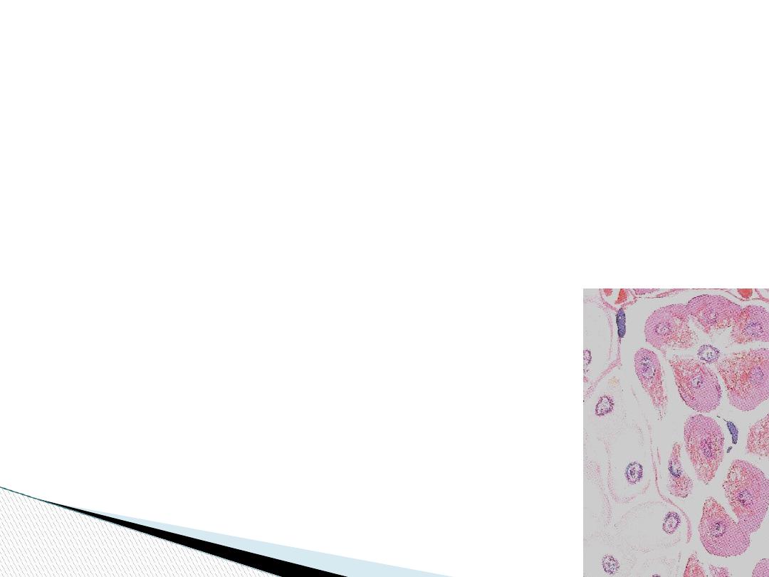
Combination of Hematoxylin and Eosin (H&E)
is used most
commonly.
Hematoxylin stains DNA of the cell nucleus and
other acidic structures (such as RNA-rich portions of the
cytoplasm and the matrix of cartilage) blue
. In contrast,
Eosin
stains other cytoplasmic components and collagen pink.

Medical applications
Biopsies are tissue samples removed during surgery or routine
medical procedures. In the operating room or medical center,
biopsies are fixed in vials of formalin for later processing and
microscopic analysis in a pathology laboratory. If results of such
analyses are required before the medical procedure is completed, for
example to know whether a growth is malignant before the patient
is closed, a much more rapid processing method is used. The biopsy
is rapidly frozen in liquid nitrogen, preserving cell structures and at
the same time making the tissue hard and ready for sectioning. The
frozen sections are placed on slides for rapid staining and
microscopic examination by a pathologist.