Lecture 6

C
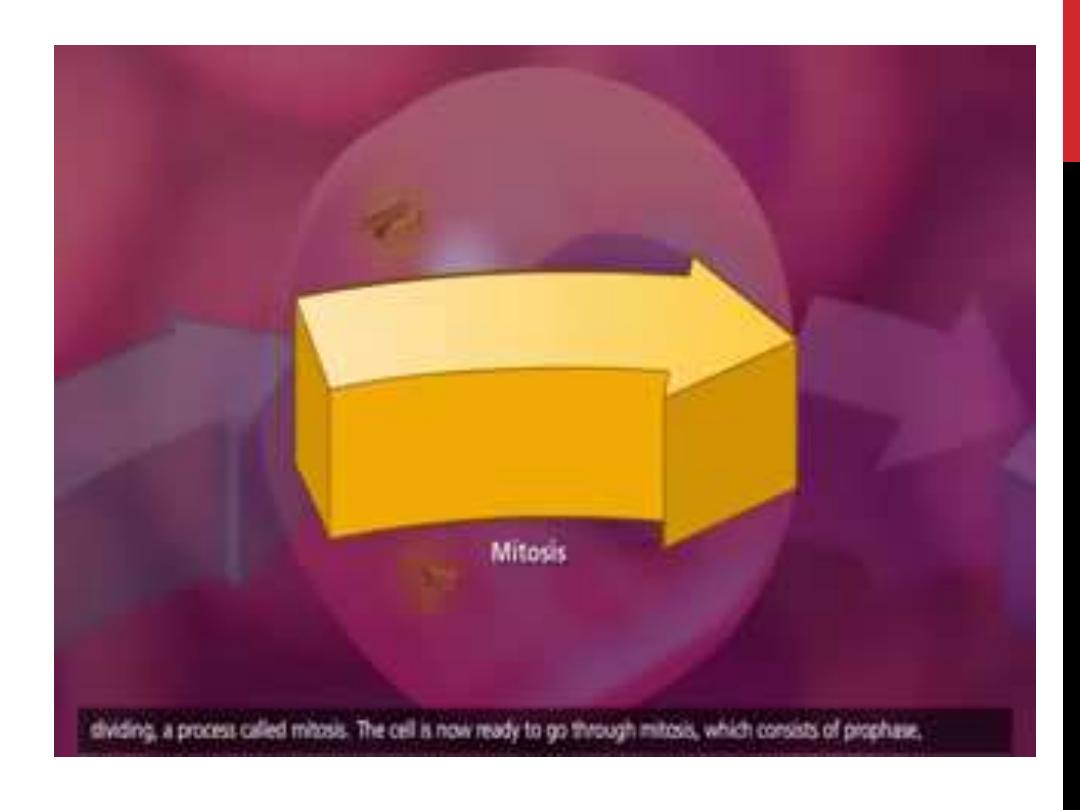
ell division, or
MITOSIS
, is the division of
somatic
cells and can be observed with the light
microscope.
During this process:
• The parent cell divides,
• Each of the daughter cells receives a
chromosomal set identical to that of the
parent cell
.

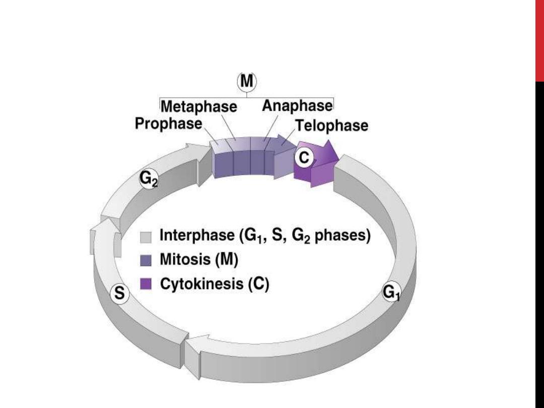
Cell cycle

•
longitudinal duplication of the chromosomes
takes place, and these chromosomes are
distributed to the daughter cells.
•
The
period
between
mitoses
is
called
Interphase
, during which the DNA is replicated
and the nucleus appears as it is most commonly
seen in histological preparations.
•
The process of mitosis is subdivided into four
phases

STAGES OF MITOSIS
Prophase
Metaphase
Anaphase
Telophase
Cell cycle
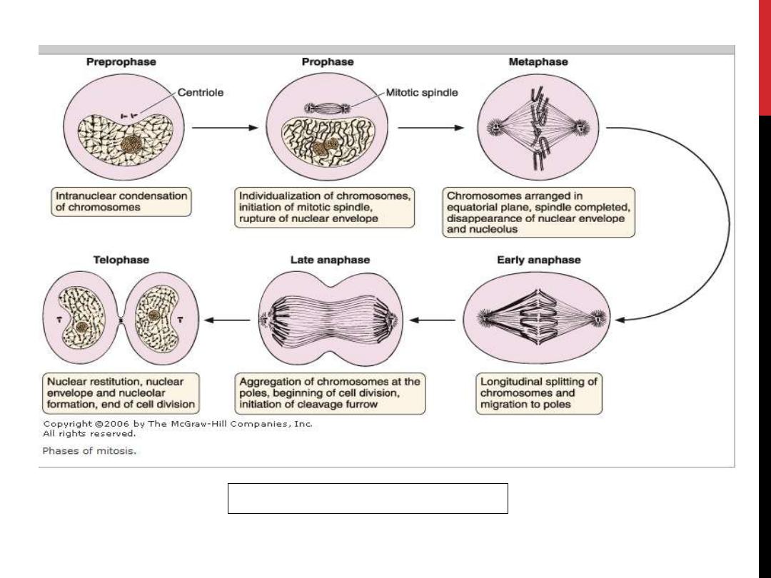
Figure 1: Phases of mitosis
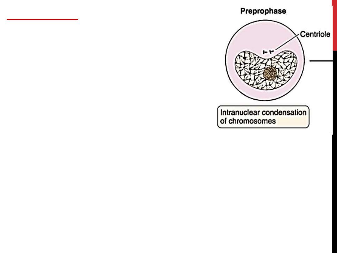
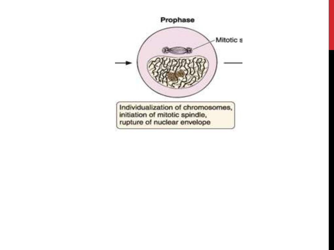
PROPHASE
1. The nuclear envelope begins to
disaggregate
2. The chromatin in the nucleus
begins to condense and becomes
visible by light microscopy as
elongated
,
spindly
chromosomes.
3. The nucleolus disappears.
4. Centrosomes begin migrating to opposite poles of the cell,
and microtubule fibers extend from centrosome to
centrosome and from centrosome to the
kinetochore
of
the centromere of each chromosome to form the mitotic
spindle.
Simultaneously with centrosome migration, the
microtubules of the mitotic spindle appear between the
two
centrosomes
, and the
nucleolus
disintegrates.
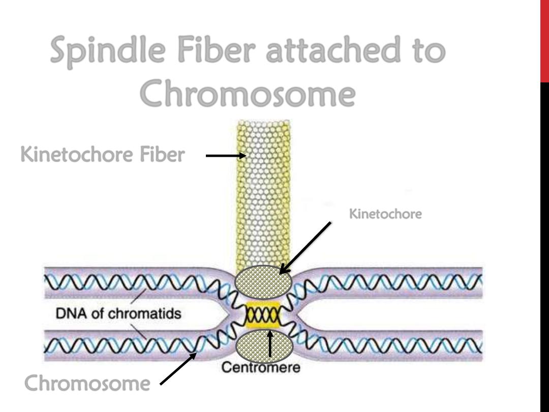
Spindle Fiber attached to
Chromosome
Kinetochore Fiber
Chromosome
Kinetochore

• Chromosomes migrate to the
equatorial plane
of
the cell,
• The chromatids attach to the microtubules of the
mitotic spindle at the
kinetochore
, located close
to the
centromere
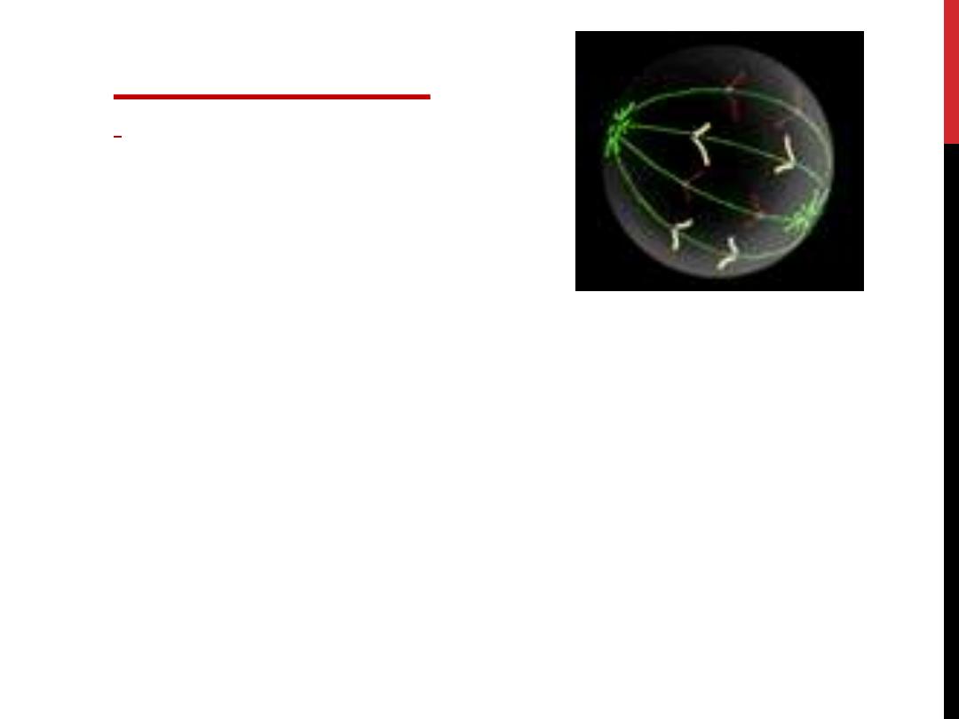
METAPHASE

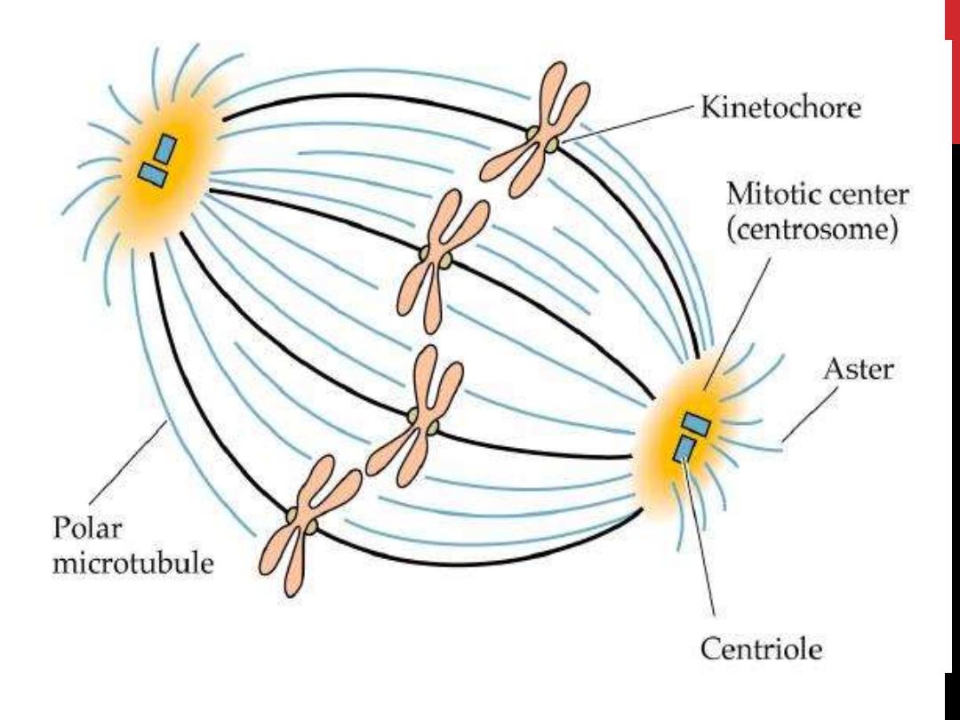
SPINDLE FIBRES
line the chromosomes along the middle
of the cell nucleus. This line is referred to as the
metaphase plate.
Polar microtubules
extend from the pole to the equator,
and typically
overlap
Kinetochore microtubules
extend from the pole to the
kinetochores
This organization helps to ensure that in the next phase,
when the chromosomes are separated, each new nucleus
will receive one copy of each chromosome.
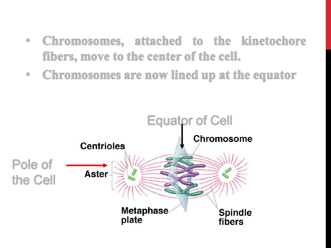
•
Chromosomes, attached to the kinetochore
fibers, move to the center of the cell.
•
Chromosomes are now lined up at the equator
14
Pole of
the Cell
Equator of Cell
ANAPHASE
1. the sister chromatids separate from each other and
migrate toward the opposite poles of the cell,
pulled by microtubules
.
2. Throughout this process, the centromeres move
away from the center, pulling the remainder of the
chromosome along.
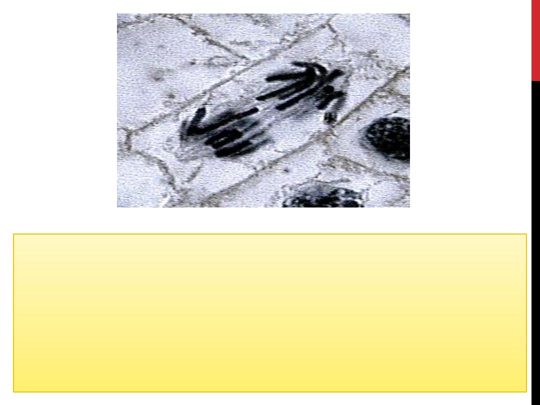
The
chromosomes
are
pulled
by
the
kinetochore
microtubules to the poles and form a "V" shape
Motion results
from a combination of kinetochore
movement along the spindle microtubules and through the
physical interaction of polar microtubules.
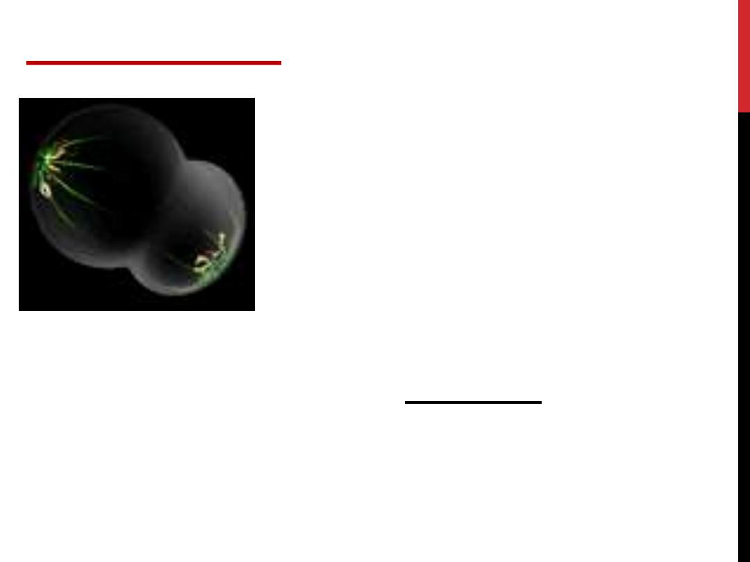
TELOPHASE
1. The reappearance of nuclei in the
daughter cells.
The spindle fibers disperse, and cytokinesis will start.
2.
The
chromosomes
revert
to
their
semidispersed
state,
and
the
nucleoli,
chromatin, and nuclear envelope reappear.

A
constriction
develops at the equatorial plane of
the parent cell and progresses until the cytoplasm
and its organelles are divided in two, this called the
cytokinesis
. In animal cells, cytokinesis results
when a fibre ring composed of a protein called
actin
around the centre of the cell contracts
dividing the cell into two daughter cells, each with
one nucleus.
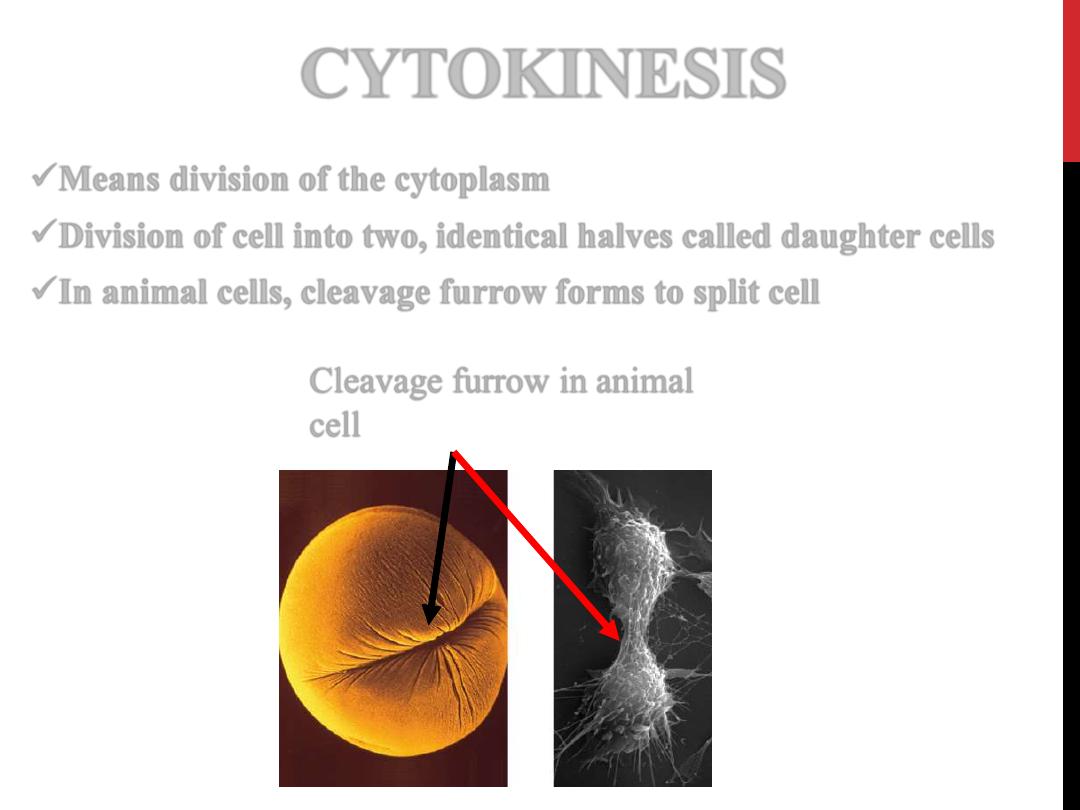
CYTOKINESIS
Means division of the cytoplasm
Division of cell into two, identical halves called daughter cells
In animal cells,
cleavage furrow
forms to split cell
19
Cleavage furrow in animal
cell
• Most tissues undergo constant cell turnover
because of continuous cell division and the
ongoing death of cells.
Nerve tissue
and
cardiac
muscle cells
are exceptions.
• The turnover rate of cells varies greatly from one
tissue to another,
rapid in the epithelium
of the
digestive tract and the epidermis and
slow in the
pancreas and the thyroid gland
.

22
Thank you