
Physiology of excitable tissues lect. 2 Asst. Prof. Dr. Zahid M. Kadhim
1
Action potential
Neurons communicate by generating electrical signals in the form of
changes in membrane potential. Some of these changes in membrane
potential trigger the release of neurotransmitter, which then carries a
signal to another cell.
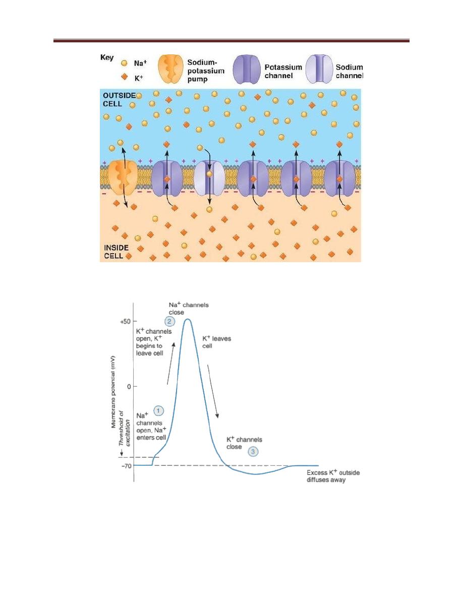
Resting membrane potential figure 1-10
a cell at rest —it is not receiving or transmitting any signals- has a
potential difference across its membrane such that the inside of the cell
is negatively charged relative to the outside. This difference is called
the resting membrane potential. The resting membrane potential of
neurons is approximately -70 mV.
In order for a potential difference to be present across a membrane
lipid bilayer, two conditions must be met. First, there must be an
unequal distribution of ions of one or more species across the
membrane (i.e., a concentration gradient). Second, the membrane
must be permeable to one or more of these ion species. The
permeability is provided by the existence of channels or pores in the bi-
layer; these channels are usually permeable to a single species of ions.
In neurons, the concentration of K is much higher inside than outside
the cell, while the reverse is the case for Na. This concentration
difference is established by Na-K-ATPase.
Action potential figure 1-11
Electrical signals occur in neurons via changes in membrane potential
that take place when certain ion channels, called gated channels, open
or close in response to particular stimuli.

Physiology of excitable tissues lect. 2 Asst. Prof. Dr. Zahid M. Kadhim
2
An action potential in a neuron consists of three distinct phases:
1. Rapid Depolarization. The first phase of an action potential is a
rapid depolarization during which the membrane potential
changes from -70 mV (rest) to +30 mV. This depolarization is
caused by a sudden and dramatic increase in permeability
to
sodium followed by an increase in the movement of sodium ions
into the cell, down sodium’s electrochemical gradient. With
permeability to sodium now greater than permeability to
potassium, the membrane potential approaches the sodium
equilibrium potential of +60 mV.
2. Repolarization. The second phase of an action potential is a
repolarization of the membrane potential during which the
membrane potential returns from +30 mV back to resting levels (-
70 mV).Within 1 msec after the increase in sodium permeability,
sodium permeability decreases rapidly, reducing the inflow of
sodium. At approximately the same time, potassium permeability
increases. Potassium then moves down its electrochemical
gradient out of the cell, repolarizing the membrane potential to
bring it back to resting levels.
3. After-hyperpolarization. The third phase of an action potential is
termed after-hyperpolarization. Potassium permeability remains
elevated for a brief time (5–15 msec) after the membrane
potential reaches the resting membrane potential, resulting in an
after-hyperpolarization. During this time, the membrane
potential is even more negative than at rest as it approaches the
potassium equilibrium potential (-94 mV).
Physiology of excitable tissues lect. 2 Asst. Prof. Dr. Zahid M. Kadhim
3
Figure 1-10 resting membrane potential
Figure 1-11 action potential of neuron