Physiology of excitable tissues lecture 4 Asst. Prof. Zahid M. Kadhim
1
-Propagation of action potential
Once an action potential is initiated in an axon, it is propagated down the length
of the axon from the trigger zone to the axon terminal without decrement. An
action potential does not actually travel down the axon; instead, an action
potential sets up electrochemical gradients in the extracellular and intracellular
fluids.
The propagation mechanisms differ, however, depending on whether the axon is
unmyelinated or myelinated.
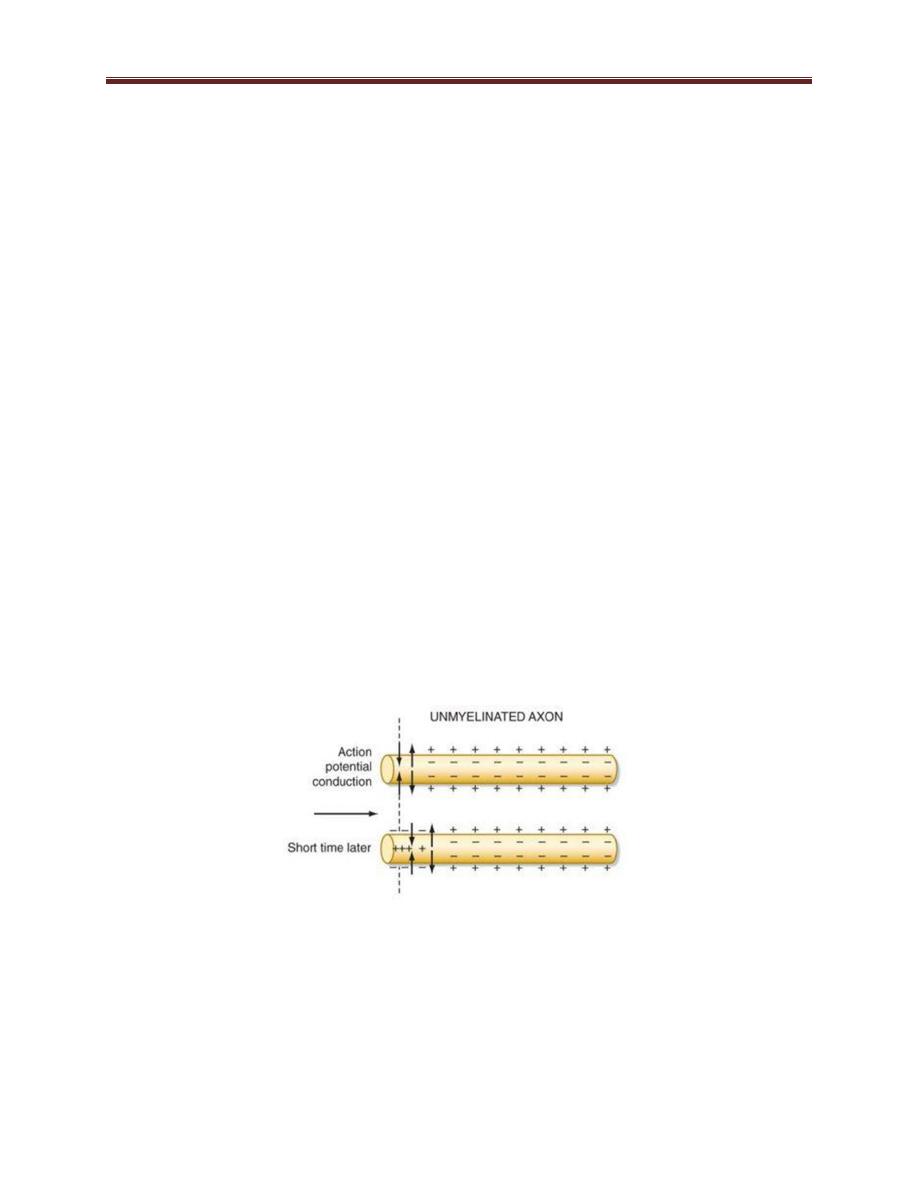
Propagation of Action Potentials in Unmyelinated Axons
Electrotonic conduction (figure 2-1) (is the passive spread of voltage changes
along a neuron, away from the site of origin) is the mechanism by which action
potentials are propagated in unmyelinated axons.
When a particular region of an axon is depolarized during an action potential, the
resulting currents travel downstream to adjacent regions of the membrane by
reversing the sign of the membrane potential, such that the inside of the cell
becomes positive and the outside becomes negative.
Figure 4-1: electrotonic conduction
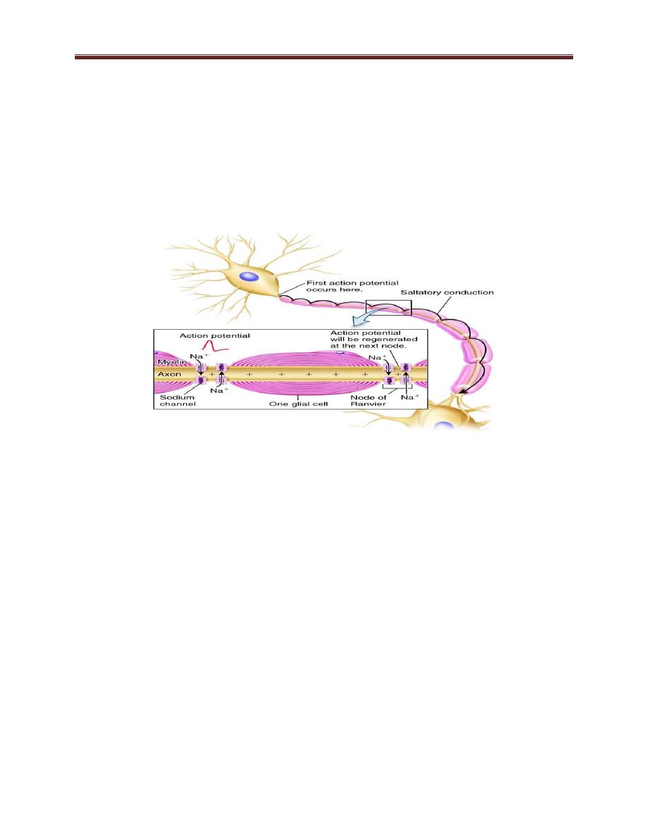
Action Potential Propagation in Myelinated Axons
In axons that are sheathed in myelin, action potentials are propagated by a
specialized type of electrotonic conduction called Saltatory conduction. Myelin
Physiology of excitable tissues lecture 4 Asst. Prof. Zahid M. Kadhim
2
provides high resistance to ion movement across the plasma membrane, but
longitudinal resistance is low. The nodes of Ranvier are gaps in the myelin where
the axon membrane lacks insulation, is exposed to the interstitial fluid, and has a
high concentration of voltage-gated sodium and potassium channels. In
myelinated fibers, action potentials are produced at the nodes of Ranvier.
Therefore, the current flow or jump from one node of Ranvier to the next until it
reach axon terminal.
Figure 2-2: salutatory conduction
Saltatory conduction (figure 2-2) is of value for two reasons. First, by causing the
depolarization process to jump long intervals along the axis of the nerve fiber, this
mechanism increases the velocity of nerve transmission in myelinated fibers as
much as 5- to 50-fold. Second, saltatory conduction conserves energy for the axon
because only the nodes depolarize, allowing perhaps 100 times less loss of ions
than would otherwise be necessary, and therefore requiring little energy
expenditure for re-establishing the sodium and potassium concentration
differences across the membrane after a series of nerve impulses.
The excellent insulation afforded by the myelin membrane and the 50-fold
decrease in membrane capacitance also allow repolarization to occur with little
transfer
Physiology of excitable tissues lecture 4 Asst. Prof. Zahid M. Kadhim
3
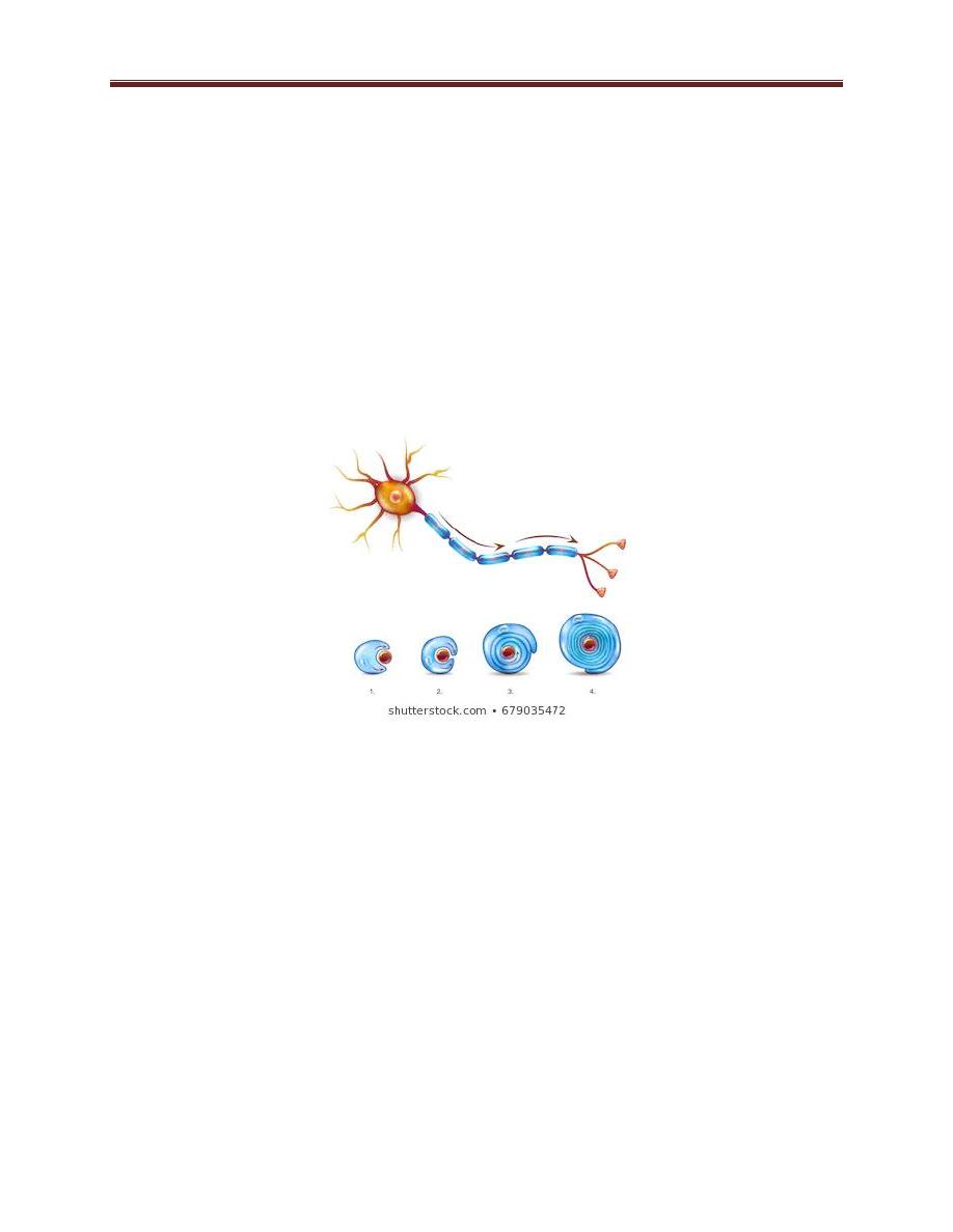
The myelin sheath is deposited around the axon by Schwann cells in the following
manner figure 3-3: The membrane of a Schwann cell first envelops the axon. The
Schwann cell then rotates around the axon many times, laying down
multiple
layers of Schwann cell membrane containing the lipid substance sphingomyelin.
This substance is an excellent electrical insulator that decreases ion flow through
the membrane about 5000-fold. At the juncture between each two successive
Schwann cells along the axon, a small uninsulated area only 2 to 3 micrometers in
length remains where ions still can flow with ease through the axon membrane
between the extracellular fluid and the intracellular fluid inside the axon. This
area is called the node of Ranvier.
Figure 3-3: formation of myelin sheath
Velocity of Conduction in Nerve Fibers. The velocity of action potential
conduction in nerve fibers varies from as little as 0.25 m/sec in small
unmyelinated fibers to as great as 100 m/sec (more than the length of a football
field in 1 second) in large myelinated fibers.
Velocity depends on many factors, the most important are
1- Heaviness of myelination, heavily myelinated nerve fibers conduct action
potential faster than lightly myelinated nerve fibers.
2- Diameter of nerve fiber (axon), the larger diameter the faster transmission
of impulses.
Physiology of excitable tissues lecture 4 Asst. Prof. Zahid M. Kadhim
4
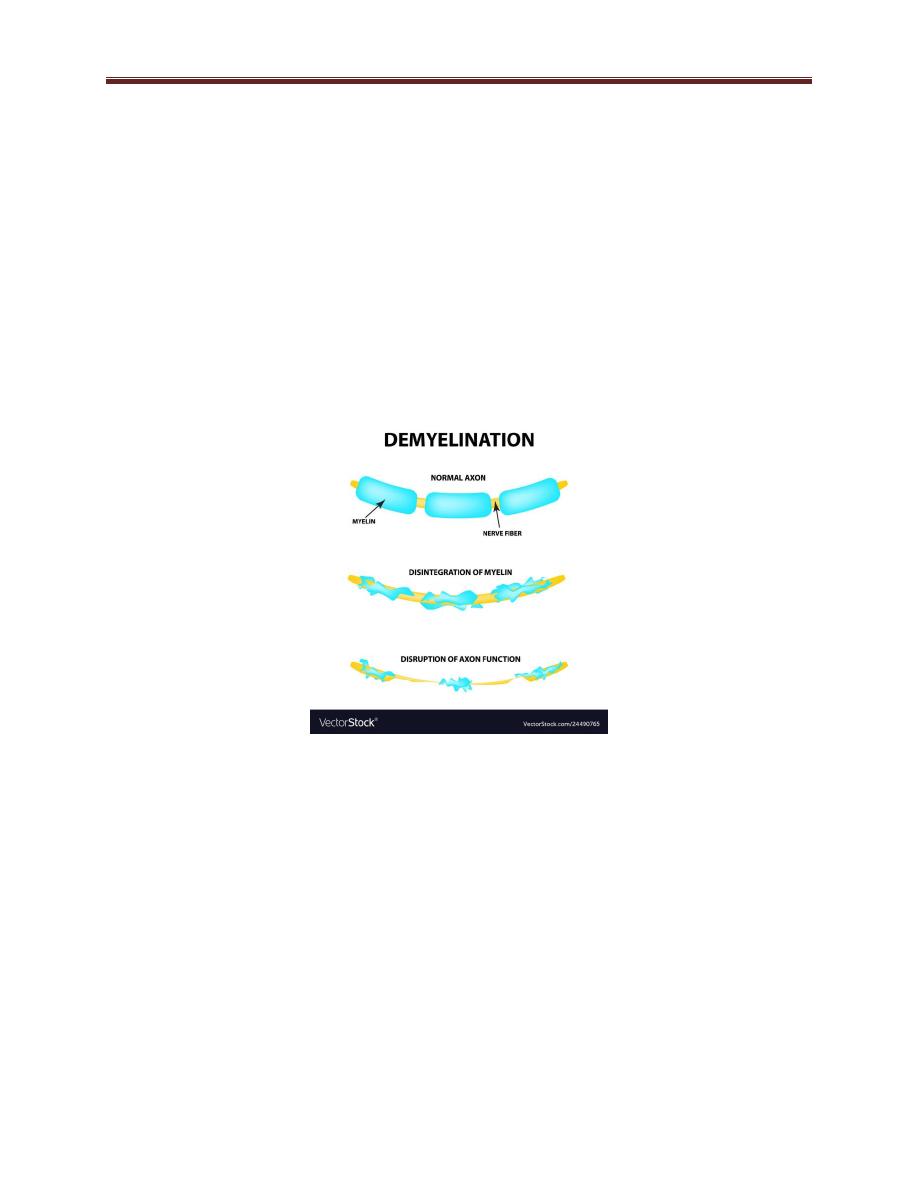
Demyelination
According to the function of myelin sheath, diseases that cause demyelination
(damage to myelin sheath), will affect transmission of nerve impulse along nerve
fiber and so if demyelination is partial there will be delay of transmission while if
there is complete demyelination (loss of entire myelin segment) there will be
block of electrical transmission.
Figure 3-4: demyelination
Guillian Barre syndrome is example of demyelinating disease that attacks
Schwann cells in the peripheral nervous system while multiple sclerosis is
example of demyelinating disease that attacks oligodendrocyte of central nervous
system.