Vesicoureteral reflux
Represents the retrograde flow of the urine from thebladder to the upper urinary tract
normally there is a functional VUJ valve prevent VUR and thus protect kidney from infection and high pressure (hydronephrosis )
The phenomenon of VUR represent balance of several factors include:
Functional integrity of the ureterAnatomic composition of the UVJ
Bladder compliance
the ureter pass obliquely through bladder wall 1-2cm
normally ratio of intera mural ureteric length to ureteric diameter is 5:1 for that reason if ureter more lateral more superior it have inadequate muscular support
A. Primary reflux
Is result from congenital abnormality of the UVJ usually involving longitudinal muscles of intramural ureterIsB –secondary reflux
either anatomic or functional
anatomic cause like:
Posterior urethral valve
Ectopic ureteral orifices
Ureterocele
Functional. Like:
-neurogenic bladder &
-bladder instability or dysfunction.
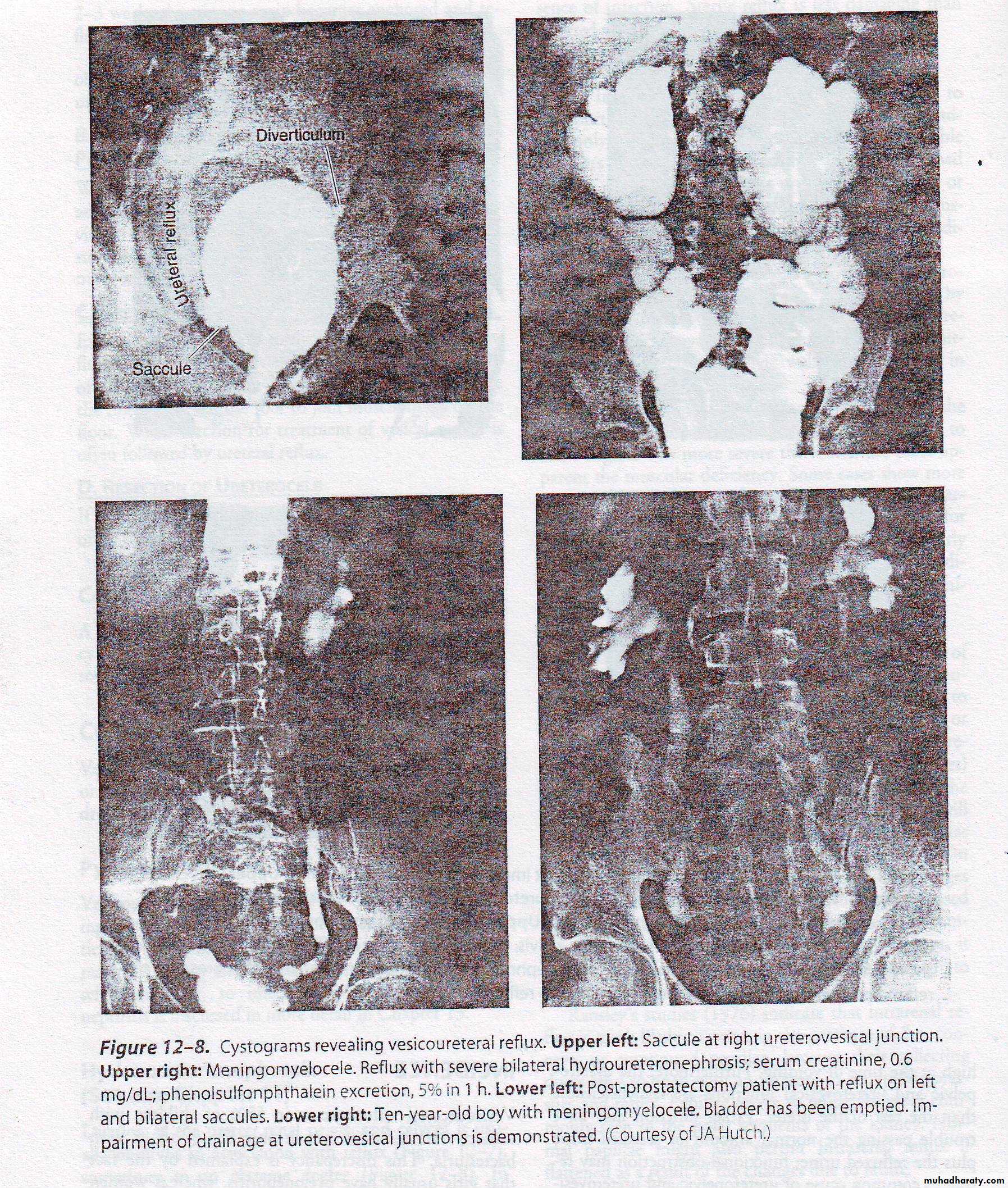
Grading of vesicoureteral reflux
Grade 1 reflux into the non dilated ureter.
Grade 2 into the pelvis & calyces without dilatation.Grade 3 mild dilatation of the ureter renal pelvis & calyces.
Grade 4 moderate dilatation of the ureter pelvis & calyces.
Grade 5 gross dilatation of ureter, pelvis & calyces.
Demography
Prevalence:It approximately 30%in children with UTI and 17% with out UTI.
Gender:
During the 1st year most are boys with posterior urtheral valves
after 1year the female: male ratio of infection with reflux is approximately 3-4:1
Rase:
10 time lower in female children of African descent
Inheritance : autosomal dominant
Diagnosis:
• Clinical findings• Symptoms related to reflux
• Symptomatic pyelonephritis
• Symptom of cystitis
• Renal pain on voiding
• Uraemia
• Hypertension
• Symptoms related to underlying disease
• Urinary tract obstruction
• Spinal cord disease
2- physical findings
During attack of acute pyelonephritis renal tenderness
Palpation and percussion of suprapubic area may reveal distended bladder
3-Lab.finding
Infection,bacteriuria,pyuria,high serum creatinine
Therefore a urine culture should be included in the evaluation of any infant or child who presents with fever & malaise
When reflux has gone undetected & renal scarring has occurred children of any age can present with
renal insufficiency,
hypertension, &
impaired somatic growth.
Complication of reflux
Pyelonephritis
hydroureteronephrosis
x-Ray finding
Plain film may reveal evidence of spina bifida or
meningomyelocele thus point to the neurologic deficit.
Excretory urograms may be
-normal, or
-dilatation of whole or part of ureter or
-hydroureteronephrosis.
Reflux is diagnosed by
voiding cystourethrography orvoiding cinefluoroscopy
Cystoscopy.
For
Morphology (stadium or horseshoe or golf hole orifice)
Position.
Treatment: medical
Maintaining urinary sterility by using single daily low dose antimicrobial prophylaxisNight time dosing allow to cover period of physiological retention
If child have infected urine then gave high dose antibiotic to sterile the urine then
continuo on low dose antibiotic
Antibiotic
Age less than 2 months we commaly use trimethoprin and amoxicillin
After 2 months antibiotic of choice is trimethoprin-sulfamethoxazole
Then follow up every 3 months by uls and urine cultures and some time need yearly radionuleotide scanning
*In toilet trained children bladder emptying by
timed voids, double voiding, help to achieve the goals
of medical management.
.
B-Surgery (ureteric Reimplantion)
Typical indication of antireflux surgery include:-1- breakthrough UTI despite prophylactic antibiotic.
2- noncompliance with medical management.3- sever reflux grade 4 or 5.
4- failure of renal growth, new scars, or deterioration of renal function on follow up ultrasound.
5- reflux persist to puberty specially in girls.
6- reflux associated with congenital abnormalities such as bladder diverticulum.
MEGAURETER
It mean a dilated ureter ,normally ureteric diameter about 5mm if it accede 7-8mm then it consider MGUsClassification
a megaureter may be obstructed, refluxing, both refluxing and obstructed, or unobstructed and not refluxing, either from a primary (idiopathic cause intrinsic to the ureter or secondary to specific pathophysiologic processes, such as outlet obstruction, neurogenic dysfunction, polyuria, or infection).
Primary (at the UVJ; adynamic aprstalitic segment) or secondary (e.g.,bladder malfunction) origins influence management and must therefore be differentiated.
Indications for correction are often driven by serially increasing pelvicalyceal dilation, increasing ureteral diameter, or pyelonephritis and ureteral pyuria.
Antibiotic prophylaxis should be used to protect the dilated ureter regardless of cause.
Many cases of antenatally diagnosed MGU will resolve spontaneously.If there is improvement in degree of hydroureteronephrosis, but not resolution, imaging at puberty is advised.