CHEST ANATOMY THI-QAR UNIVERSITY
COLLEGE OF MEDICINE
LECTURE 3 2019/2020
Dr. Rafid AL-Temimi ; Clinical radiology ( CABM)
Page
1
Dr. Ahmed Abdulameer Daffar ; Thoracic & Vascular Surgeon ( FIBMS )
THE CHEST
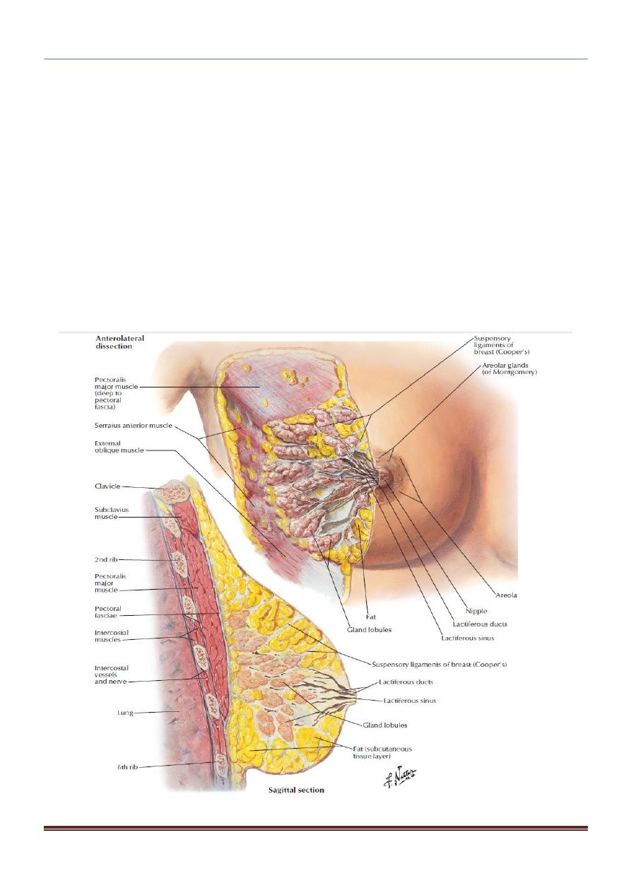
THE BREASTS:
The breasts are specialized accessory glands of the skin that secrete milk
.
The nipples are small and surrounded by a colored area of skin called the areola.
The base of the breast extends from the 2
nd
to 6th rib and from the lateral margin of
the sternum to the midaxillary line.
The greater part of the gland lies in the superficial fascia. A small part, called the
axillary tail , extends upward and laterally.
Each breast consists of 15 to 20 lobes, which radiate out from the nipple.
The lobes of the gland are separated by fibrous septa that serve as suspensory
ligaments.
Behind the breasts is a space filled by loose connective tissue called the
retromammary space
CHEST ANATOMY THI-QAR UNIVERSITY
COLLEGE OF MEDICINE
LECTURE 3 2019/2020
Dr. Rafid AL-Temimi ; Clinical radiology ( CABM)
Page
2
Dr. Ahmed Abdulameer Daffar ; Thoracic & Vascular Surgeon ( FIBMS )
o
Blood Supply
Arteries:
The branches to the breasts include the perforating branches of the internal thoracic artery
and the intercostal arteries.
The axillary artery also supplies the gland via its lateral thoracic and thoracoacromial
branches.
Veins:
The veins correspond to the arteries.
MEDIASTINUM
The mediastinum, though thick, is a movable partition that extends superiorly to the
thoracic outlet and the root of the neck and inferiorly to the diaphragm.
It extends anteriorly to the sternum and posteriorly to the vertebral column.
The mediastinum is divided into superior and inferior mediastina by an imaginary
plane passing from the sternal angle anteriorly to the lower border of the body of the
4
th
thoracic vertebra posteriorly.

CHEST ANATOMY THI-QAR UNIVERSITY
COLLEGE OF MEDICINE
LECTURE 3 2019/2020
Dr. Rafid AL-Temimi ; Clinical radiology ( CABM)
Page
3
Dr. Ahmed Abdulameer Daffar ; Thoracic & Vascular Surgeon ( FIBMS )
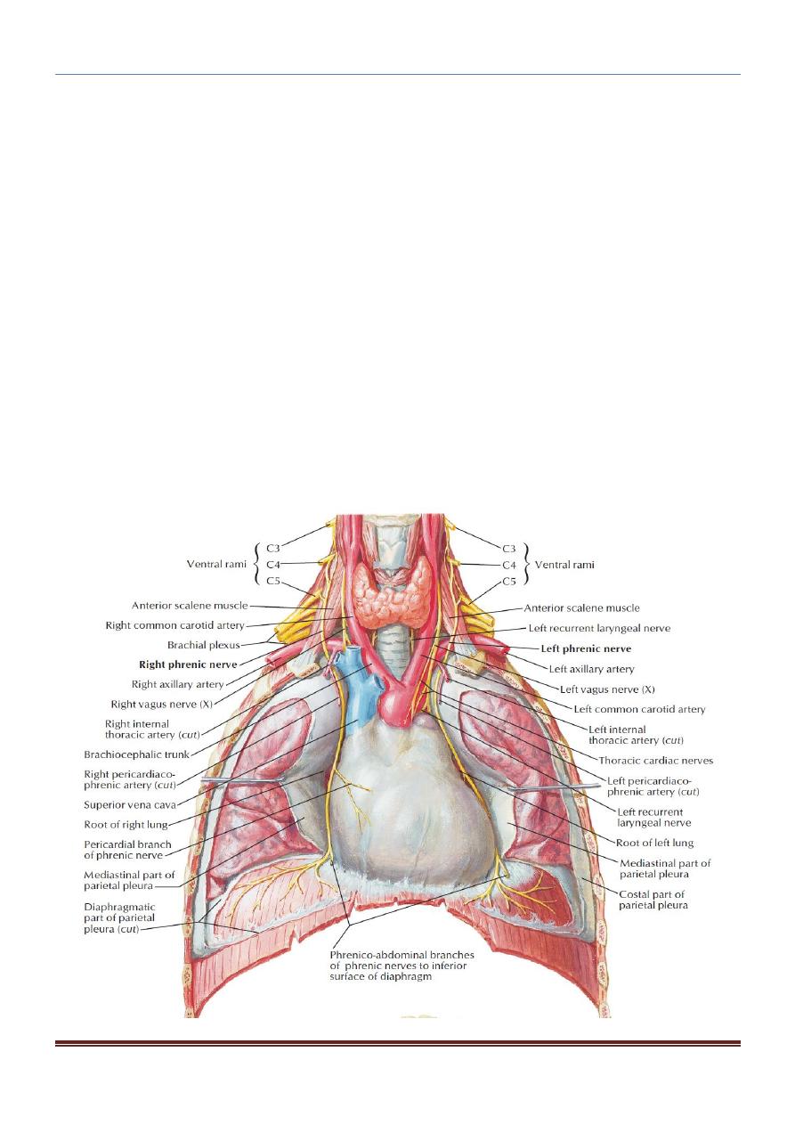
Superior Mediastinum: the contents
1. Thymus.
2. Large veins. ( brachiocephalic veins ; superior vena cava )
3. Large arteries. (Arch of aorta & its branches).
4. Sympathetic trunks & nerves (phrenic; vagus and left recurrent largngeal nerves).
5. Trachea.
6. Esophagus.
7. Thoracic duct; lymph nodes.
Inferior Mediastinum: the contents
a) Thymus ; lymph nodes
b) Heart within the pericardium with the phrenic nerves on each side
c) Esophagus.
d) Thoracic duct.
e) Descending aorta.
f) Sympathetic trunks & nerves.
g) Azygous & hemiazygous venous system.
The inferior mediastinum is further subdivided into the middle mediastinum, which
consists of the pericardium and heart; the anterior mediastinum, which is a space
between the pericardium and the sternum; and the posterior mediastinum, which
lies between the pericardium and the vertebral column.
Anterior Mediastinum: the contents
1. Thymus
2. Lymph nodes.
Middle Mediastinum: the contents
1. Heart & Pericardium.
2. Great vessels.
3. Trachea bifurcation and both main bronchi.
Posterior Mediastinum: the contents
1. Descending aorta.
2. Esophagus.
3. Thoracic duct & lymph node.
4. Azygous & hemiazygous venous system.
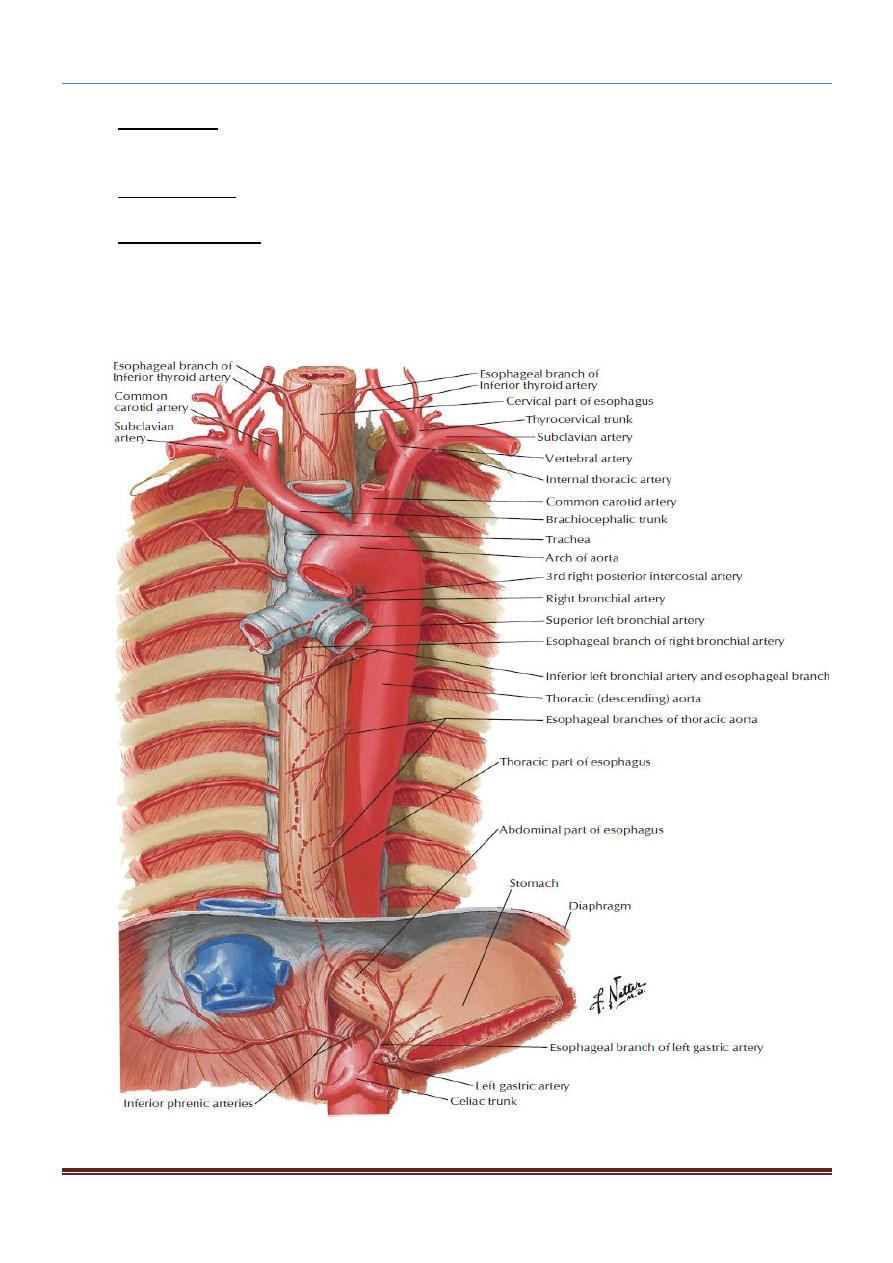
Esophagus
The esophagus is a tubular structure about 10 in. (25 cm) long that is continuous above
with the laryngeal part of the pharynx opposite the sixth cervical vertebra.
It passes through the diaphragm at the level of the 10th thoracic vertebra to join the
stomach.
CHEST ANATOMY THI-QAR UNIVERSITY
COLLEGE OF MEDICINE
LECTURE 3 2019/2020
Dr. Rafid AL-Temimi ; Clinical radiology ( CABM)
Page
4
Dr. Ahmed Abdulameer Daffar ; Thoracic & Vascular Surgeon ( FIBMS )
In the neck, the esophagus lies in front of the vertebral column; laterally, it is related
to the lobes of the thyroid gland; and anteriorly, it is in contact with the trachea and
the recurrent laryngeal nerves .
In the thorax, it passes downward and to the left through the superior and then the
posterior mediastinum.
In the abdomen, the esophagus descends for about 0.5 in. (1.3 cm) and then enters
the stomach. It is related to the left lobe of the liver anteriorly and to the left crus of
the diaphragm posteriorly.

CHEST ANATOMY THI-QAR UNIVERSITY
COLLEGE OF MEDICINE
LECTURE 3 2019/2020
Dr. Rafid AL-Temimi ; Clinical radiology ( CABM)
Page
5
Dr. Ahmed Abdulameer Daffar ; Thoracic & Vascular Surgeon ( FIBMS )
Blood Supply of the Esophagus:
o
The upper third of the esophagus is supplied by the inferior thyroid artery.
o
The middle third by branches from the descending thoracic aorta.
o
The lower third by branches from the left gastric artery.
The veins from the upper third drain into the inferior thyroid veins, from the middle third
into the azygos veins, and from the lower third into the left gastric vein, a tributary of the
portal vein.
Thymus:
The thymus is a flattened, bilobed structure; lying between the sternum and the
pericardium in the anterior mediastinum.
Blood Supply
The blood supply of the thymus is from the inferior thyroid and internal thoracic arteries
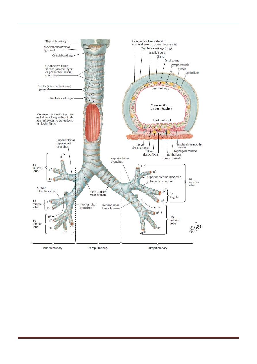
Trachea:
The trachea begins in the neck as a continuation of the larynx at the lower border of the
cricoid cartilage at the level of the 6th cervical vertebra.
It descends in the midline of the neck.
In the thorax, the trachea ends below at the carina by dividing into right and left
principal (main) bronchi at the level of the sternal angle.
Blood Supply of the Trachea
The upper two thirds are supplied by the inferior thyroid arteries and the lower third is
supplied by the bronchial arteries.
CHEST ANATOMY THI-QAR UNIVERSITY
COLLEGE OF MEDICINE
LECTURE 3 2019/2020
Dr. Rafid AL-Temimi ; Clinical radiology ( CABM)
Page
6
Dr. Ahmed Abdulameer Daffar ; Thoracic & Vascular Surgeon ( FIBMS )