1l
BONE
Is mineralized and specialized connective tissue confers mechanical and
metabolic functions to the skeleton
Bone is composed of intercellular calcified material, (matrix),
and 3 cell types:
osteocytes (Gr. Osteon, bone +cytos, cell), which are found in
cavities (lacunae) within the matrix,
osteoblasts (Osteon, bone + blasts, germ), which synthesize the
organic components of the matrix,
osteoclast (Osteon, bone + klastos, broken), which are
multinucleated giant cells involved in the resorption and
remodeling of bone tissue.
Bone functions are:
1. Supports fleshy structures.
2. Protects vital organs such as those in the cranial and thoracic
cavities.
3. Harbors the bone marrow, where blood cells are formed.
4. Acts as a reservoir of calcium, phosphate, and other ions that can be
released or stored in a controlled fusion to maintain constant
concentrations of these important ions in body fluids.
5. Form a system of levers that multiply the forces generated during
skeletal muscle contraction and transform them into bodily
movement.
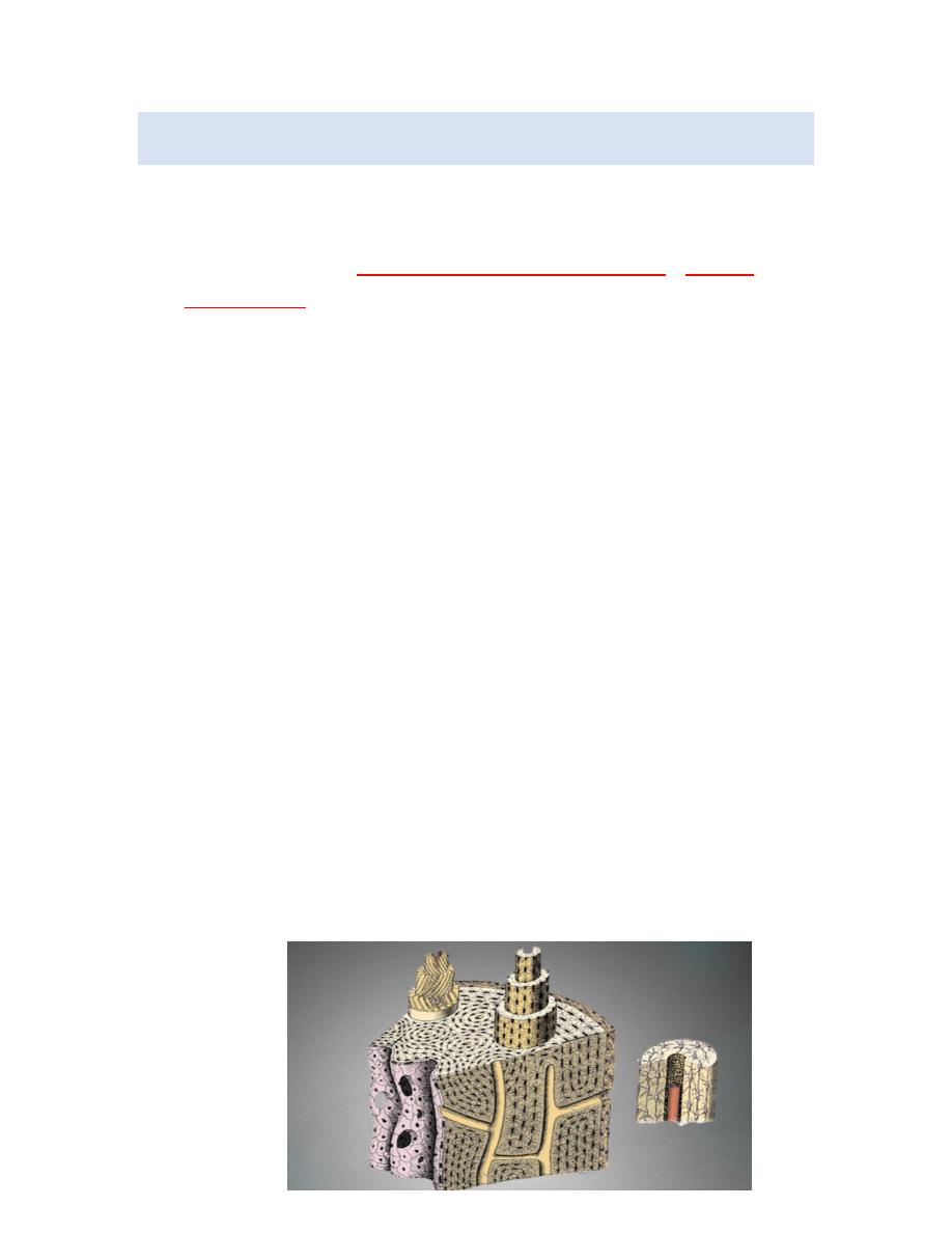
2l
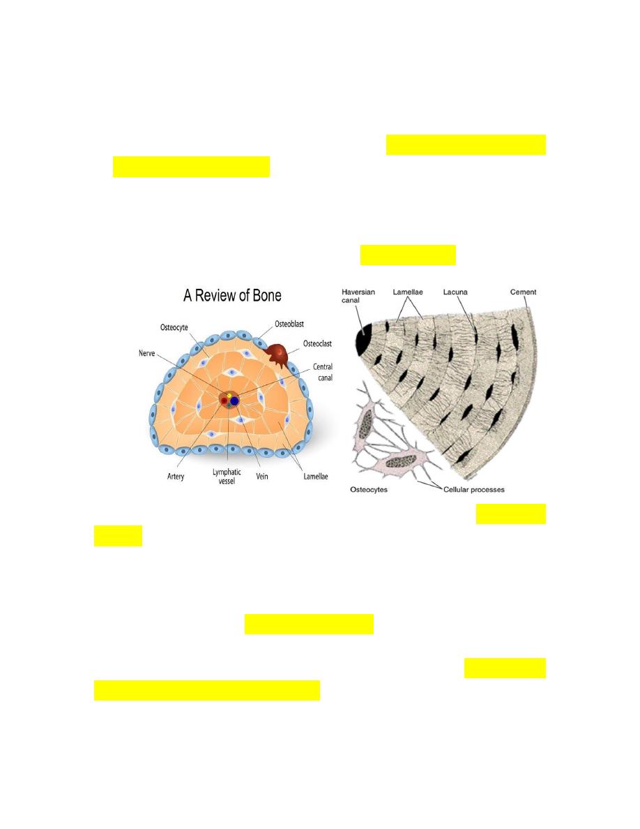
Schematic drawing of the wall of a long-bone diaphysis showing
3 types of lamellar bone: Haversian system and outer and inner
circumferential lamellae. The protruding Haversian system on the
left shows the orientation of collagen fibers in each lamella. At
the right is a Haversian system showing lamellae, a central blood
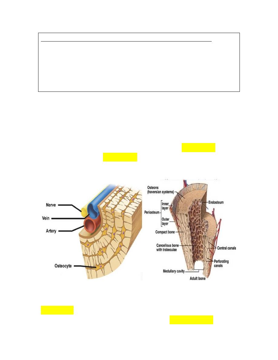
capillary, and many osteocytes with their processes.
metabolites are unable to diffuse through the calcified matrix of
the bone, the exchanges between osteocytes and blood capillaries
depend on communication through the canaliculi (L. canalis,
canal), which are thin, cylindrical spaces that perforate the
matrix. All bones are lined on both internal and external surfaces
by layers of tissue containing osteogenic cells
endosteum on the
internal surface and periosteum on the external surface
.
because if its hardness, bone is difficult to section with the
microtome, and special technique that permits the observation of
the cells and organic matrix is based on the decalcification of bone
3l
preserved by standard fixatives. The mineral is removed by
immersion in solution containing calcium-chelating substance
(Ethyline diamine tetra acetic acid)
[EDTA] the decalcified tissue is then
embedded, sectioned and stained.
4l
BONE CELLS
Osteoblasts: osteoblasts are responsible for the synthesis of
the organic component of bone matrix (type collagen,
proteoglycans, and glycoproteins).
Depositions of the inorganic component of bone depend on
the presence of osteoblasts.
Osteoblasts are exclusively located at the surfaces of bone
tissue, side by side, in a way that resembles simple
epithelium. When they are actively engaged in matrix
synthesis.
Osteoblasts have cuboidal to columnar shape and basophilic
cytoplasm when their synthesizing activity declines, they
flatten and cytoplasmic basophilia declines.
Some osteoblasts are gradually surrounded by newly
formed matrix and become osteocytes. During this process a
space called a lacuna is formed.
lacuna are occupied by osteocytes and their extension along
with a small amount of extra cellular no calcified matrix.
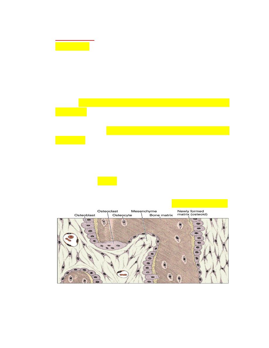
5l
Osteocytes
Osteocytes which derive from osteoblast lie in the lacunae
situated between lamella of matrix. Only one osteocyte is
found in each lacuna, the thin cylindrical matrix canaliculi
house cytoplasmic processes of osteocytes processes of
adjacent cells make contact via gap junction and molecules are
passed via this structures from cell to cell
.
this exchange can
provide nourishment for a chain of about 15 cells
Schematic drawing of 2 osteocytes and part of a Haversian
system. Collagen fibers of contiguous lamellae are sectioned at
different angles. Note the numerous canaliculi that permit
communication between lacunae and with the Haversian canals.
Each lamella consists of multiple parallel arrays of collagen fibers.
In adjacent lamellae, the collagen fibers are oriented in different
directions. The presence of large numbers of lamellae with
differing fiber orientations provides the bone with great
strength,despite its light weight.
When compared with osteoblasts the flat, almond-shaped
osteocyte exhibit a significantly reduce rough endoplasmic
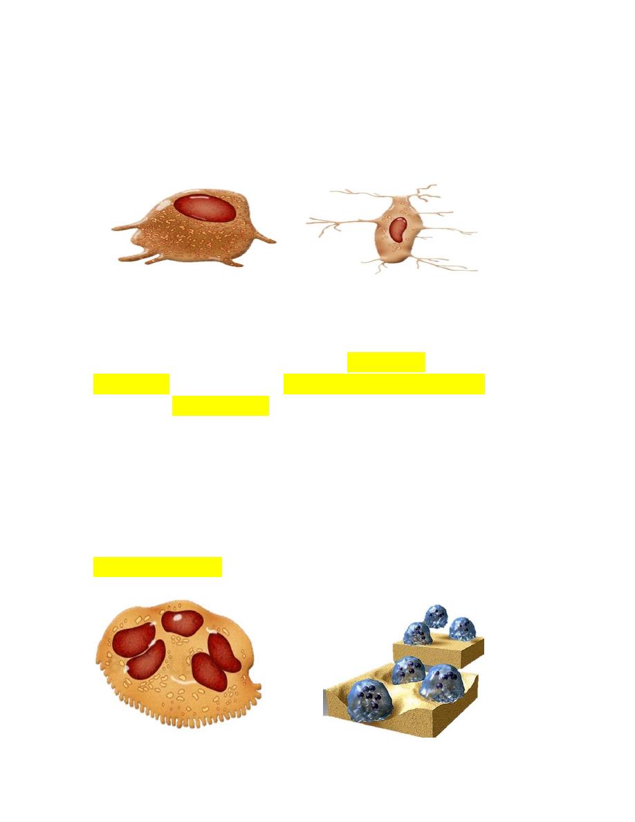
6l
reticulum and Golgi complex and more condensed nuclear
chromatin.
These cells are actively involved in the maintenance of bony
matrix and their death is followed by resorption of this matrix
Osteoclasts
While bone matrix is deposited by osteoblasts, it is eroded by
osteoclasts, these large (20 to 100Mm in diameter) multi-
nucleated (2 to 50 nuclei), cells are a type of macrophage. Like
other microphages they develop from monocytes that originated
in the hemopoitic tissue of the bone marrow.
These precursor cells are released into the bloodstream and
collect at sites of bone reabsoption where they fuse to form the
multinucleated osteoclasts. They are found close association with
the surface of bone, often in shallow excavations known as
Howships lacunae.

7l
Pdf by zahraa jalil