CHEST ANATOMY THI-QAR UNIVERSITY
COLLEGE OF MEDICINE
LECTURE 5 2019/2020
Dr. Rafid AL-Temimi ; Clinical radiology ( CABM
Page
1
Dr. Ahmed Abdulameer Daffar ; Thoracic & Vascular Surgeon ( FIBMS )
THE CHEST
HEART
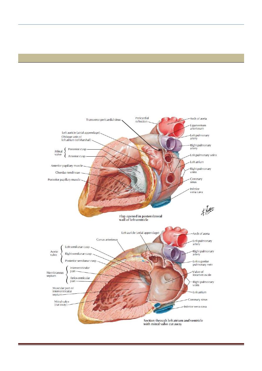
3. Left Atrium:
Similar to the right atrium, the left atrium consists of a main cavity and a left
auricle.
The left atrium is situated behind the right atrium and forms the greater part of
the base or the posterior surface of the heart.
The interior of the left atrium is smooth, but the left auricle possesses muscular
ridges as in the right auricle.
CHEST ANATOMY THI-QAR UNIVERSITY
COLLEGE OF MEDICINE
LECTURE 5 2019/2020
Dr. Rafid AL-Temimi ; Clinical radiology ( CABM
Page
2
Dr. Ahmed Abdulameer Daffar ; Thoracic & Vascular Surgeon ( FIBMS )
Openings into the Left Atrium:
The four pulmonary veins, two from each lung, open through the posterior wall and have
no valves.
The left atrioventricular orifice is guarded by the mitral valve.
4. Left Ventricle:
The left ventricle communicates with the left atrium through the atrioventricular
orifice and with the aorta through the aortic orifice.
The walls of the left ventricle are three times thicker than those of the right
ventricle. (The left intraventricular blood pressure is six times higher than that
inside the right ventricle.)
In cross section, the left ventricle is circular; the right is crescentic because of the
bulging of the ventricular septum into the cavity of the right ventricle .
The part of the ventricle below the aortic orifice is called the aortic vestibule.
The mitral valve guards the atrioventricular orifice. It consists of two cusps, one
anterior and one posterior, which have a structure similar to that of the cusps of
the tricuspid valve.
The attachment of the chordae tendineae to the cusps and the papillary muscles is
similar to that of the tricuspid valve.
The aortic valve guards the aortic orifice and is precisely similar in structure to
the pulmonary valve.
Behind each cusp, the aortic wall bulges to form an aortic sinus. The anterior
aortic sinus gives origin to the right coronary artery, and the left posterior sinus
gives origin to the left coronary artery.
Structure of the Heart:
- The walls of the heart are composed of a thick layer of cardiac muscle, the
myocardium, covered externally by the epicardium and lined internally by the
endocardium.
- The atrial portion of the heart has relatively thin walls and is divided by the atrial
(interatrial) septum into the right and left atria.

CHEST ANATOMY THI-QAR UNIVERSITY
COLLEGE OF MEDICINE
LECTURE 5 2019/2020
Dr. Rafid AL-Temimi ; Clinical radiology ( CABM
Page
3
Dr. Ahmed Abdulameer Daffar ; Thoracic & Vascular Surgeon ( FIBMS )
- The septum runs from the anterior wall of the heart backward and to the right. The
ventricular portion of the heart has thick walls and is divided by the ventricular
(interventricular) septum into the right and left ventricles.
- The septum is placed obliquely, with one surface facing forward and to the right and
the other facing backward and to the left.
- Its position is indicated on the surface of the heart by the anterior and posterior
interventricular grooves. The lower part of the septum is thick and formed of
muscle.
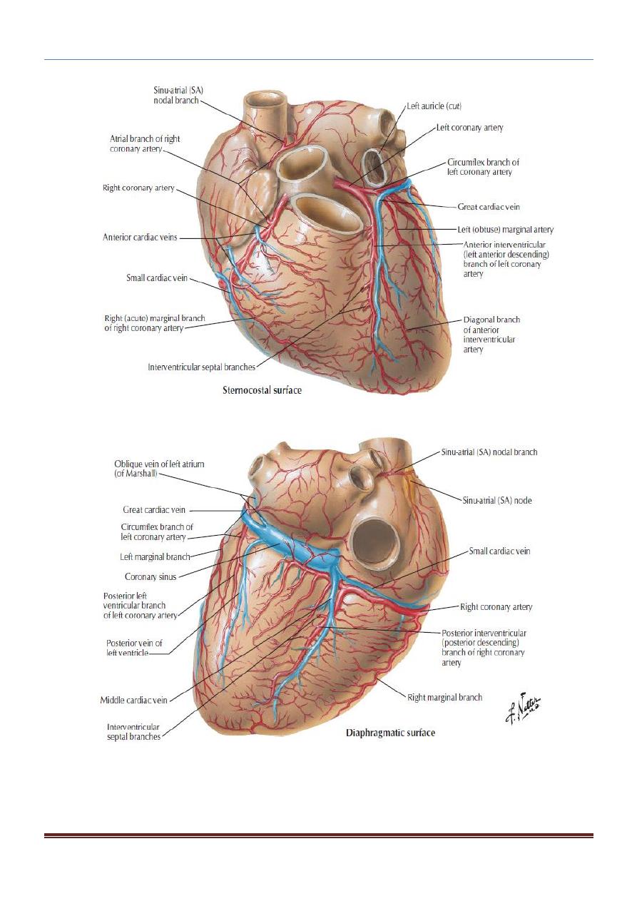
The Arterial Supply of the Heart:
The arterial supply of the heart is provided by the right and left coronary arteries, which
arise from the ascending aorta immediately above the aortic valve.
The right coronary artery :
Branches
1. The right conus artery supplies the anterior surface of the pulmonary conus
(infundibulum of the right ventricle)and the upper part of the anterior wall of the
right ventricle.
2. The anterior ventricular branches are two or three in number and supply the
anterior surface of the right ventricle.
The marginal branch is the largest and runs along the lower margin of the costal
surface to reach the apex.
3. The posterior ventricular branches are usually two in number and supply the
diaphragmatic surface of the right ventricle.
4. The posterior interventricular (descending) artery runs toward the apex in the
posterior interventricular groove. It gives off branches to the right and left
ventricles, including its inferior wall. A large septal branch supplies the
atrioventricular node.

CHEST ANATOMY THI-QAR UNIVERSITY
COLLEGE OF MEDICINE
LECTURE 5 2019/2020
Dr. Rafid AL-Temimi ; Clinical radiology ( CABM
Page
4
Dr. Ahmed Abdulameer Daffar ; Thoracic & Vascular Surgeon ( FIBMS )
5. The atrial branches supply the anterior and lateral surfaces of the right atrium. One
branch supplies the posterior surface of both the right and left atria.
The artery of the sinuatrial node supplies the node and the right and left atria; in
35% of individuals it arises from the left coronary artery.
The left coronary artery:
Branches
1. The anterior interventricular (descending) branch In one third of individuals, it
ends at the apex of the heart.
2. A small left conus artery supplies the pulmonary conus.
3. The circumflex artery is the same size as the anteriorinterventricular artery. It
winds around the left margin of the heart in the atrioventricular groove. A left
marginal artery is a large branch that supplies the left margin of the left ventricle
down to the apex.
4. Anterior ventricular and posterior ventricular branches supply the left ventricle.
Atrial branches supply the left atrium.
The surface markings of the heart valves are as following:
The tricuspid valve lies behind the right half of the sternum opposite the 4th
intercostal space.
The mitral valve lies behind the left half of the sternum opposite the 4th costal
cartilage.
The pulmonary valve lies behind the medial end of the third left costal cartilage
and the adjoining part of the sternum.
The aortic valve lies behind the left half of the sternum opposite the 3rd
intercostal space.
CHEST ANATOMY THI-QAR UNIVERSITY
COLLEGE OF MEDICINE
LECTURE 5 2019/2020
Dr. Rafid AL-Temimi ; Clinical radiology ( CABM
Page
5
Dr. Ahmed Abdulameer Daffar ; Thoracic & Vascular Surgeon ( FIBMS )