Done by
Dr.Rafid Remthan Al-Temimi
Clinical Radiology
CAMB,DMRD,M.B.Ch.B.,.
المرحلة
:
الثانية
المادة
:
التشريح
ج
امعة ذي قار
/
كلية الطب
الدكتور
رافد
رمثان التميمي
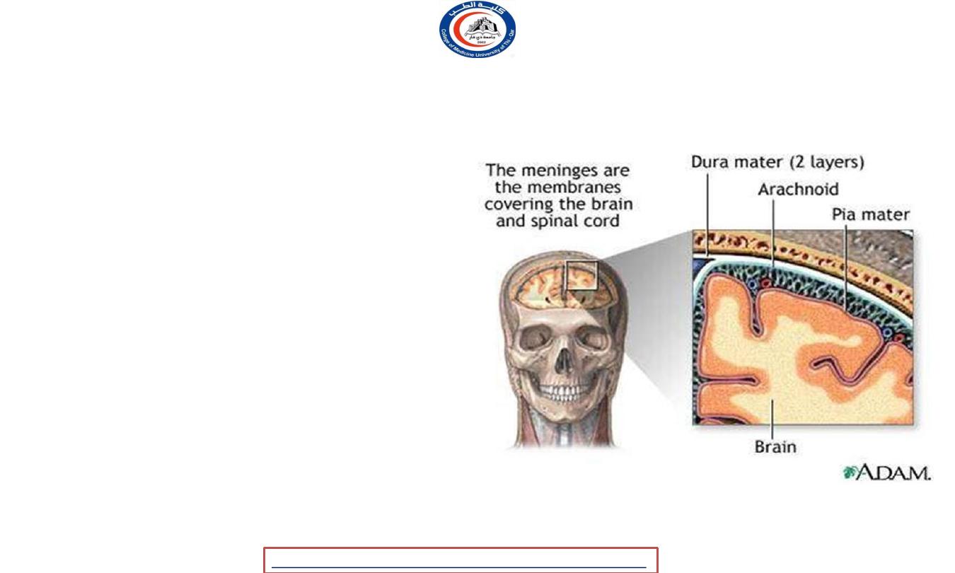
The Meninges
• The Meninges are the
membrane covering the brain and
spinal cord.
• The Meninges consist of three
membranes:
1. The dura mater,
2. The arachnoid mater,
3. The pia mater.
University Of Thi-Qar
College Of medicine
Dr.Rafid Remthan AL-Temimi,Clinical Radiology,CAMB, 2020
Anatomy lecture . 2
nd
stage
Dr.Rafid Al-Temimi
2
The Meninges
1.
Dura mater
-
strong, "Tough mother"
a. Falx cerebri
b. Falx cerebelli
c. Tentorium cerebelli
d. Diaphragma sella
2.
Arachnoid
-
spidery, holds
blood vessels
3.
Pia mater
-
"delicate
mother"
University Of Thi-Qar
College Of medicine
Dr.Rafid Remthan AL-Temimi,Clinical Radiology,CAMB, 2020
Anatomy lecture . 2
nd
stage
Dr.Rafid Al-Temimi
3
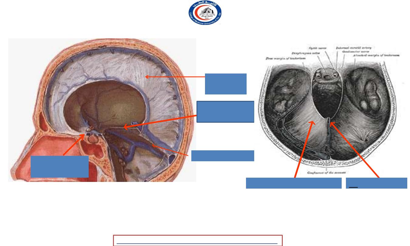
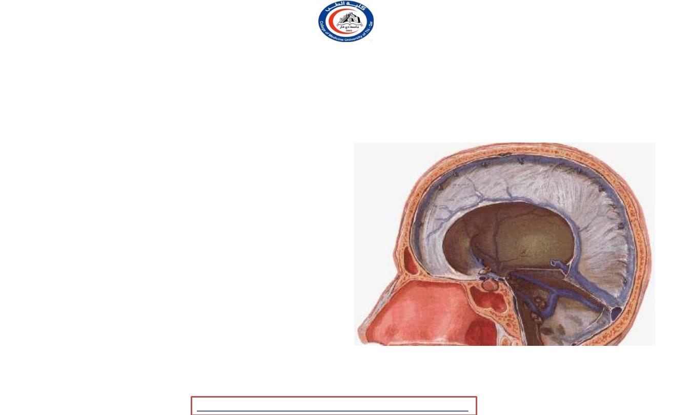
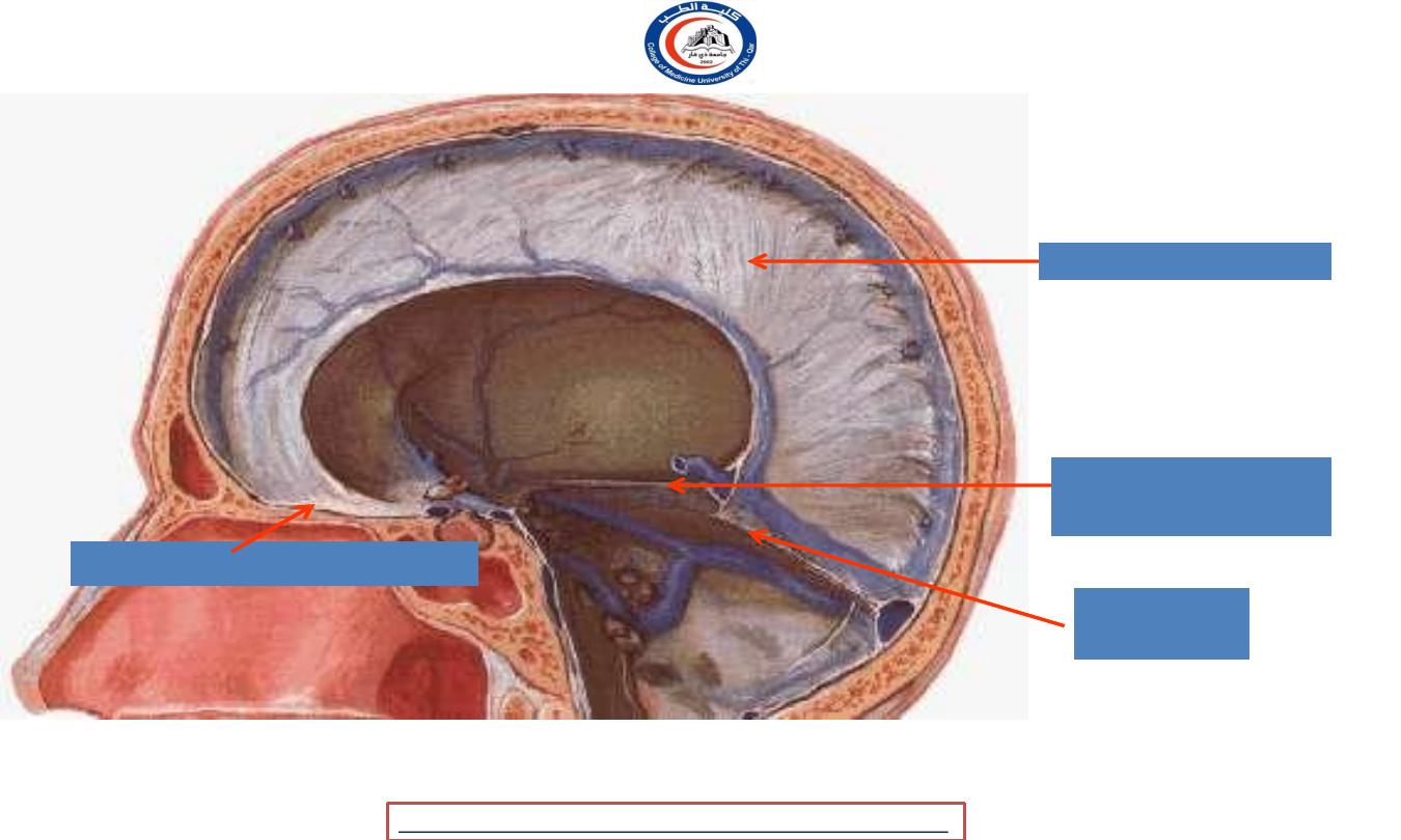
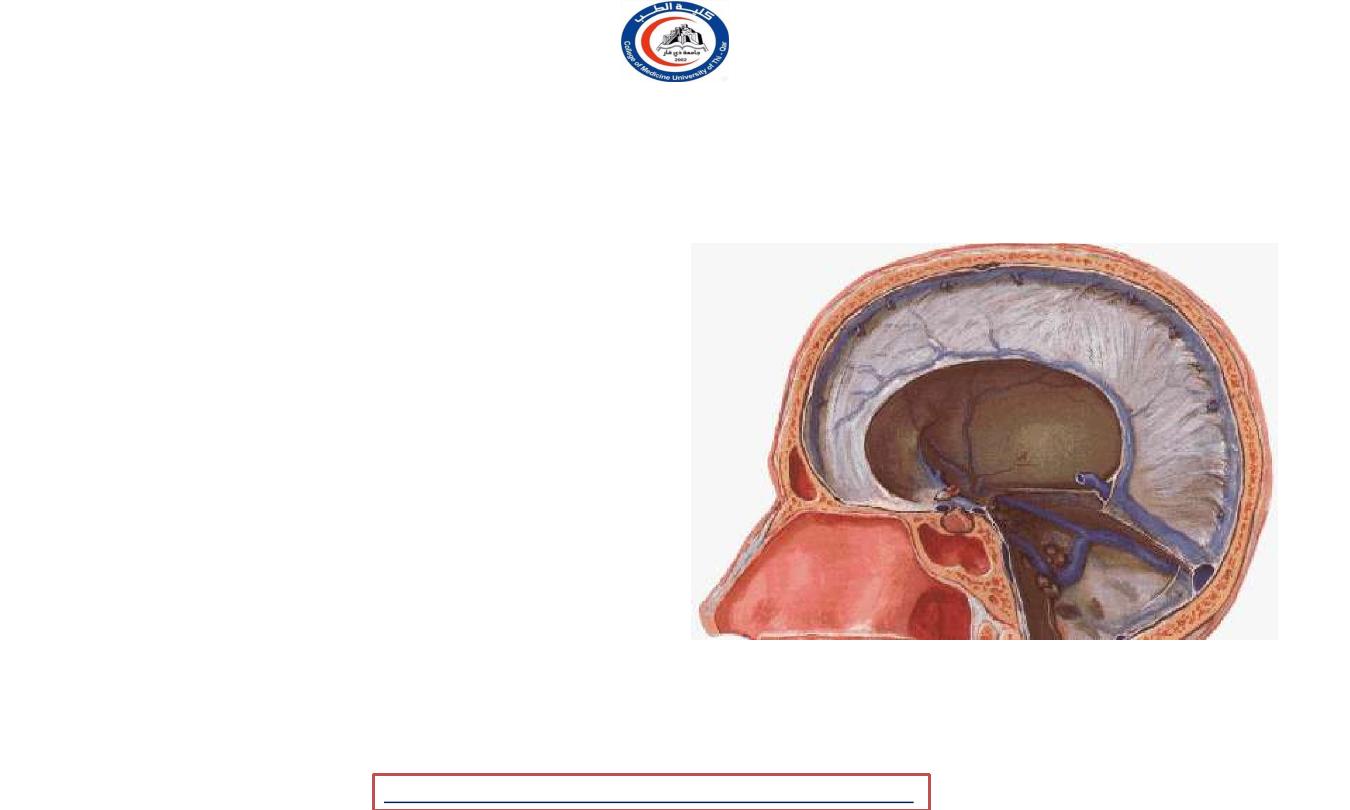
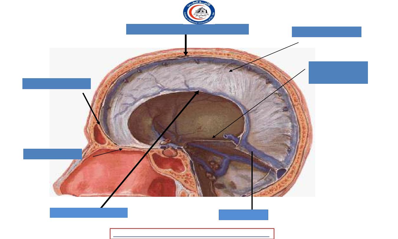
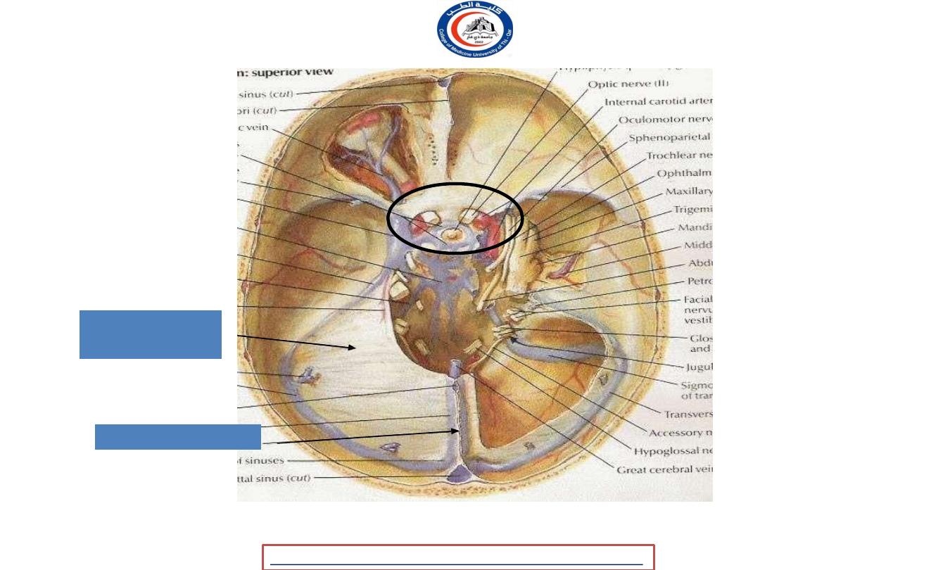
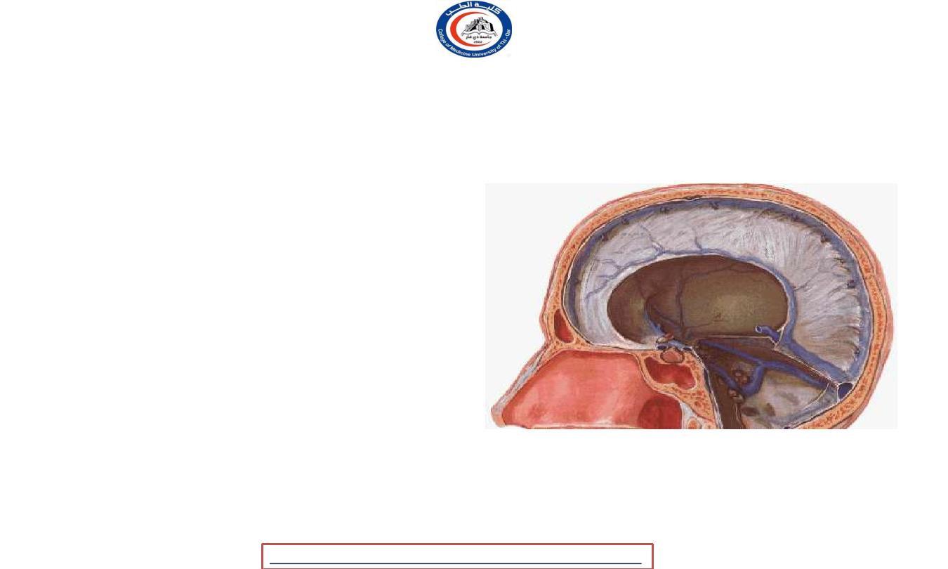
Sagittal section showing the duramater
1) Falx
cerebri
2) Tentorium
cerebelli
3) Falx cerebelli
4) Diaphragma
sellae
1) Falx cerebri
2)
Tentorium cerebelli
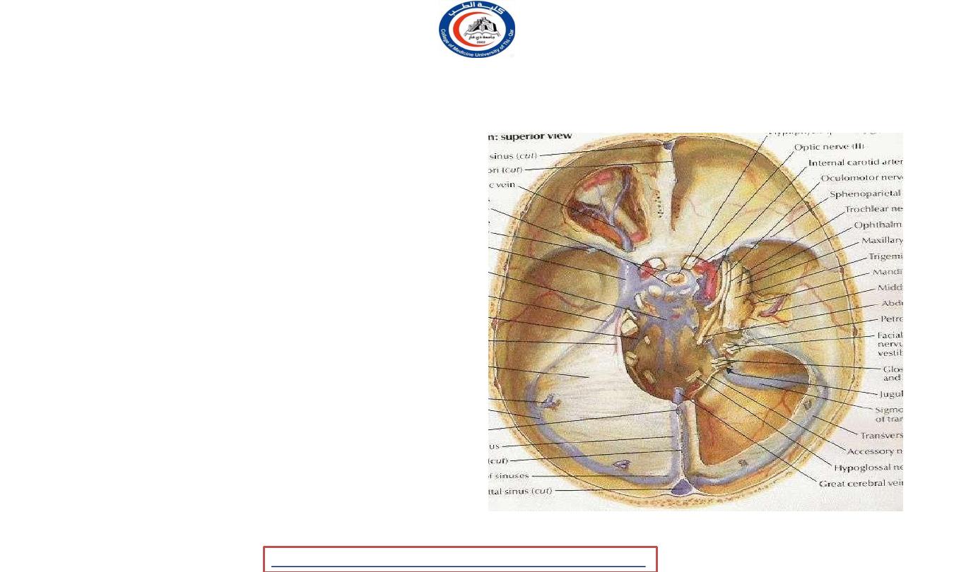
Superior view showing the duramater
University Of Thi-Qar
College Of medicine
Dr.Rafid Remthan AL-Temimi,Clinical Radiology,CAMB, 2020
Anatomy lecture . 2
nd
stage
Dr.Rafid Al-Temimi
4

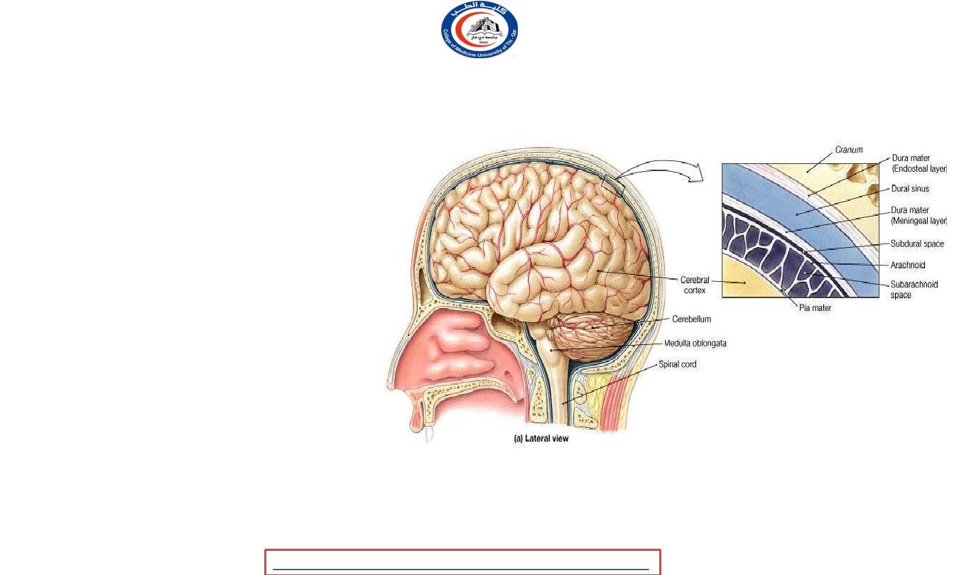
DURA MATER
Thick dense inelastic membrane and the
outermost layer of the meninges
Bilaminar:
Endosteal layer (outer)
Meningeal layer (inner)
These are closely united except along
certain lines, where they separate to form
venous sinuses.
University Of Thi-Qar
College Of medicine
Dr.Rafid Remthan AL-Temimi,Clinical Radiology,CAMB, 2020
Anatomy lecture . 2
nd
stage
Dr.Rafid Al-Temimi
5
DURA MATER
Endosteal layer ;
o
Periosteum - inner surface of the
skull bones
o
Not continuous with dura mater of spinal
cord
Meningeal layer ;
o Dura mater proper
o Covering the brain
o Continuous with dura mater of spinal
cord
o Folded inwards as 4 septa between
part of the brain
o The function of these septa is to restrict the
rotatory displacement of the brain.
University Of Thi-Qar
College Of medicine
Dr.Rafid Remthan AL-Temimi,Clinical Radiology,CAMB, 2020
Anatomy lecture . 2
nd
stage
Dr.Rafid Al-Temimi
6
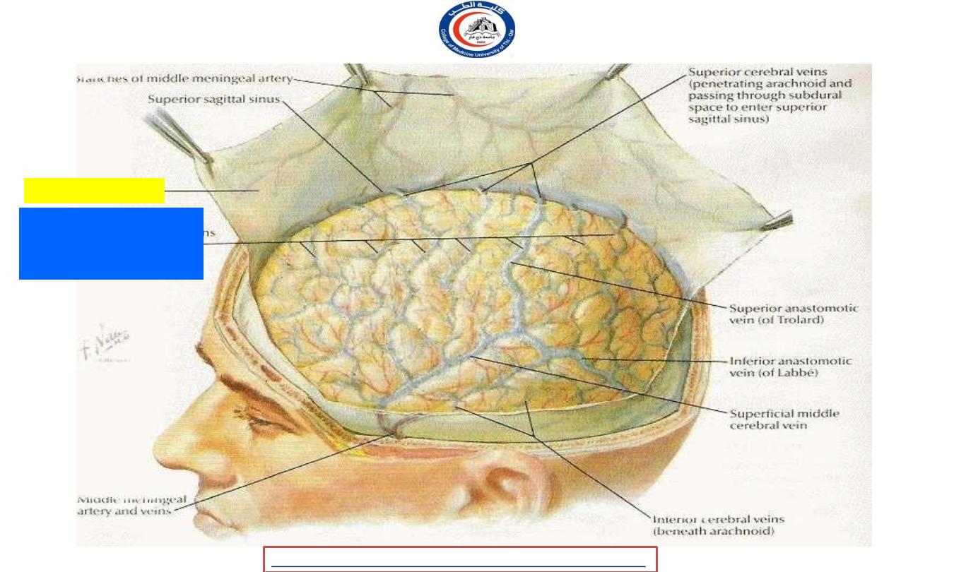
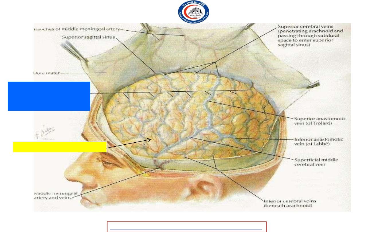
duramater
Superior cerebral
veins beneath
arachnoid
University Of Thi-Qar
College Of medicine
Dr.Rafid Remthan AL-Temimi,Clinical Radiology,CAMB, 2020
Anatomy lecture . 2
nd
stage
Dr.Rafid Al-Temimi
7
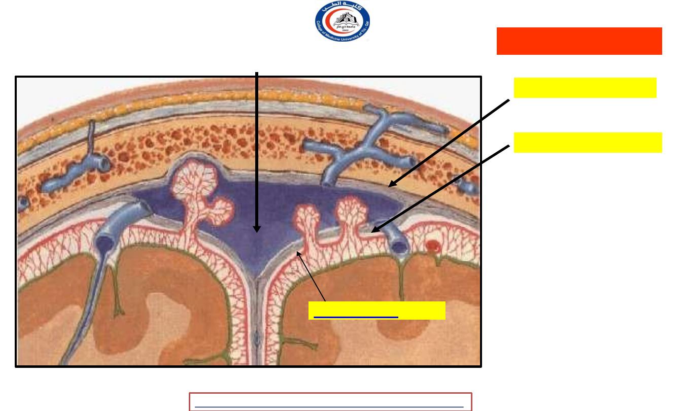
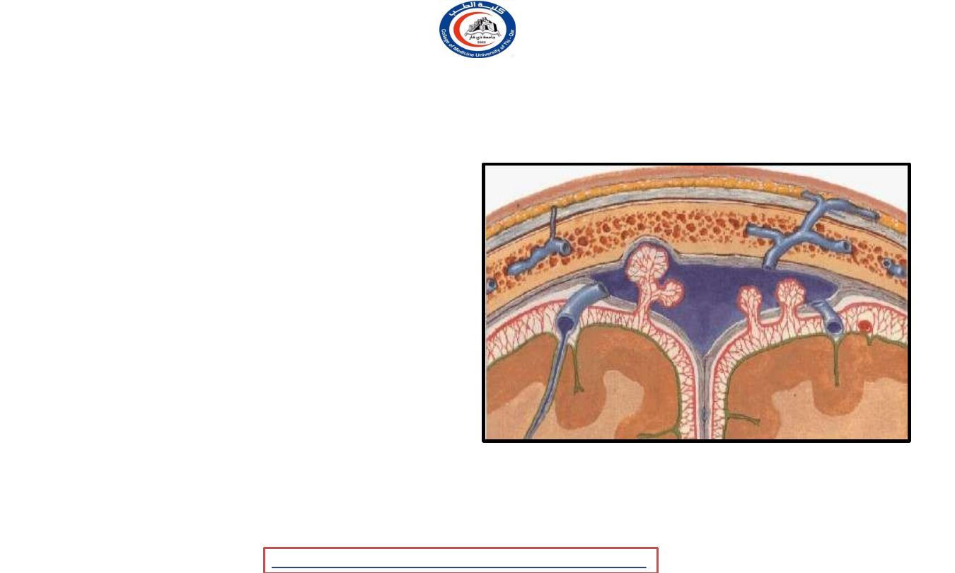
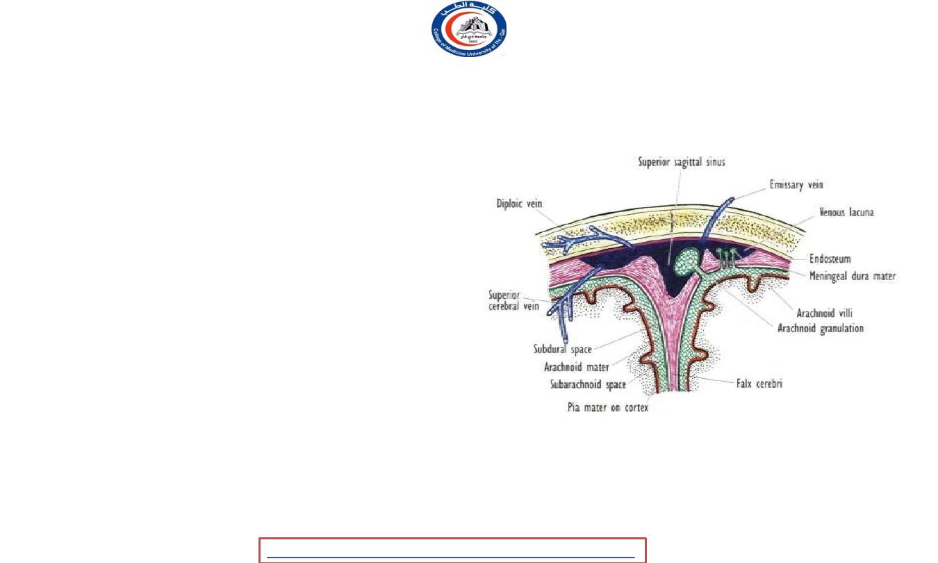
Coronal section of the upper part of the head
Endosteal layer
Meningeal layer
They are closely united
except along certain
lines; they are
separated to form
venous sinuses
Superior sagittal sinus (Dural venous sinus)
Dura mater
Subdural space
University Of Thi-Qar
College Of medicine
Dr.Rafid Remthan AL-Temimi,Clinical Radiology,CAMB, 2020
Anatomy lecture . 2
nd
stage
Dr.Rafid Al-Temimi
8
DURA MATER
Dura mater septa:
1. Falx cerebri
2. Falx cerebelli
3. Tentorium cerebelli
4. Diaphragma sella
University Of Thi-Qar
College Of medicine
Dr.Rafid Remthan AL-Temimi,Clinical Radiology,CAMB, 2020
Anatomy lecture . 2
nd
stage
Dr.Rafid Al-Temimi
9
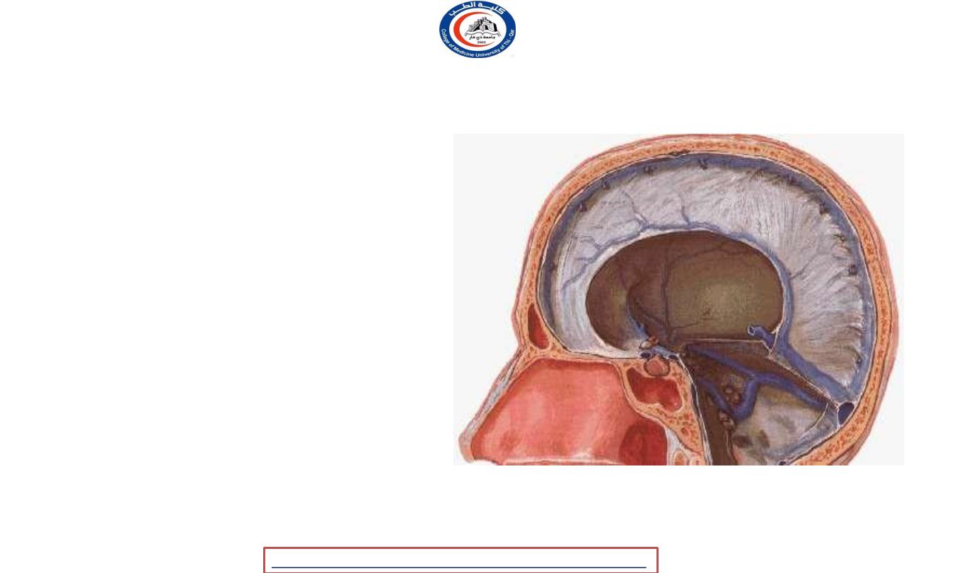
Sagittal section showing the duramater
1) Falx cerebri
2) Tentorium
cerebelli
3) Falx
cerebelli
4) Diaphragma sellae
University Of Thi-Qar
College Of medicine
Dr.Rafid Remthan AL-Temimi,Clinical Radiology,CAMB, 2020
Anatomy lecture . 2
nd
stage
Dr.Rafid Al-Temimi
10
The Falx Cerebri
•
It is a sickle-shaped fold of dura mater that lies in the
midline between the two cerebral hemispheres.
•
Its narrow end in front is attached to the internal frontal
crest and the crista galli.
•
Its broad posterior part blends in the midline with the
upper surface of the tentorium cerebelli.
•
The superior sagittal sinus runs
in its upper fixed margin,
the inferior sagittal sinus
runs in its lower concave free
margin, and the straight sinus runs along its attachment to
the tentorium cerebelli.
University Of Thi-Qar
College Of medicine
Dr.Rafid Remthan AL-Temimi,Clinical Radiology,CAMB, 2020
Anatomy lecture . 2
nd
stage
Dr.Rafid Al-Temimi
11
Falx cerebri
Superior sagittal sinus
Inferior sagittal sinus
Tentorium
cerebelli
*
Frontal crest
Crista galli
Straight sinus
University Of Thi-Qar
College Of medicine
Dr.Rafid Remthan AL-Temimi,Clinical Radiology,CAMB, 2020
Anatomy lecture . 2
nd
stage
Dr.Rafid Al-Temimi
12
The Tentorium Cerebelli
• The tentorium cerebelli is a
crescent-shaped fold of dura
mater that roofs over the
posterior cranial fossa.
• It covers the upper surface
of the cerebellum and
supports the occipital lobes
of the cerebral
hemispheres.
University Of Thi-Qar
College Of medicine
Dr.Rafid Remthan AL-Temimi,Clinical Radiology,CAMB, 2020
Anatomy lecture . 2
nd
stage
Dr.Rafid Al-Temimi
13
Tentorium
cerebelli
Falx cerebri
University Of Thi-Qar
College Of medicine
Dr.Rafid Remthan AL-Temimi,Clinical Radiology,CAMB, 2020
Anatomy lecture . 2
nd
stage
Dr.Rafid Al-Temimi
14
The Falx Cerebelli
• The falx cerebelli is a small, sickle-
shaped fold of
dura mater that is attached to the
internal occipital crest and projects
forward between the two
cerebellar hemispheres.
• Its posterior fixed margin contains
the occipital sinus.
University Of Thi-Qar
College Of medicine
Dr.Rafid Remthan AL-Temimi,Clinical Radiology,CAMB, 2020
Anatomy lecture . 2
nd
stage
Dr.Rafid Al-Temimi
15

The Diaphragma Sellae
• The diaphragma sellae is a
small circular fold of dura
mater that forms the roof for
the sella turcica.
• A small opening in its
center allows passage of
the stalk of the pituitary
gland
University Of Thi-Qar
College Of medicine
Dr.Rafid Remthan AL-Temimi,Clinical Radiology,CAMB, 2020
Anatomy lecture . 2
nd
stage
Dr.Rafid Al-Temimi
16

Dural Nerve Supply
• Branches of the trigeminal, vagus, and first three
cervical nerves and branches from the sympathetic
system pass to the dura.
• The dura is sensitive to stretching, which produces
the sensation of headache.
University Of Thi-Qar
College Of medicine
Dr.Rafid Remthan AL-Temimi,Clinical Radiology,CAMB, 2020
Anatomy lecture . 2
nd
stage
Dr.Rafid Al-Temimi
17

Dural Blood Supply
Dural Arterial Supply
• The dura mater’s arteries supply
from the internal carotid, maxillary,
ascending pharyngeal, occipital,
and vertebral arteries.
• From a clinical standpoint, the
most important is the middle
meningeal artery, which is
commonly damaged in head
injuries.
Dural Venous Drainage
• The meningeal veins lie in the
endosteal layer of dura.
• The middle meningeal vein
follows the branches of the
middle meningeal artery and
drains into the pterygoid venous
plexus or the sphenoparietal
sinus.
• The veins lie lateral to the
arteries.
University Of Thi-Qar
College Of medicine
Dr.Rafid Remthan AL-Temimi,Clinical Radiology,CAMB, 2020
Anatomy lecture . 2
nd
stage
Dr.Rafid Al-Temimi
18
Arachnoid Mater
Delicate, impermeable & avascular
membrane covering the brain
Lying between Pia mater
(internally) & dura
Mater(externally)
Separated from dura mater by a
potential space, the subdural space
(filled by a film of fluid)
Separated from pia mater by the
subarachnoid space (filled with CSF)
The outer and inner surfaces covered
with flattened mesothelial cells
University Of Thi-Qar
College Of medicine
Dr.Rafid Remthan AL-Temimi,Clinical Radiology,CAMB, 2020
Anatomy lecture . 2
nd
stage
Dr.Rafid Al-Temimi
19
Superior cerebral
veins beneath
arachnoid
Arachnoid mater
University Of Thi-Qar
College Of medicine
Dr.Rafid Remthan AL-Temimi,Clinical Radiology,CAMB, 2020
Anatomy lecture . 2
nd
stage
Dr.Rafid Al-Temimi
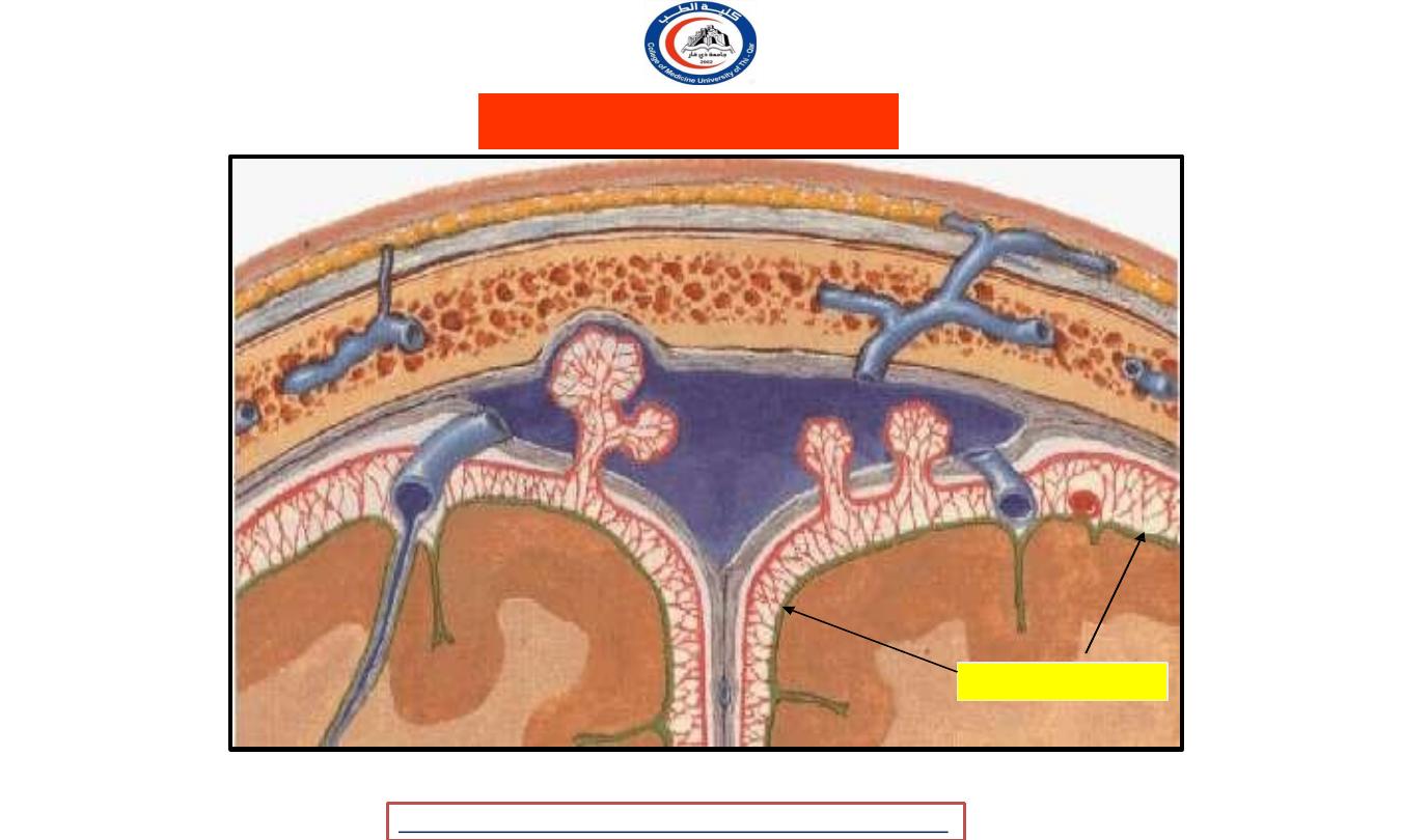
20
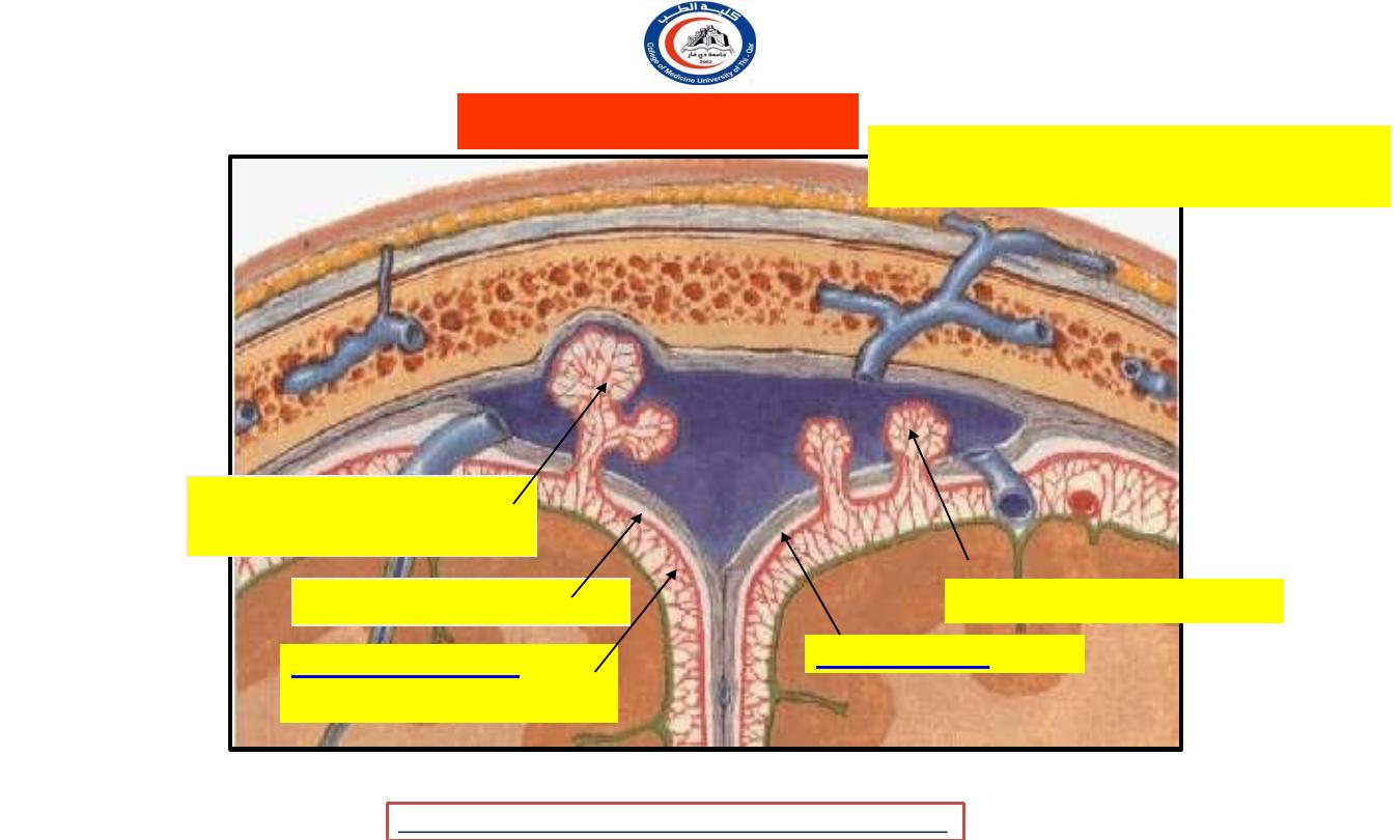
Arachnoid mater
Arachnoid villi
Arachnoid mater
Subarachnoid space
Arachnoid
granulations
Subdural space
Arachnoid projects into venous sinuses
- sites for CSF diffuses into bloodstream
University Of Thi-Qar
College Of medicine
Dr.Rafid Remthan AL-Temimi,Clinical Radiology,CAMB, 2020
Anatomy lecture . 2
nd
stage
Dr.Rafid Al-Temimi
21
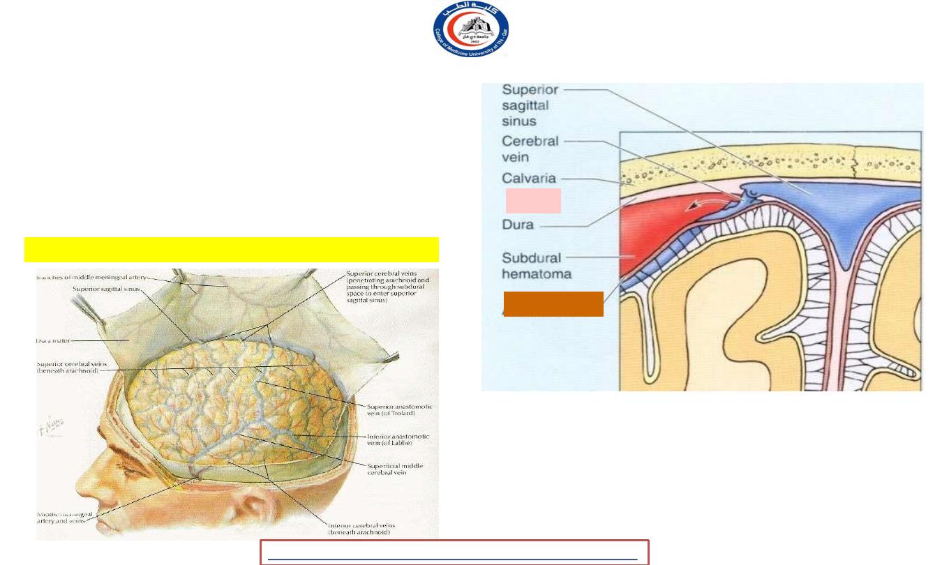
• Superior cerebral veins, traverse
the
subdural space to reach the superior
sagittal sinus and its lacunae
SUBDURAL SPACE :
Superior cerebral veins beneath arachnoid
Subdural haematoma
*
Dura
Arachnoid
University Of Thi-Qar
College Of medicine
Dr.Rafid Remthan AL-Temimi,Clinical Radiology,CAMB, 2020
Anatomy lecture . 2
nd
stage
Dr.Rafid Al-Temimi
22
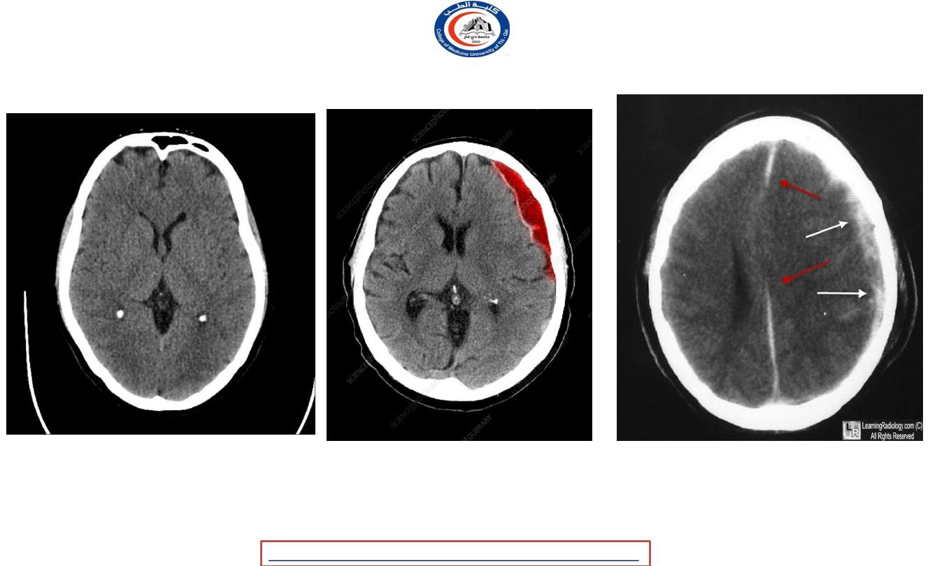
Subdural haematoma
Subdural haematoma
Normal CT
University Of Thi-Qar
College Of medicine
Dr.Rafid Remthan AL-Temimi,Clinical Radiology,CAMB, 2020
Anatomy lecture . 2
nd
stage
Dr.Rafid Al-Temimi
23

Subarachnoid Space (SP) :
Relatively narrow over the surface of cerebral hemisphere, but
sometimes becomes much wider in areas at the base of the brain,
the widest space is called subarachnoid cisterns
The cisterna cerebellomedularis lies between inferior surface of
the cerebellum and roof of 4
th
ventricle
The cisterna interpeduncularis lies between 2 cerebral hemispheres.
All the cisternae are in free communication with one
another & with the remainder of subarachnoid space
University Of Thi-Qar
College Of medicine
Dr.Rafid Remthan AL-Temimi,Clinical Radiology,CAMB, 2020
Anatomy lecture . 2
nd
stage
Dr.Rafid Al-Temimi
24
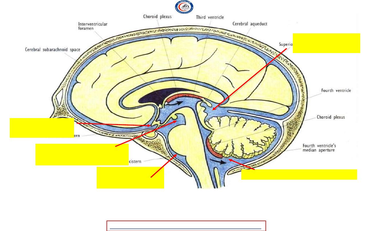
Median sagittal section to show the subarachnoid cisterns & circulation of CSF
Superior
cistern
Interpeduncular
cistern
Cerebellomedullary cistern
Chiasmatic
cistern
Pontine
cistern
University Of Thi-Qar
College Of medicine
Dr.Rafid Remthan AL-Temimi,Clinical Radiology,CAMB, 2020
Anatomy lecture . 2
nd
stage
Dr.Rafid Al-Temimi
25
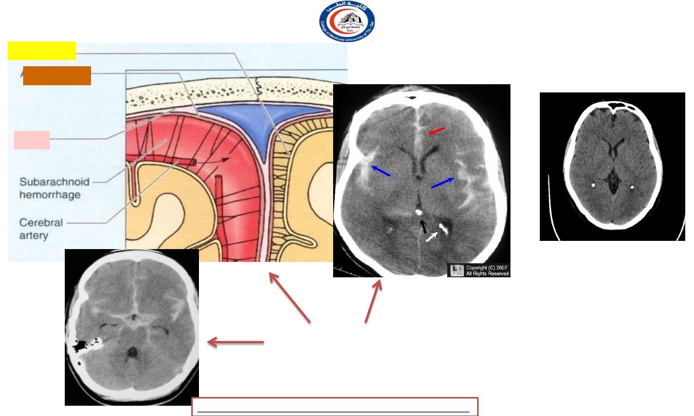
*
Subarachnoid haemorrage
Dura
Arachnoid
Pia mater
NORMAL CT
University Of Thi-Qar
College Of medicine
Dr.Rafid Remthan AL-Temimi,Clinical Radiology,CAMB, 2020
Anatomy lecture . 2
nd
stage
Dr.Rafid Al-Temimi
26
Pia Mater
• Pia Mater is a vascular
membrane
covered
by
mesothelial cells.
• Closely invests the brain,
covering the gyri,
descending into the
deepest sulci & closely
applied to the cortical
surface.
University Of Thi-Qar
College Of medicine
Dr.Rafid Remthan AL-Temimi,Clinical Radiology,CAMB, 2020
Anatomy lecture . 2
nd
stage
Dr.Rafid Al-Temimi
27
Pia mater
Pia mater
University Of Thi-Qar
College Of medicine
Dr.Rafid Remthan AL-Temimi,Clinical Radiology,CAMB, 2020
Anatomy lecture . 2
nd
stage
Dr.Rafid Al-Temimi
28

Pia Mater
It extends out over the cranial nerves & fuses with their
epineurium
The cerebral arteries entering the substance of the brain,
carry a sheath of pia mater with them
The pia mater forms the TELA CHOROIDAE .
The tela choroidae fuse with ependyma to form the choroid
plexus
Choroid plexus forms CSF
University Of Thi-Qar
College Of medicine
Dr.Rafid Remthan AL-Temimi,Clinical Radiology,CAMB, 2020
Anatomy lecture . 2
nd
stage
Dr.Rafid Al-Temimi
29
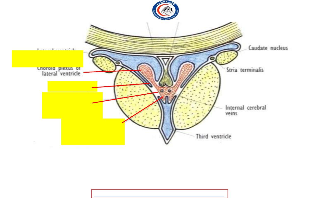
Coronal section of the interventricular foramen showing the choroid plexus of 3
rd
& lateral ventricles
Ependyma
Choroid plexus
of
3
rd
ventricle
Choroid plexus of
lateral ventricle
Pia mater of tela
choroidae
University Of Thi-Qar
College Of medicine
Dr.Rafid Remthan AL-Temimi,Clinical Radiology,CAMB, 2020
Anatomy lecture . 2
nd
stage
Dr.Rafid Al-Temimi
30

University Of Thi-Qar
College Of medicine
Anatomy lecture . 2
nd
stage
Dr.Rafid Al-Temimi
Dr.Rafid Remthan AL-Temimi,Clinical Radiology,CAMB, 2020
31