الدكتور
رافد رمثان حسين
التميمي
دكتوراه أشعة تشخيصية
Spinal Cord
Blood Supply Anatomy
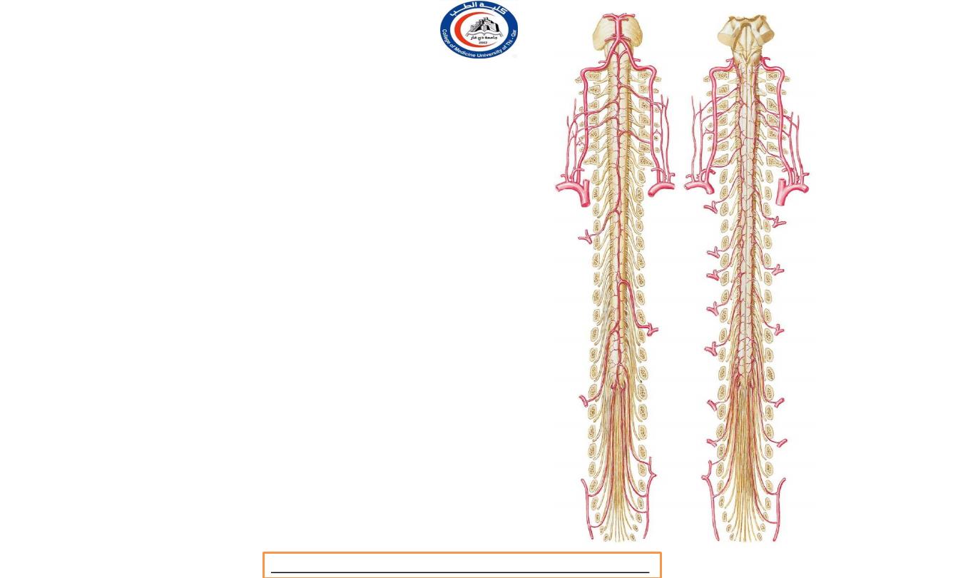
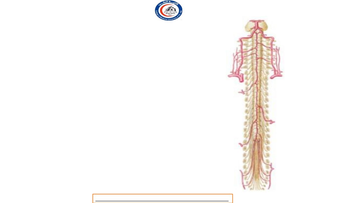
Blood supply
• The spinal cord is supplied by the
(single)
anterior
and
(right and left) posterior spinal
arteries
which descend from the level of the
foramen magnum and form
three longitudinal
channels
from which branches enter the cord.
• They are supplemented at variable levels by
anastomoses with a variable number of
radicular arteries.
• The main arteries supply the spinal cord lies on
pia mater whereas their small branches
evaginate it.
Dr.Rafid Remthan AL-Temimi,Clinical Radiology,CAMB, 2020
University Of Thi-Qar
College Of medicine
Anatomy lecture . 2
nd
stage
Dr.Rafid Al-Temimi
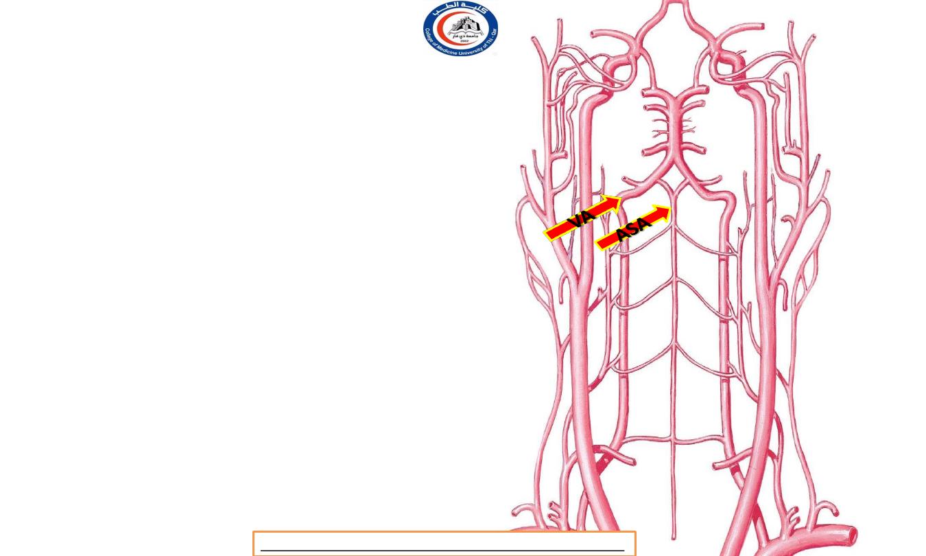
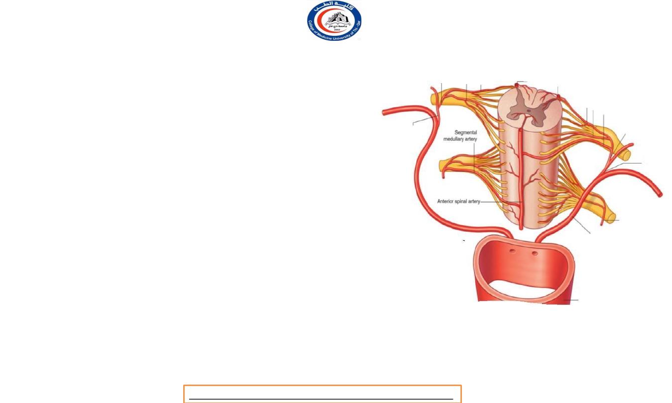
Anterior spinal artery
• Origion :
union of the two anterior spinal
branches, each given off by a vertebral artery
above the foramen magnum.
• Supply : ( 4 areas ) It supplies the
whole cord
anterior to the posterior grey columns
, i.e. the
lateral grey and white columns
and the
anterior
grey and white columns
of both sides.
• The anterior spinal artery is a midline vessel that
lies on the anterior median fissure.
** ASA = Anterior Spinal Artery
VA = Vertebral Artery
Dr.Rafid Remthan AL-Temimi,Clinical Radiology,CAMB, 2020
University Of Thi-Qar
College Of medicine
Anatomy lecture . 2
nd
stage
Dr.Rafid Al-Temimi
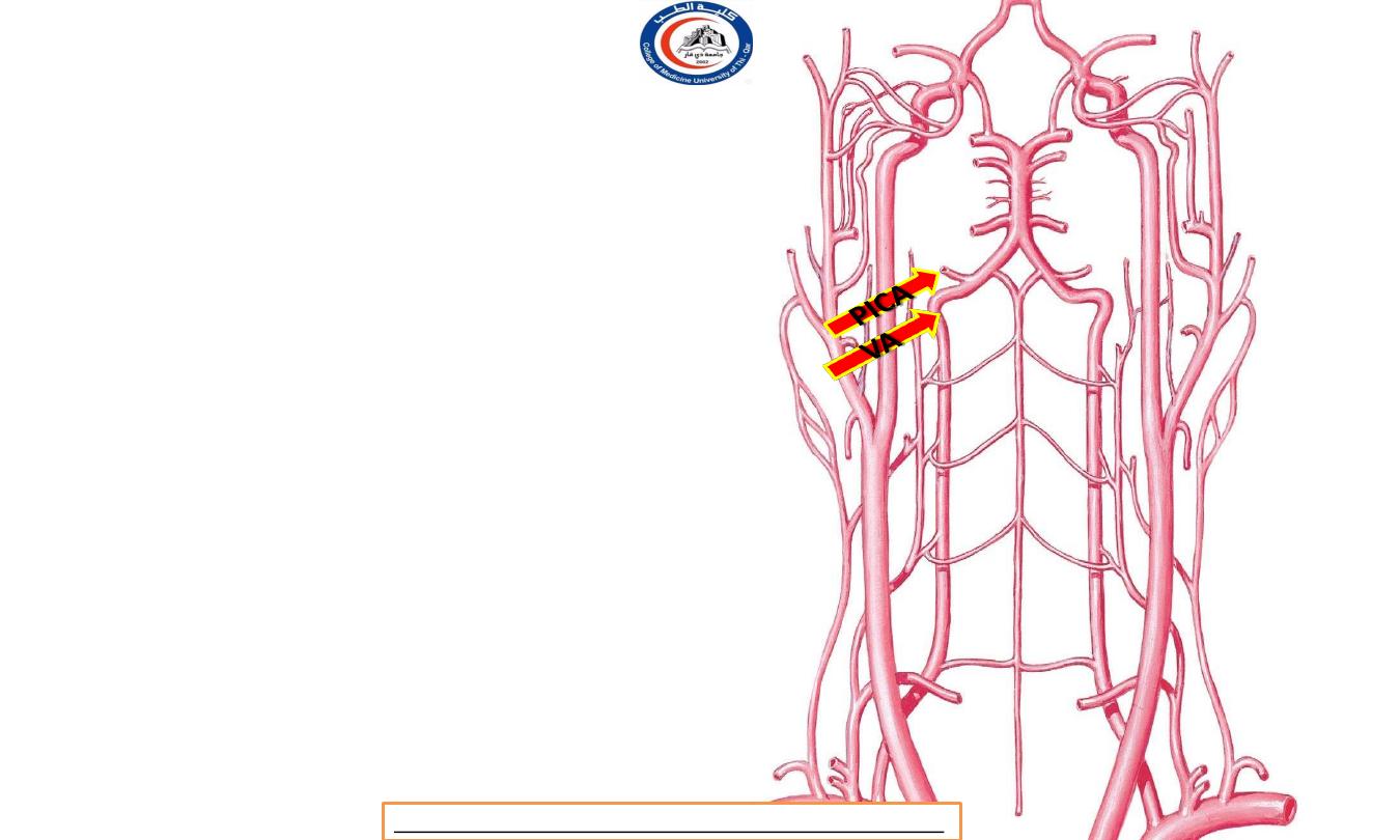
Posterior spinal artery
• Origion :
The posterior spinal artery on each side arises
from the posterior inferior cerebellar or vertebral artery
above the foramen magnum.
• Supply :
The posterior spinal artery supplies the grey and
white posterior columns of its own side.
• Posterior spinal a. is NOT shown.
• PSA is forming longitudinal trunks that run through and
behind the posterior nerve rootlets for the whole length
of the cord.
• There is some anastomosis between the vessels of the
two sides, with rather scanty connections with the
anterior spinal artery,
except at the lower end of the cord
where there are often good anastomoses.
** PICA = Posterior Inferior Cerebellar
Artery
VA = Vertebral Artery
Dr.Rafid Remthan AL-Temimi,Clinical Radiology,CAMB, 2020
University Of Thi-Qar
College Of medicine
Anatomy lecture . 2
nd
stage
Dr.Rafid Al-Temimi
Radicular arteries
• At one stag during embryonic development every segment of
the cord receives a radicular vessel on both sides; they enter
through the intervertebral foramina as spinal arteries to
penetrate the meninges and run along the nerve roots.
•
They are derived from various parent vessels depending on
the level:
costocervical,
posterior
lumbar,
and lateral
vertebral,
intercostals,
sacral.
• As fetal growth proceeds, most of the radicular arteries disappear.
• Their most characteristic feature is
1.
their variability in number and position.
2.
blood from them may flow up and/or down the cord.
Dr.Rafid Remthan AL-Temimi,Clinical Radiology,CAMB, 2020
University Of Thi-Qar
College Of medicine
Anatomy lecture . 2
nd
stage
Dr.Rafid Al-Temimi
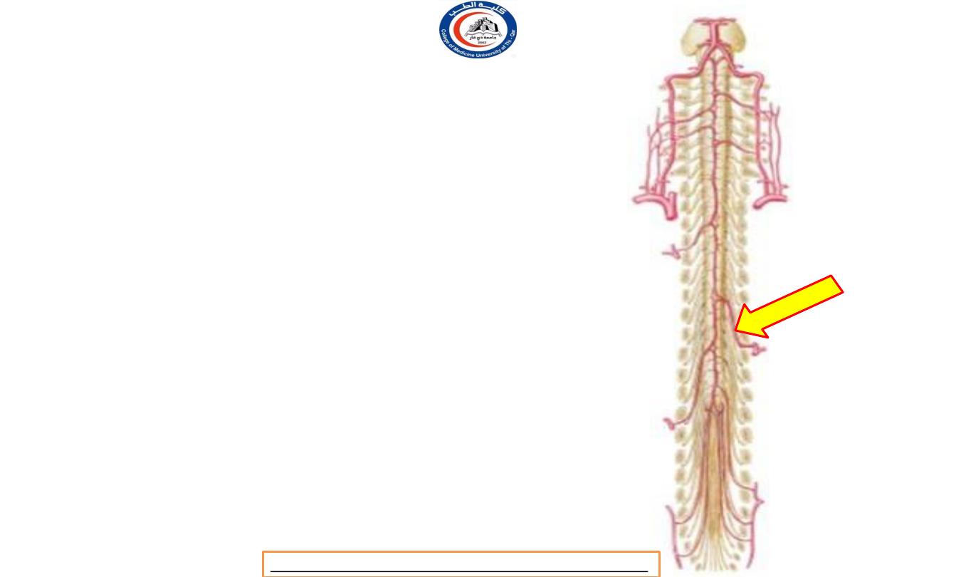
Adamkiewicz artery
• arteria
radicularismagna
(of
Adamkiewicz), usually
arises from a lowe
intercostal or upper lumbar branch of the aorta
on the left side.
• Operations on the vertebral column or adjacen
structures (such as aortic aneurysms) that
interfere with the parent stem of a major
radicular vessel may seriously impair the blood
supply to the cord.
Dr.Rafid Remthan AL-Temimi,Clinical Radiology,CAMB, 2020
University Of Thi-Qar
College Of medicine
Anatomy lecture . 2
nd
stage
Dr.Rafid Al-Temimi
Anastomosis
• The anastomotic connections on the surface of
the cord (deep to the pia mater) between the
anterior and posterior spinal and radicular
vessels provide
very small pial arteries that are
capable of supplying peripheral areas of the
cord.
• This is important with respect to the
lateral
corticospinal
and
anterolateral tracts
whose
fibres are laminated, with sacral fibres lying
nearest to the surface.
• Interference with the anterior spinal supply
may
eliminate the function of these tracts, except for
the sacral fibres which remain supplied by the
pial vessels
(‘sacral sparing’).
Dr.Rafid Remthan AL-Temimi,Clinical Radiology,CAMB, 2020
University Of Thi-Qar
College Of medicine
Anatomy lecture . 2
nd
stage
Dr.Rafid Al-Temimi
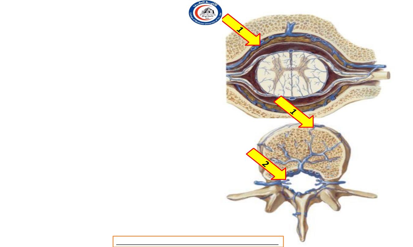
Venous drainage
• The
spinal veins
form loose-knit
there
are an
plexuses in which
anterior
and a posterior
midline
longitudinal vein
, and on each side a
pair of longitudinal veins posterior to
• the anterior and posterior nerve roots.
•
These veins drain to
the internal vertebral
venous plexus (1) ( between dural and bony
canal )
, and thence
plexus (2)
to the segmental
via
the external vertebral venous
veins:
vertebral in the neck; azygos in the thorax;
lumbar in the lumbar region; and lateral sacral
in the sacral region.
• At the foramen magnum they communicate with
the
veins of the medulla.
Dr.Rafid Remthan AL-Temimi,Clinical Radiology,CAMB, 2020
University Of Thi-Qar
College Of medicine
Anatomy lecture . 2
nd
stage
Dr.Rafid Al-Temimi
Anatomy lecture . 2
nd
stage
Dr.Rafid Al-Temimi
University Of Thi-Qar
College Of medicine
Anatomy lecture . 2
nd
stage
Dr.Rafid Al-Temimi
Thank you