CHEST ANATOMY THI-QAR UNIVERSITY
COLLEGE OF MEDICINE
LECTURE 7 2019/2020
Dr. Rafid AL-Temimi ; Clinical radiology ( CABM)
Page
1
Dr. Ahmed Abdulameer Daffar ; Thoracic & Vascular Surgeon ( FIBMS )
THE CHEST
Lungs
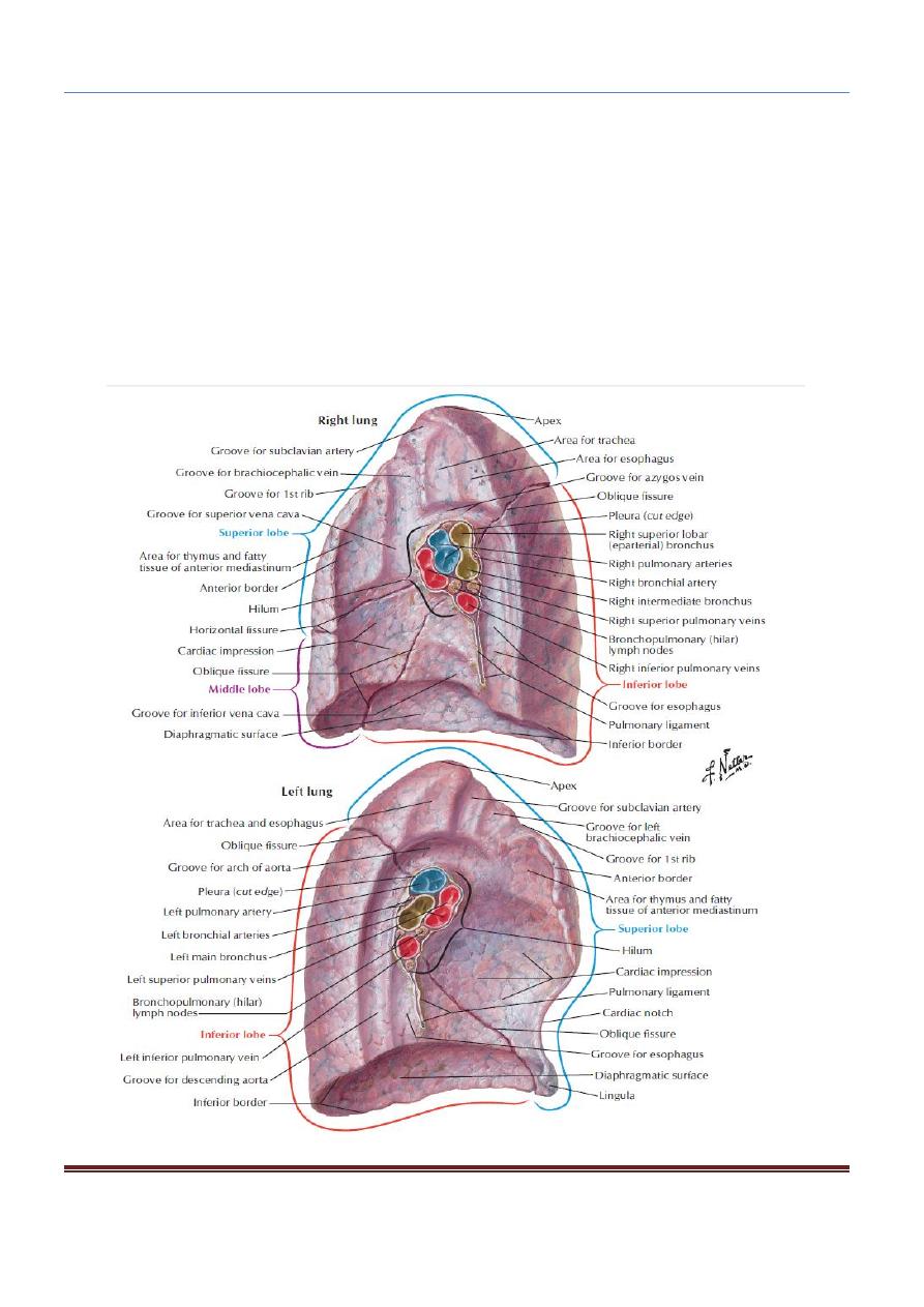
Each lung is conical, covered with visceral pleura, and suspended free in its own
pleural cavity, being attached to the mediastinum only by its root (hilum).
The anterior border is thin and overlaps the heart; it is here on the left lung that the
cardiac notch is found. The posterior border is thick and lies beside the vertebral
column.

CHEST ANATOMY THI-QAR UNIVERSITY
COLLEGE OF MEDICINE
LECTURE 7 2019/2020
Dr. Rafid AL-Temimi ; Clinical radiology ( CABM)
Page
2
Dr. Ahmed Abdulameer Daffar ; Thoracic & Vascular Surgeon ( FIBMS )
The contents of hilum:
1. Bronchi.
2. Pulmonary artery and vein.
3. Bronchial artery and vein.
4. Lymphatics.
5. Nerves.
Lobes and Fissures:
Right Lung:
The right lung is slightly larger than the left and is divided by the oblique and
horizontal fissures into three lobes: the upper, middle, and lower lobes.
The middle lobe is bounded by the horizontal and oblique fissures.
Left Lung:
The left lung is divided by a similar oblique fissure into two lobes: the upper and
lower lobes. There is no horizontal fissure in the left lung.
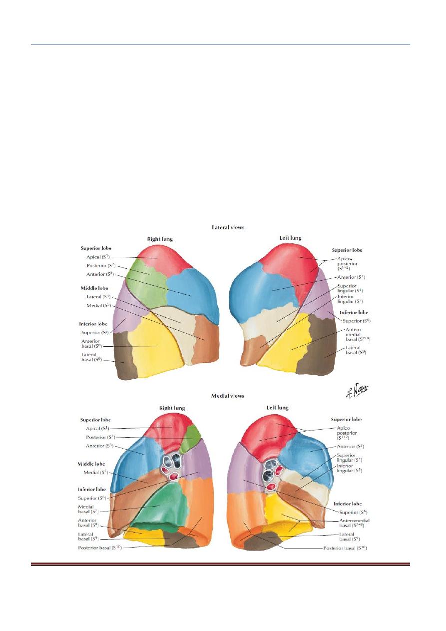
Bronchopulmonary Segment:
It is pyramid shaped, with its apex toward the lung root. It is surrounded by connective
tissue. It has a segmental bronchus, a segmental artery, lymph vessels, and autonomic
nerves. The segmental vein lies in the connective tissue between adjacent
bronchopulmonary segments.
The bronchopulmonary segments are the anatomic, functional, and surgical units
of the lungs.
Each lobar (secondary) bronchus, which passes to a lobe of the lung, gives off
branches called segmental (tertiary) bronchi.
Each segmental bronchus passes to a structurally and functionally independent
unit of a lung lobe called a bronchopulmonary segment, which is surrounded
by connective tissue.
CHEST ANATOMY THI-QAR UNIVERSITY
COLLEGE OF MEDICINE
LECTURE 7 2019/2020
Dr. Rafid AL-Temimi ; Clinical radiology ( CABM)
Page
3
Dr. Ahmed Abdulameer Daffar ; Thoracic & Vascular Surgeon ( FIBMS )
The bronchioles then divide and give rise to terminal bronchioles lead to
respiratory bronchioles.
The respiratory bronchioles end by branching into alveolar ducts, which lead
into tubular passages with numerous thin-walled outpouching called alveolar
sacs which contain several alveoli opening into a single chamber.
Each alveolus is surrounded by a rich network of blood capillaries. Gaseous
exchange takes place between the air in the alveolar lumen through the alveolar
wall into the blood within the surrounding capillaries.

CHEST ANATOMY THI-QAR UNIVERSITY
COLLEGE OF MEDICINE
LECTURE 7 2019/2020
Dr. Rafid AL-Temimi ; Clinical radiology ( CABM)
Page
4
Dr. Ahmed Abdulameer Daffar ; Thoracic & Vascular Surgeon ( FIBMS )
The main bronchopulmonary segments are as follows:
Right lung:
1. Superior lobe: Apical, posterior, anterior
2.
Middle lobe: Lateral, medial
3. Inferior lobe: Superior (apical), medial basal, anterior basal, lateral basal,
posterior basal
Left lung:
1. Superior lobe: Apical, posterior, anterior, superior lingular, inferior lingular
2. Inferior lobe: Superior (apical), medial basal, anterior basal, lateral basal,
posterior basal
The root of the lung is formed of structures that are entering or leaving the lung. It is made
up of the bronchi, pulmonary artery and veins, lymph vessels, bronchial vessels, and
nerves.
The root is surrounded by a tubular sheath of pleura, which joins the mediastinal parietal
pleura to the visceral pleura covering the lungs.
Blood Supply of the Lungs:
The bronchi, the connective tissue of the lung, and the visceral pleura receive their
blood supply from the bronchial arteries, which are branches of the descending aorta.
The bronchial veins (which communicate with the pulmonary veins) drain into the
azygos and hemiazygos veins.
The alveoli receive deoxygenated blood from the terminal branches of the
pulmonary arteries.
The oxygenated blood leaving the alveolar capillaries drains into the tributaries of
the pulmonary veins. Two pulmonary veins leave each lung root to empty into the
left atrium of the heart.