
CHEST ANATOMY THI-QAR UNIVERSITY
COLLEGE OF MEDICINE
LECTURE 8 2019/2020
Dr. Rafid AL-Temimi ; Clinical radiology ( CABM)
Page
1
Dr. Ahmed Abdulameer Daffar ; Thoracic & Vascular Surgeon ( FIBMS )
THE CHEST
Diaphragm
The diaphragm is a thin muscular and tendinous septum that separates the chest
cavity above from the abdominal cavity below.
It is pierced by the structures that pass between the chest and the abdomen.
The diaphragm is the most important muscle of respiration.
It is dome shaped and consists of a peripheral muscular part, which arises from
the margins of the thoracic opening, and a centrally placed tendon.
The origin of the diaphragm can be divided into three parts:
1. A sternal part arising from the posterior surface of the xiphoid process.
2. A costal part arising from the deep surfaces of the lower six ribs and their costal
cartilages.
3. A vertebral part arising by vertical columns or crura and from the arcuate
ligaments.
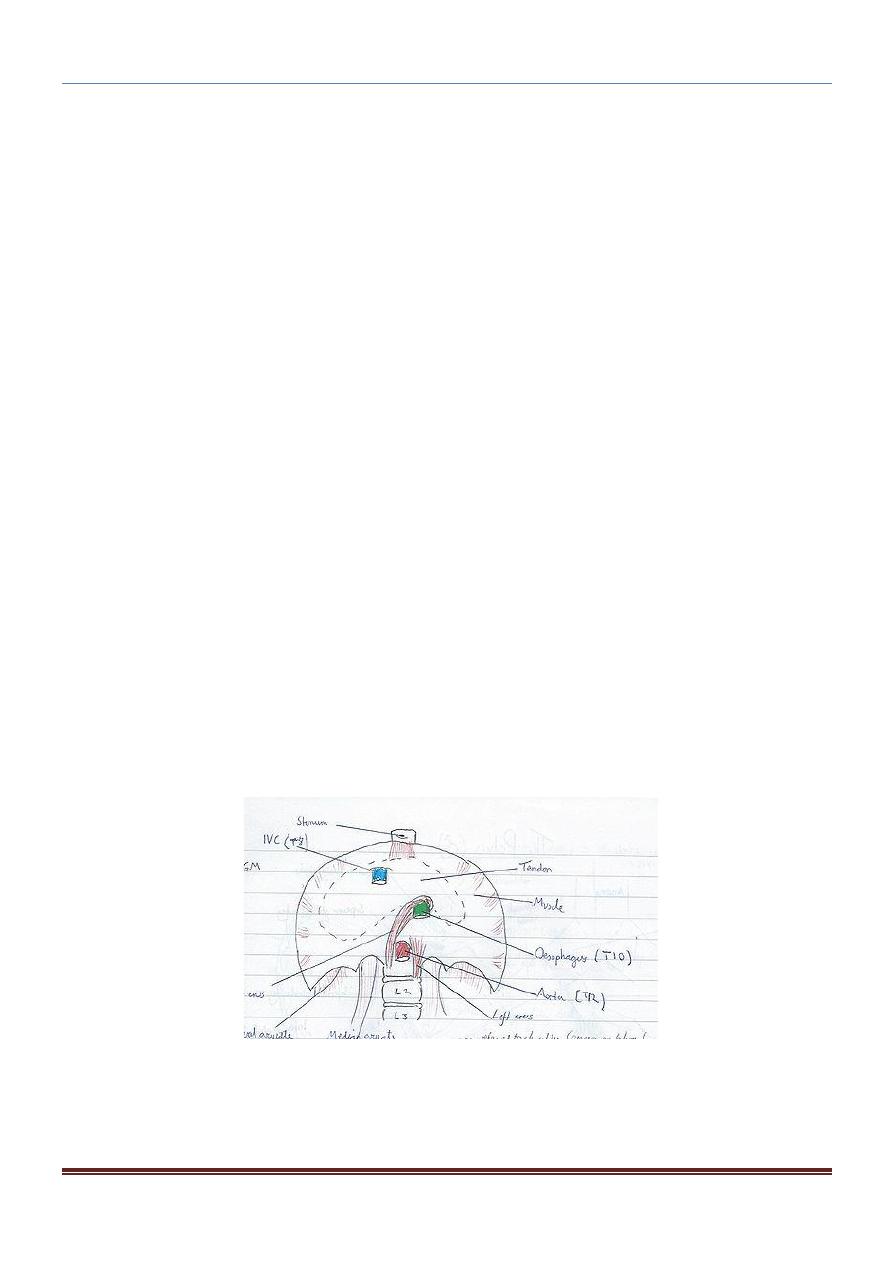
The right crus arise from the sides of the bodies of the first three lumbar
vertebrae and the intervertebral discs.
The left crus arise from the sides of the bodies of the first two lumbar vertebrae
and the intervertebral disc.
Lateral to the crura the diaphragm arises from the medial and lateral arcuate
ligaments.
The medial arcuate ligament extends from the side of the body of the second
lumbar vertebra to the tip of the transverse process of the first lumbar vertebra.
The lateral arcuate ligament extends from the tip of the transverse process of
the first lumbar vertebra to the lower border of the 12th rib.
The diaphragm is inserted into a central tendon, which is shaped like three leaves.
The superior surface of the tendon is partially fused with the inferior surface of the
fibrous pericardium.
Some of the muscle fibers of the right crus pass up to the left and surround the
esophageal orifice in a slinglike loop.
These fibers appear to act as a sphincter and possibly assist in the prevention of
regurgitation of the stomach contents into the thoracic part of the esophagus.
CHEST ANATOMY THI-QAR UNIVERSITY
COLLEGE OF MEDICINE
LECTURE 8 2019/2020
Dr. Rafid AL-Temimi ; Clinical radiology ( CABM)
Page
2
Dr. Ahmed Abdulameer Daffar ; Thoracic & Vascular Surgeon ( FIBMS )
Shape of the Diaphragm:
As seen from in front, the diaphragm curves up into right and left domes.
The right dome reaches as high as the upper border of the 5th rib, and the left
dome may reach the lower border of the 5th rib.
(The right dome lies at a higher level, because of the large size of the right
lobe of the liver.)
The central tendon lies at the level of the xiphisternal joint.
The domes support the right and left lungs, whereas the central tendon
supports the heart.
The levels of the diaphragm vary with the phase of respiration, the posture,
and the degree of distention of the abdominal viscera.
Nerve Supply of the Diaphragm:
Motor nerve supply: The right and left phrenic nerves (C3, 4, 5).
Sensory nerve supply: The parietal pleura and peritoneum covering the central
surfaces of the diaphragm are from the phrenic nerve and the periphery of the
diaphragm is from the lower six intercostal nerves.

CHEST ANATOMY THI-QAR UNIVERSITY
COLLEGE OF MEDICINE
LECTURE 8 2019/2020
Dr. Rafid AL-Temimi ; Clinical radiology ( CABM)
Page
3
Dr. Ahmed Abdulameer Daffar ; Thoracic & Vascular Surgeon ( FIBMS )
Openings in the Diaphragm:
The diaphragm has three main openings:
1. The
aortic opening
lies anterior to the body of the 12
th
thoracic vertebra
between the crura. It transmits:
1. The aorta.
2. The thoracic duct.
3. The azygos vein.
2. The
esophageal opening
lies at the level of the 10th thoracic vertebra in a
sling of muscle fibers derived from the right crus. It transmits:
1) The esophagus.
2) The right and left vagus nerves.
3) The esophageal branches of the left gastric vessels.
4) The lymphatics from the lower third of the esophagus.
3. The caval opening lies at the level of the 8th thoracic vertebra in the central
tendon.
It transmits:
1) The inferior vena cava.
2) Terminal branches of the right phrenic nerve.
The diaphragm also has minor openings:
a. The sympathetic splanchnic nerves pierce the crura; the sympathetic
trunks pass posterior to the medial arcuate ligament on each side.
b. The superior epigastric vessels pass between the sternal and costal origins
of the diaphragm on each side.
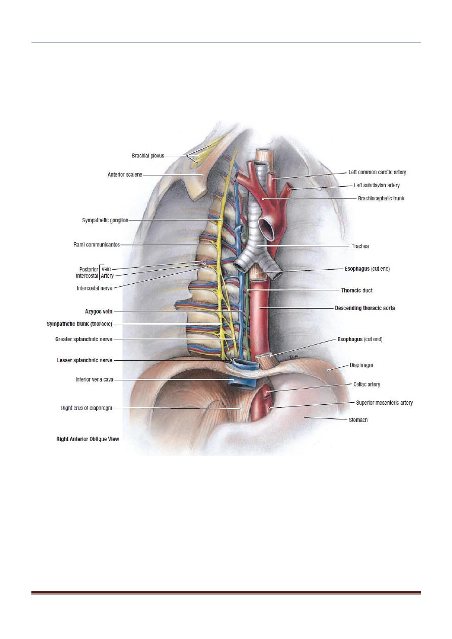
Internal Thoracic Artery:
The internal thoracic artery supplies the anterior wall of the body from the
clavicle to the umbilicus.
It is a branch of the first part of the subclavian artery in the neck.

CHEST ANATOMY THI-QAR UNIVERSITY
COLLEGE OF MEDICINE
LECTURE 8 2019/2020
Dr. Rafid AL-Temimi ; Clinical radiology ( CABM)
Page
4
Dr. Ahmed Abdulameer Daffar ; Thoracic & Vascular Surgeon ( FIBMS )
It descends vertically on the pleura behind the costal cartilages, a
fingerbreadth lateral to the sternum, and ends in the sixth intercostal space
by dividing into the superior epigastric and musculophrenic arteries.
Branches of
Internal
Thoracic Artery:
1. Two anterior intercostal arteries for the upper six intercostal spaces.
2. Perforating arteries, which accompany the terminal branches of the
corresponding intercostal nerves
3. The pericardiacophrenic artery, which accompanies the phrenic nerve and
supplies the pericardium
4. Mediastinal arteries to the contents of the anterior mediastinum (e.g., the
thymus)
5. The superior epigastric artery, which enters the rectus sheath of the anterior
abdominal wall and supplies the rectus muscle as far as the umbilicus
6. The musculophrenic artery, which runs around the costal margin of the
diaphragm and supplies the lower intercostal spaces and the diaphragm
Nerves of the Thorax
Vagus Nerves:
The right vagus &The left vagus nerve :: they form pulmonary plexus
&esophageal plexus
Branches
Both vagi supply the lungs and esophagus. The right vagus gives off cardiac
branches, and the left vagus gives origin to the left recurrent laryngeal nerve. (The
right recurrent laryngeal nerve arises from the right vagus in the neck
Phrenic Nerves:
The phrenic nerves arise from the neck from the anterior rami of the 3rd, 4th, and
5th cervical nerve
.
CHEST ANATOMY THI-QAR UNIVERSITY
COLLEGE OF MEDICINE
LECTURE 8 2019/2020
Dr. Rafid AL-Temimi ; Clinical radiology ( CABM)
Page
5
Dr. Ahmed Abdulameer Daffar ; Thoracic & Vascular Surgeon ( FIBMS )