CHEST ANATOMY THI-QAR UNIVERSITY
COLLEGE OF MEDICINE
LECTURE 9 2019/2020
Dr. Rafid AL-Temimi ; Clinical radiology ( CABM)
Page
1
Dr. Ahmed Abdulameer Daffar ; Thoracic & Vascular Surgeon ( FIBMS )
THE CHEST
Large Arteries of the Thorax
1) Aorta
The aorta is the main arterial trunk that delivers oxygenated blood from the left
ventricle of the heart to the tissues of the body.
It is divided for purposes of description into the following parts: ascending aorta, arch
of the aorta, descending thoracic aorta, and abdominal aorta.
i. Ascending Aorta:
The ascending aorta begins at the base of the left ventricle and runs upward and
forward to come to lie behind the right half of the sternum where it becomes
continuous with the arch of the aorta.
The ascending aorta lies within the fibrous pericardium and is enclosed with the
pulmonary trunk in a sheath of serous pericardium.
At its root, it possesses three bulges, the sinuses of the aorta, one behind each aortic
valve cusp.
Branches:
Right coronary artery & Left coronary artery
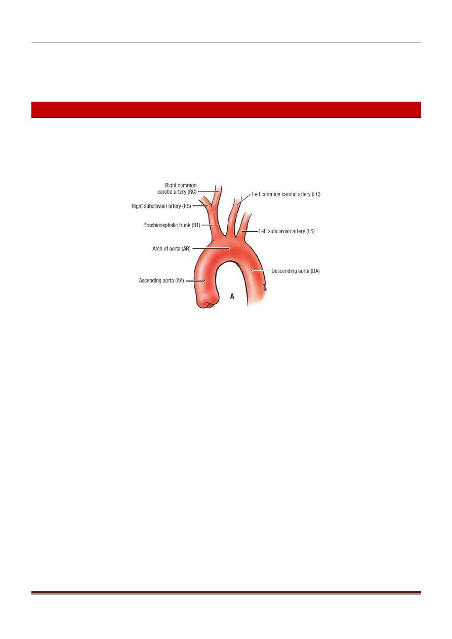
ii. Arch of the Aorta:
The arch of the aorta is a continuation of the ascending aorta. It lies behind the
manubrium sterni and arches upward, backward, and to the left in front of the trachea
(its main direction is backward).
It then passes downward to the left of the trachea and, at the level of the sternal angle,
becomes continuous with the descending aorta.

CHEST ANATOMY THI-QAR UNIVERSITY
COLLEGE OF MEDICINE
LECTURE 9 2019/2020
Dr. Rafid AL-Temimi ; Clinical radiology ( CABM)
Page
2
Dr. Ahmed Abdulameer Daffar ; Thoracic & Vascular Surgeon ( FIBMS )
Branches:
1. The brachiocephalic artery which gives rise to right subclavian and right
common carotid arteries behind the right sternoclavicular joint.
2. The left common carotid arteryIt runs upward and to the left of the trachea
and enters the neck behind the left sternoclavicular joint.
3. The left subclavian artery arises from the aortic arch behind the left common
carotid artery. It arches over the apex of the left lun
g.
iii. Descending Thoracic Aorta:
The descending thoracic aorta lies in the posterior mediastinum and begins as a
continuation of the arch of the aorta on the left side opposite the sternal angle.
At the level of the 12th thoracic vertebra, it passes behind the diaphragm ( through
the aortic opening ) in the midline and becomes continuous with the abdominal aorta.
Branches
1. Posterior intercostal arteries are given off to the lower nine intercostal spaces
on each side.
2. Subcostal arteries run along the lower border of the 12th rib.
3. Pericardial, esophageal, and bronchial arteries.
2) Pulmonary Trunk:
The pulmonary trunk conveys deoxygenated blood from the right ventricle of the
heart to the lungs.
It is about 5 cm ) long and terminates in the concavity of the aortic arch by dividing
into right and left pulmonary arteries
Together with the ascending aorta, it is enclosed in the fibrous pericardium and a
sheath of serous pericardium.
The right pulmonary artery runs to the right behind the ascending aorta and superior
vena cava to enter the root of the right lung.
The left pulmonary artery runs to the left in front of the descending aorta to enter
the root of the left lung.

CHEST ANATOMY THI-QAR UNIVERSITY
COLLEGE OF MEDICINE
LECTURE 9 2019/2020
Dr. Rafid AL-Temimi ; Clinical radiology ( CABM)
Page
3
Dr. Ahmed Abdulameer Daffar ; Thoracic & Vascular Surgeon ( FIBMS )
Large Veins of the Thorax:
1. Brachiocephalic Veins:
The right brachiocephalic vein : is formed at the root of the neck by the union of
the right subclavian and the right internal jugular veins.
The left brachiocephalic vein has a similar origin.It passes obliquely downward and
to the right behind the manubrium sterni and in front of the large branches of the
aortic arch.
It joins the right brachiocephalic vein to form the superior vena cava.
2.
Superior Vena Cava:
The superior vena cava contains all the venous blood from the head and neck and both
upper limbs and is formed by the union of the two brachiocephalic veins.
It passes downward to end in the right atrium of the heart .
The vena azygos joins the posterior aspect of the superior vena cava just before it
enters the pericardium.
3. Azygos Veins:
a) Azygos Vein:
The origin of the azygos vein is variable. It is often formed by the union of the right
ascending lumbar vein and the right subcostal vein.
It ascends through the aortic opening in the diaphragm on the right side of the aorta
to the level sterna angle.
Here it arches forward above the root of the right lung to empty into the posterior
surface of the superior vena cava.
The azygos vein has numerous tributaries, including the eight lower right intercostal
veins, the right superior intercostal vein, the superior and inferior hemiazygos
veins, and numerous mediastinal veins.
CHEST ANATOMY THI-QAR UNIVERSITY
COLLEGE OF MEDICINE
LECTURE 9 2019/2020
Dr. Rafid AL-Temimi ; Clinical radiology ( CABM)
Page
4
Dr. Ahmed Abdulameer Daffar ; Thoracic & Vascular Surgeon ( FIBMS )
b) Inferior Hemiazygos Vein
The inferior hemiazygos vein is often formed by the union of the left ascending
lumbar vein and the left subcostal vein.
It ascends through the left crus of the diaphragm and, at about the level of the eighth
thoracic vertebra, turns to the right and joins the azygos vein.
It receives as tributaries some lower left intercostal veins and mediastinal veins.
c) Superior Hemiazygos Vein
The superior hemiazygos vein is formed by the union of the fourth to the eighth
intercostal veins.
It joins the azygos vein at the level of the seventh thoracic vertebra.
4. Inferior Vena Cava
The inferior vena cava pierces the central tendon of the diaphragm opposite the eighth
thoracic vertebra and almost immediately enters the lowest part of the right atrium .
5. Pulmonary Veins
Two pulmonary veins leave each lung carrying oxygenated blood to the left atrium of the
heart.
Lymph Nodes and Vessels of the Thorax:
Thoracic Duct:
The thoracic duct begins below in the abdomen as a dilated sac, the cisterna chyli. It
ascends through the aortic opening in the diaphragm.
It enter the beginning of the left brachiocephalic vein.
The thoracic duct thus conveys to the blood all lymph from the lower limbs, pelvic
cavity, abdominal cavity, left side of the thorax, and left side of the head, neck, and
left arm .
Right Lymphatic Duct:
The right jugular, subclavian, and bronchomediastinal trunks, which drain the right
side of the head and neck, the right upper limb, and the right side of the thorax,
respectively, may join to form the right lymphatic duct.
This duct opens into the beginning of the right brachiocephalic vein.