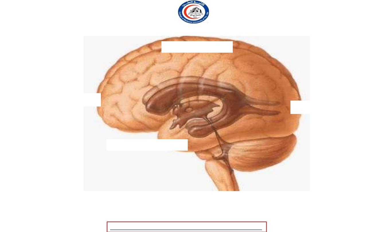
VENTRICLES
Done by
Dr.Rafid Remthan Al-Temimi
Clinical Radiology
CAMB,DMRD,M.B.Ch.B.,.
المرحلة
:
الثانية
المادة
:
التشريح
ج
امعة ذي قار
/
كلية الطب
الدكتور
رافد
رمثان التميمي
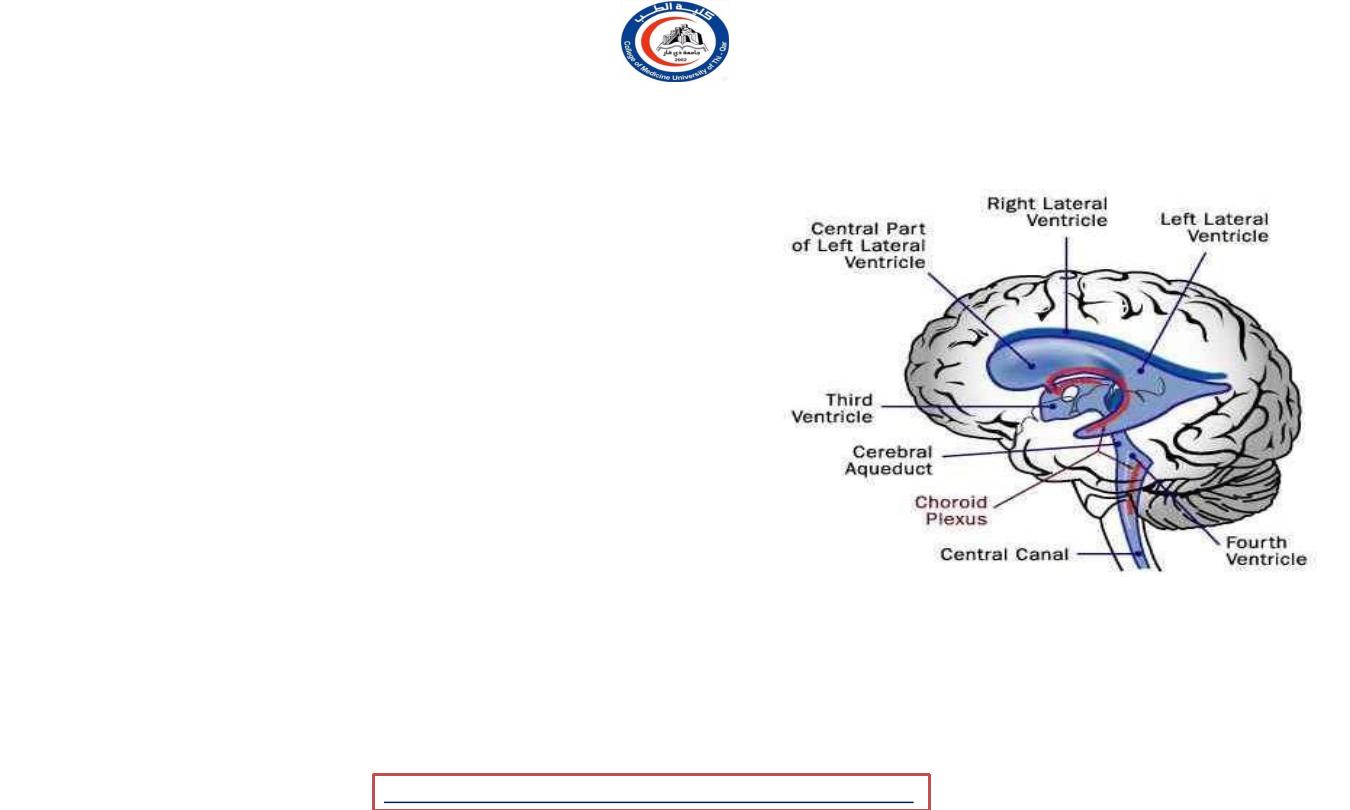
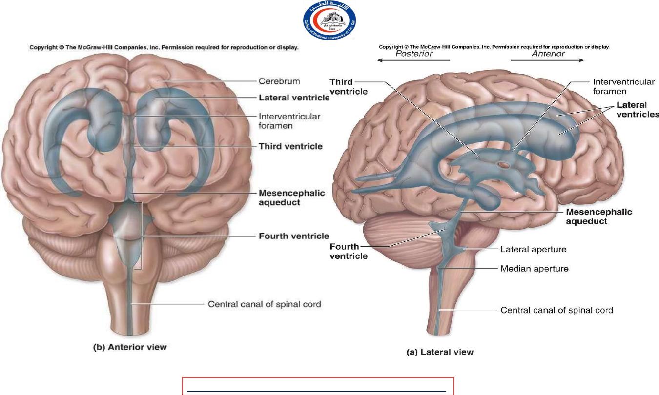
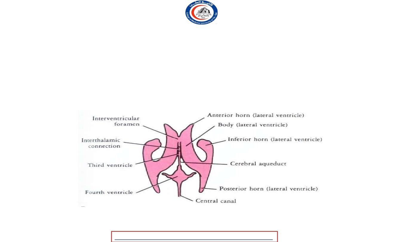
VENTRICLES(Ventricular System)
• A ventricle is an internal cavity of the brain.
Within the brain, which is filled with
cerebrospinal fluid(CSF).
• The ventricular system is composed of two
lateral ventricles
and two midline ventricles(
third and fourth ventricles
).
• The chambers are connected to allow the flow
of cerebrospinal fluid via two
interventricular
foramen
(referred to as the
foramen of Monro
)
and the
cerebral aqueduct
(referred to as the
aqueduct of Sylvius
).
University Of Thi-Qar
College Of medicine
Dr.Rafid Remthan AL-Temimi,Clinical Radiology,CAMB, 2020
Anatomy lecture . 2
nd
stage
Dr.Rafid Al-Temimi
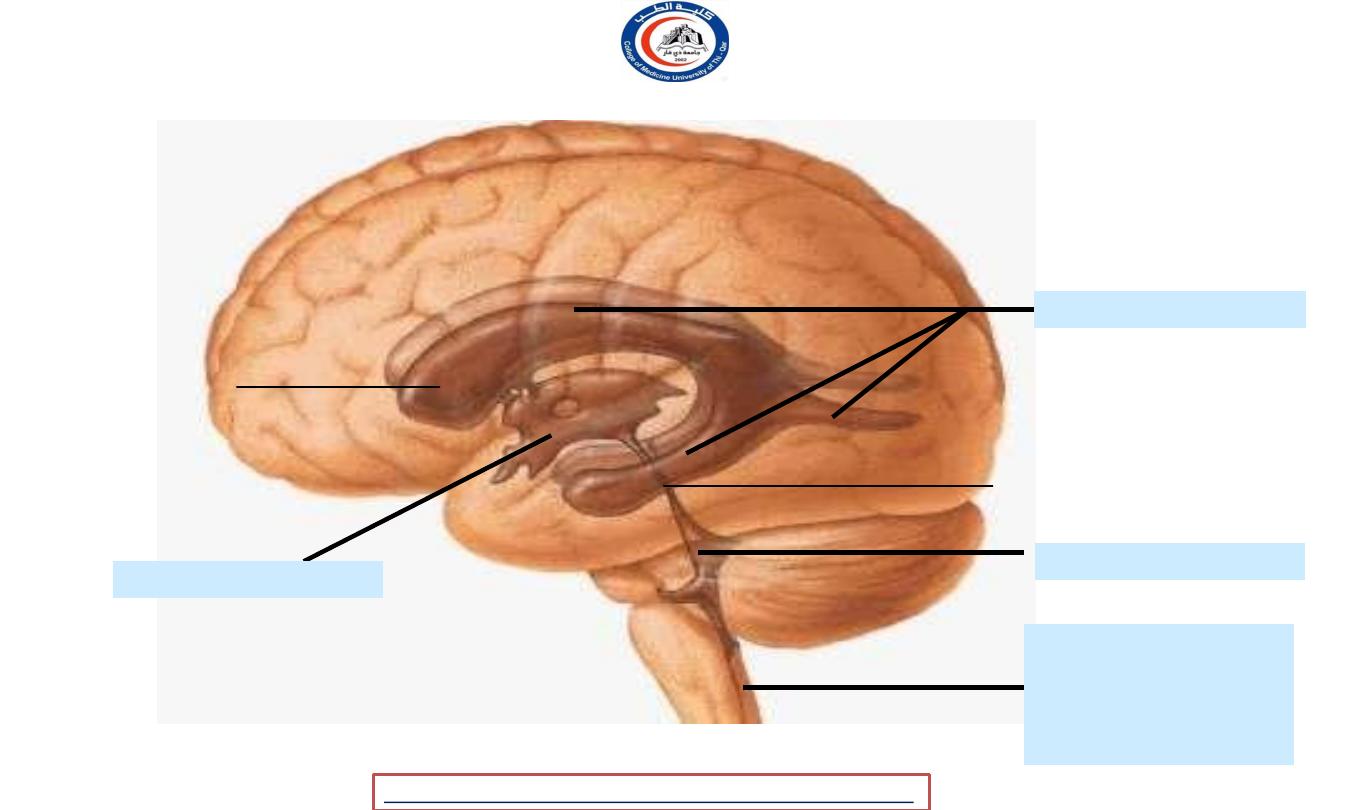
Central canal of
medulla oblongata
& spinal cord
Fourth ventricle
Lateral ventricle
Third ventricle
Interventricular
foramen (Monro)
Cerebral
aqueduct
Lateral view to show the ventricular system of the CNS
University Of Thi-Qar
College Of medicine
Dr.Rafid Remthan AL-Temimi,Clinical Radiology,CAMB, 2020
Anatomy lecture . 2
nd
stage
Dr.Rafid Al-Temimi
VENTRICLES(Ventricular System)
CONSISTS OF :
1) Lateral ventricle
2) Third ventricle
3) Fourth ventricle
4) Central canal of the
medulla oblongata &
spinal cord
University Of Thi-Qar
College Of medicine
Dr.Rafid Remthan AL-Temimi,Clinical Radiology,CAMB, 2020
Anatomy lecture . 2
nd
stage
Dr.Rafid Al-Temimi
University Of Thi-Qar
College Of medicine
Dr.Rafid Remthan AL-Temimi,Clinical Radiology,CAMB, 2020
Anatomy lecture . 2
nd
stage
Dr.Rafid Al-Temimi
University Of Thi-Qar
College Of medicine
Dr.Rafid Remthan AL-Temimi,Clinical Radiology,CAMB, 2020
Anatomy lecture . 2
nd
stage
Dr.Rafid Al-Temimi
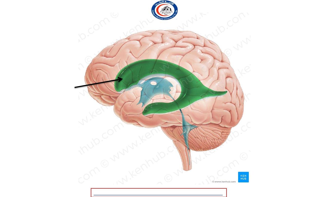
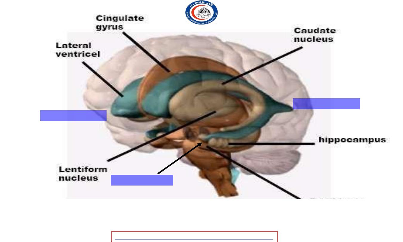
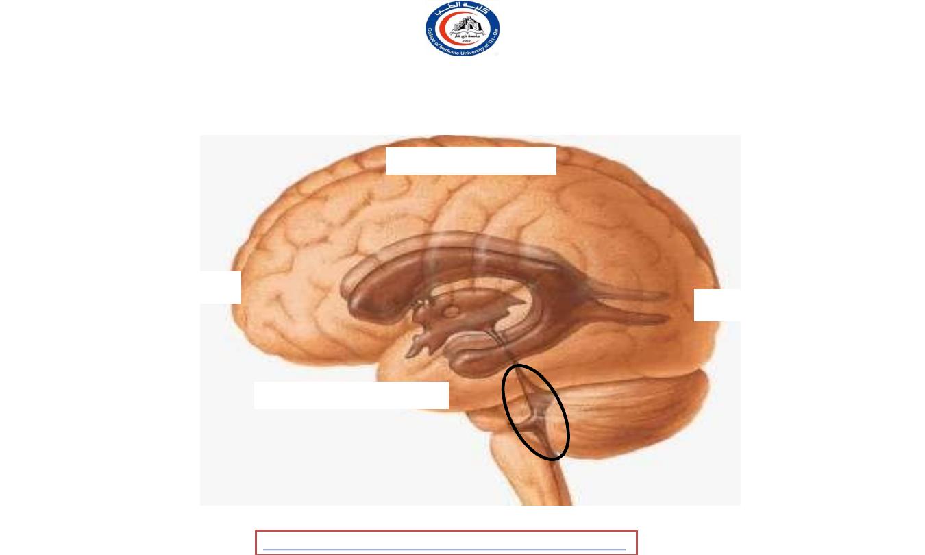
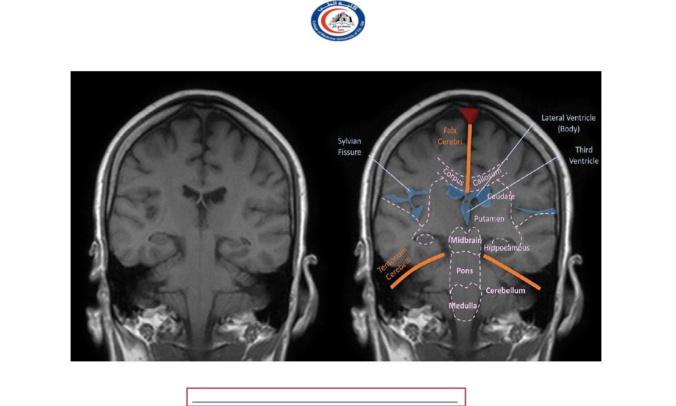
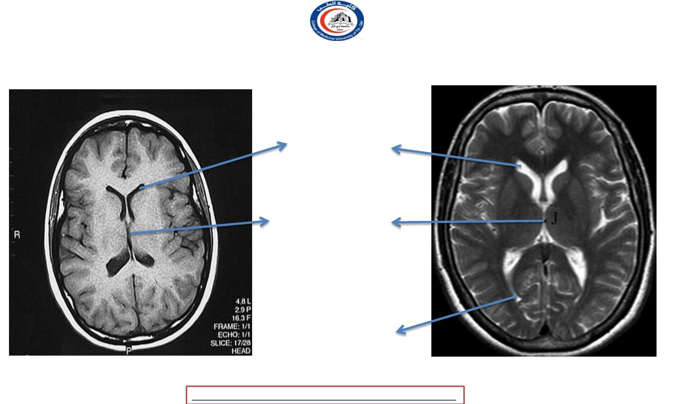
Lateral Ventricle
Lateral Ventricles
• The lateral ventricles are two
curved shaped cavities
located within the
cerebrum.
• The lateral ventricles are
separated by the
septum
pellucidum
and do not
communicate directly
University Of Thi-Qar
College Of medicine
Dr.Rafid Remthan AL-Temimi,Clinical Radiology,CAMB, 2020
Anatomy lecture . 2
nd
stage
Dr.Rafid Al-Temimi
Lateral ventricle
Frontal lobe
Parietal lobe
Temporal lobe
Occipital lobe
University Of Thi-Qar
College Of medicine
Dr.Rafid Remthan AL-Temimi,Clinical Radiology,CAMB, 2020
Anatomy lecture . 2
nd
stage
Dr.Rafid Al-Temimi
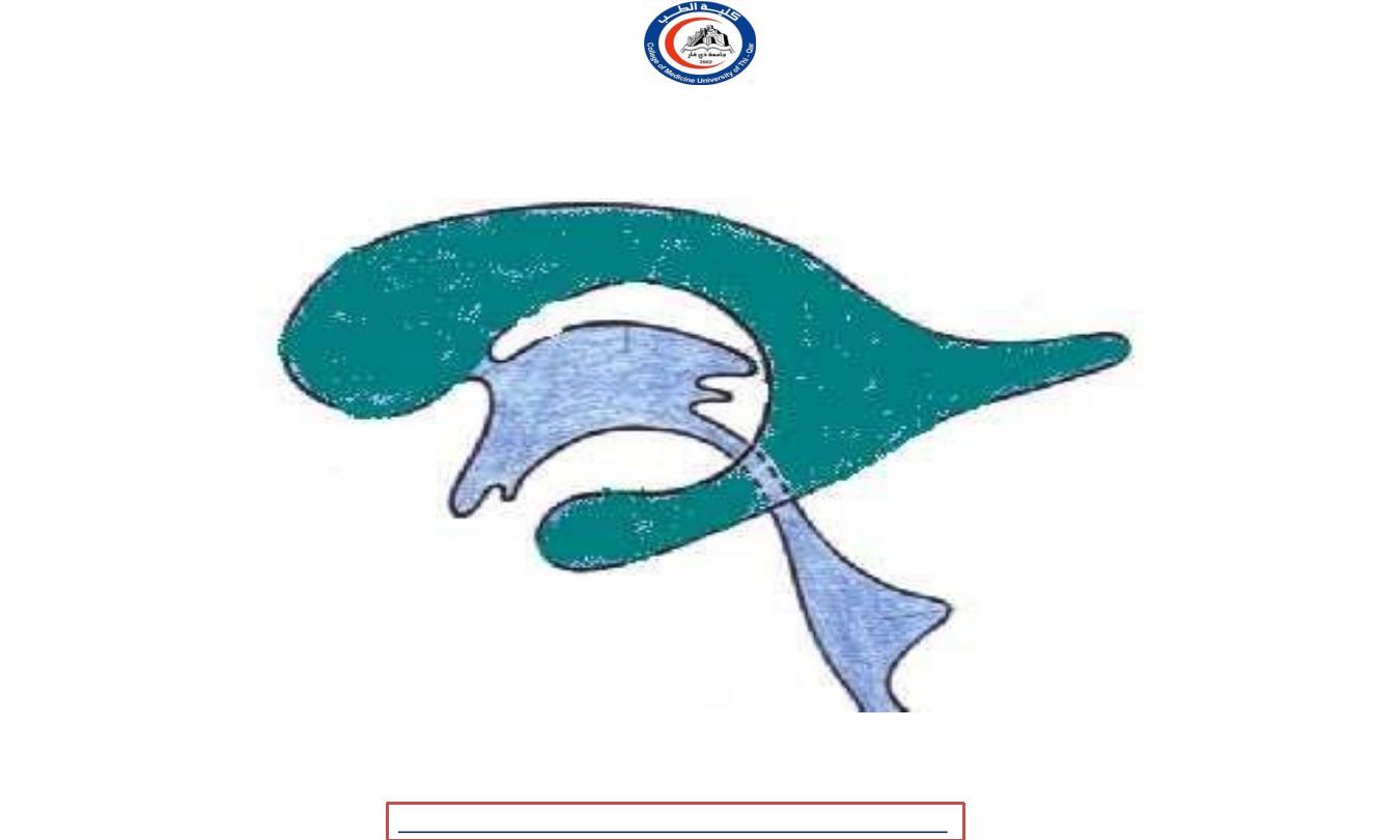
C-shaped cavity & may be divided into
:
2. Anterior
horn
1. Body
3. Posterior
horn
Third
ventricle
4. Inferior horn
Fourth
ventricle
Lateral ventricle
Lateral view of the ventricular cavities of the brain
University Of Thi-Qar
College Of medicine
Dr.Rafid Remthan AL-Temimi,Clinical Radiology,CAMB, 2020
Anatomy lecture . 2
nd
stage
Dr.Rafid Al-Temimi
Anterior horn
Inferior horn
Posterior horn
Lateral view to show the ventricular system of the CNS
University Of Thi-Qar
College Of medicine
Dr.Rafid Remthan AL-Temimi,Clinical Radiology,CAMB, 2020
Anatomy lecture . 2
nd
stage
Dr.Rafid Al-Temimi
University Of Thi-Qar
College Of medicine
Dr.Rafid Remthan AL-Temimi,Clinical Radiology,CAMB, 2020
Anatomy lecture . 2
nd
stage
Dr.Rafid Al-Temimi
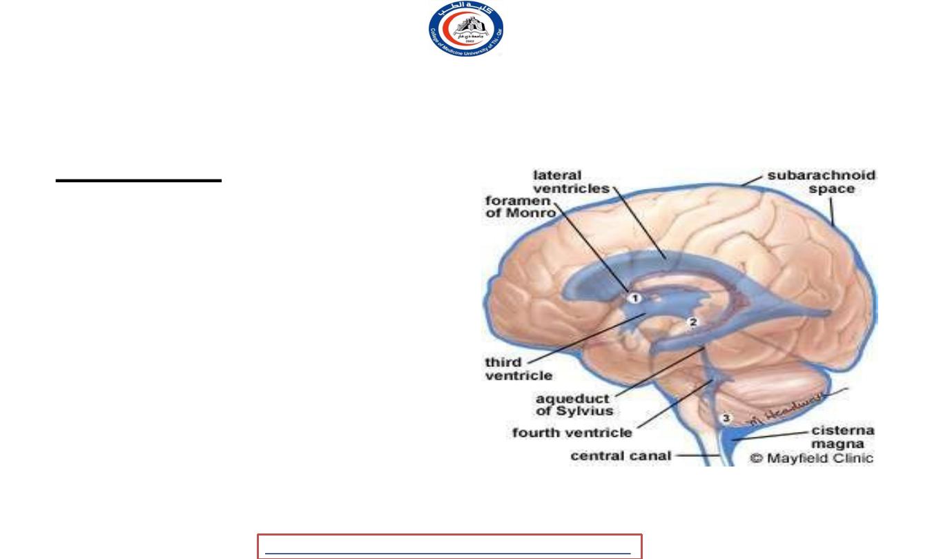
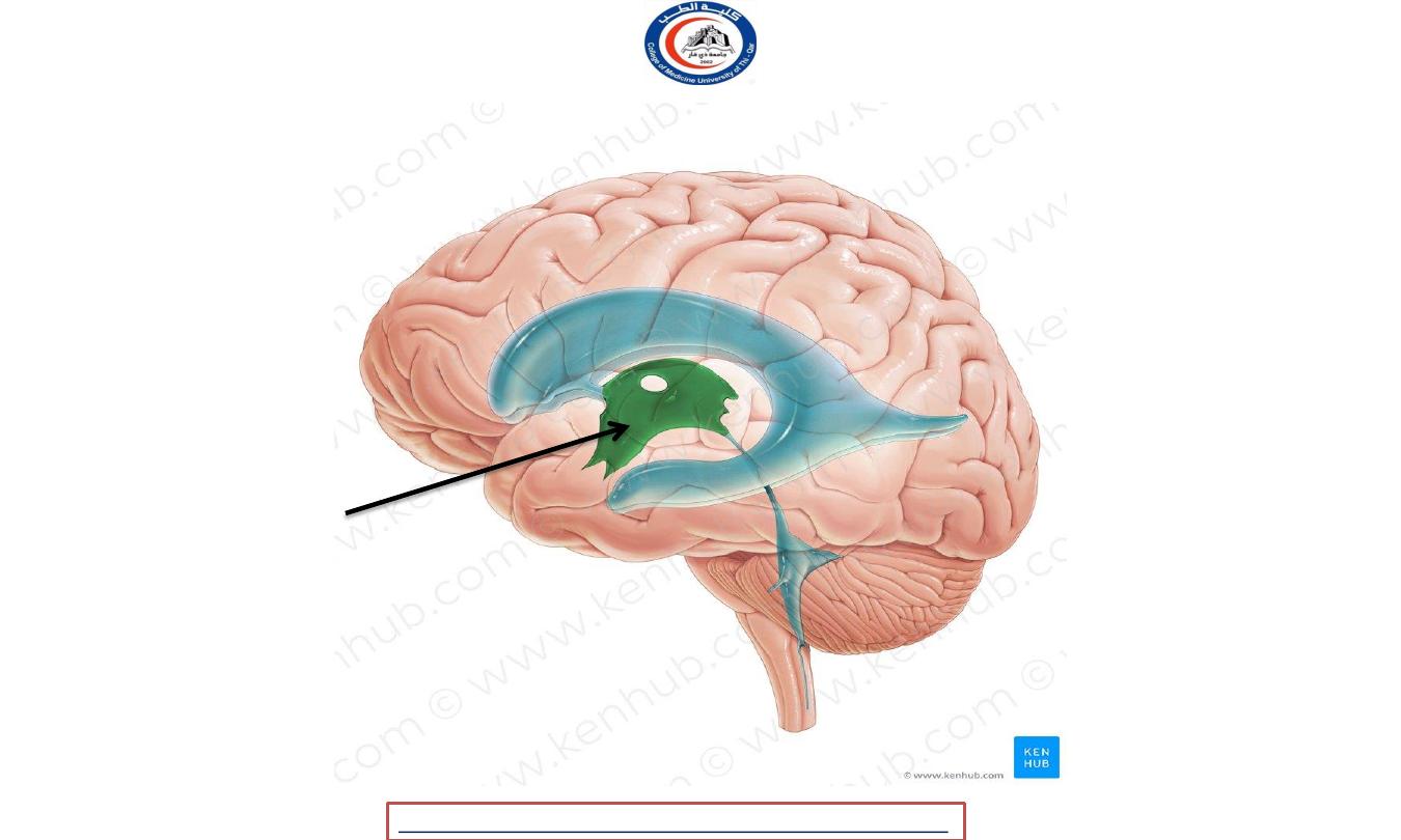
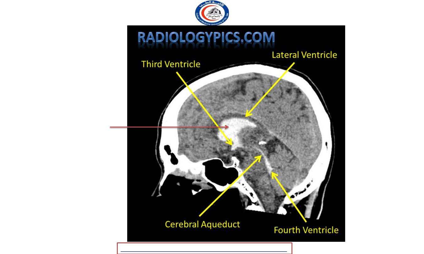
Third ventricle
The third ventricle is a narrow cavity or a slitlike cleft between the 2 thalamus
Communicates ;
•
Anteriorly with lateral ventricles through
interventricular foramina (of monro)
•
Posteriorly with fourth ventricle through
cerebral aqueduct (of sylvius)
Posterior view to show the ventricular system of the CNS
Third ventricle
University Of Thi-Qar
College Of medicine
Dr.Rafid Remthan AL-Temimi,Clinical Radiology,CAMB, 2020
Anatomy lecture . 2
nd
stage
Dr.Rafid Al-Temimi
Frontal lobe
Parietal lobe
Temporal lobe
Occipital lobe
Third ventricle
University Of Thi-Qar
College Of medicine
Dr.Rafid Remthan AL-Temimi,Clinical Radiology,CAMB, 2020
Anatomy lecture . 2
nd
stage
Dr.Rafid Al-Temimi
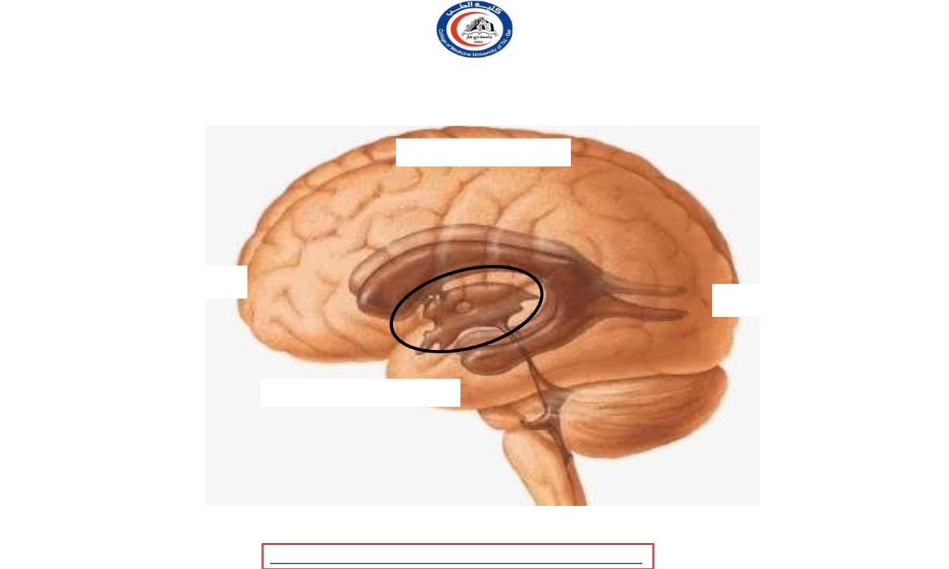
Third
ventricle
Hypothalamus
Coronal section of the brain (posterior view)
Third ventricle
Thalamus
ROOF
FLOOR
Lateral wall
Body of fornix
University Of Thi-Qar
College Of medicine
Dr.Rafid Remthan AL-Temimi,Clinical Radiology,CAMB, 2020
Anatomy lecture . 2
nd
stage
Dr.Rafid Al-Temimi
University Of Thi-Qar
College Of medicine
Dr.Rafid Remthan AL-Temimi,Clinical Radiology,CAMB, 2020
Anatomy lecture . 2
nd
stage
Dr.Rafid Al-Temimi
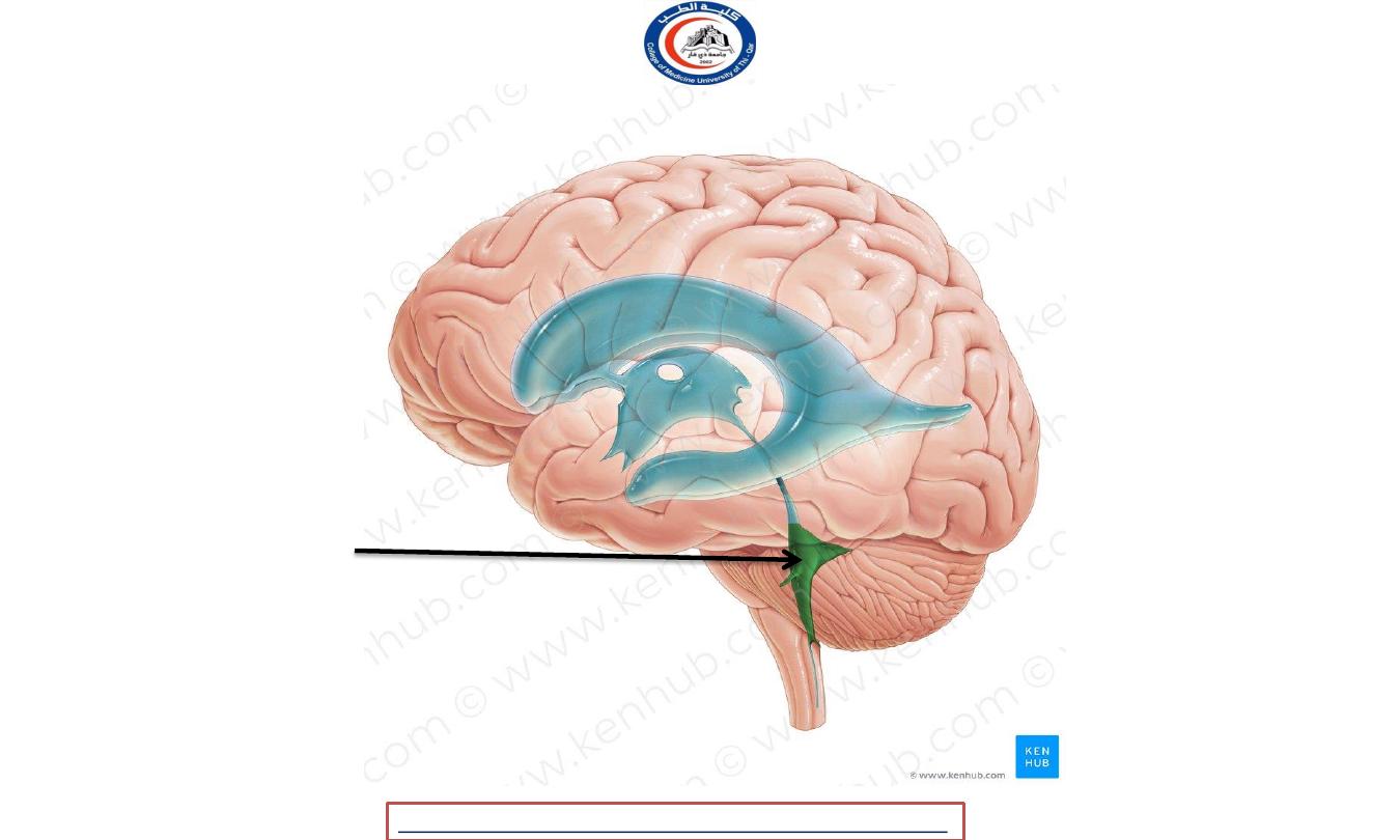
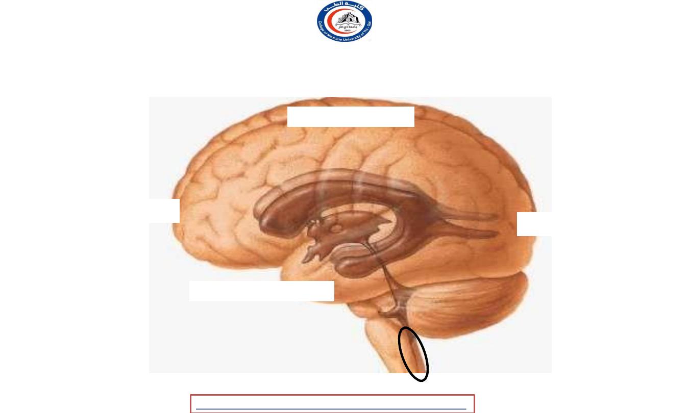
Fourth ventricle
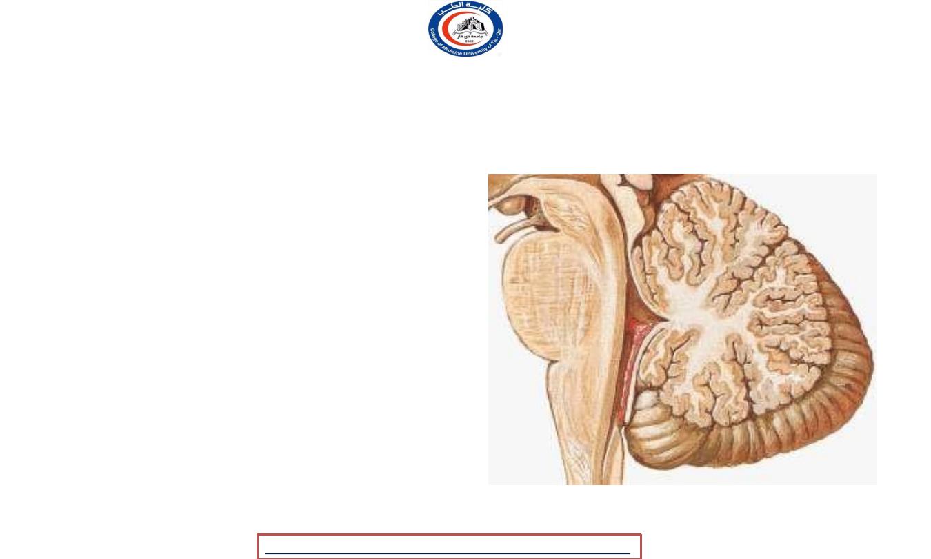
Fourth ventricle
• The fourth ventricle Is a
rhomboid or diamond shaped
cavity.
• It is a wide and flattened
space located just anterior to
the cerebellum and posterior
to the
upper, or superior, half of the
medulla oblongata and the
pons.
University Of Thi-Qar
College Of medicine
Dr.Rafid Remthan AL-Temimi,Clinical Radiology,CAMB, 2020
Anatomy lecture . 2
nd
stage
Dr.Rafid Al-Temimi
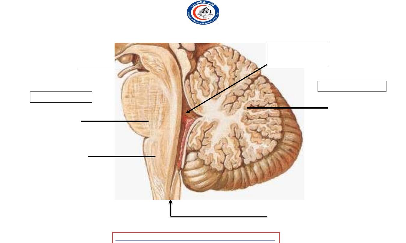
Cerebellum
Pons
Medulla
oblongata
(superior
half)
Sagittal section of the 4
th
ventricle
Cerebral
aqueduct
Fourth ventricle
Fourth
ventricle
ANTERIOR
POSTERIOR
Central canal (spinal cord)
University Of Thi-Qar
College Of medicine
Dr.Rafid Remthan AL-Temimi,Clinical Radiology,CAMB, 2020
Anatomy lecture . 2
nd
stage
Dr.Rafid Al-Temimi
Frontal lobe
Parietal lobe
Temporal lobe
Occipital lobe
Fourth ventricle
University Of Thi-Qar
College Of medicine
Dr.Rafid Remthan AL-Temimi,Clinical Radiology,CAMB, 2020
Anatomy lecture . 2
nd
stage
Dr.Rafid Al-Temimi
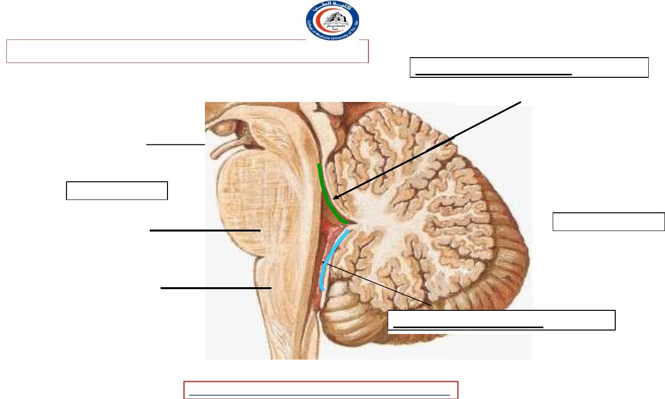
Pons
Medulla
oblongata
(superior
half)
Fig. : Sagittal section of the 4
th
ventricle
Cerebral
aqueduct
ANTERIOR
POSTERIOR
Superior part of the roof ;
Superior medullary velum
Inferior part of the roof ;
Inferior medullary velum
Roof or posterior wall of fourth ventricle:
University Of Thi-Qar
College Of medicine
Dr.Rafid Remthan AL-Temimi,Clinical Radiology,CAMB, 2020
Anatomy lecture . 2
nd
stage
Dr.Rafid Al-Temimi
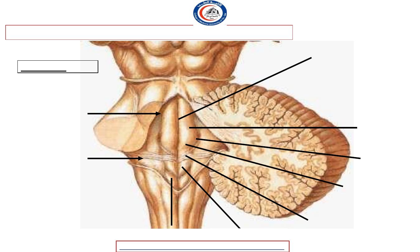
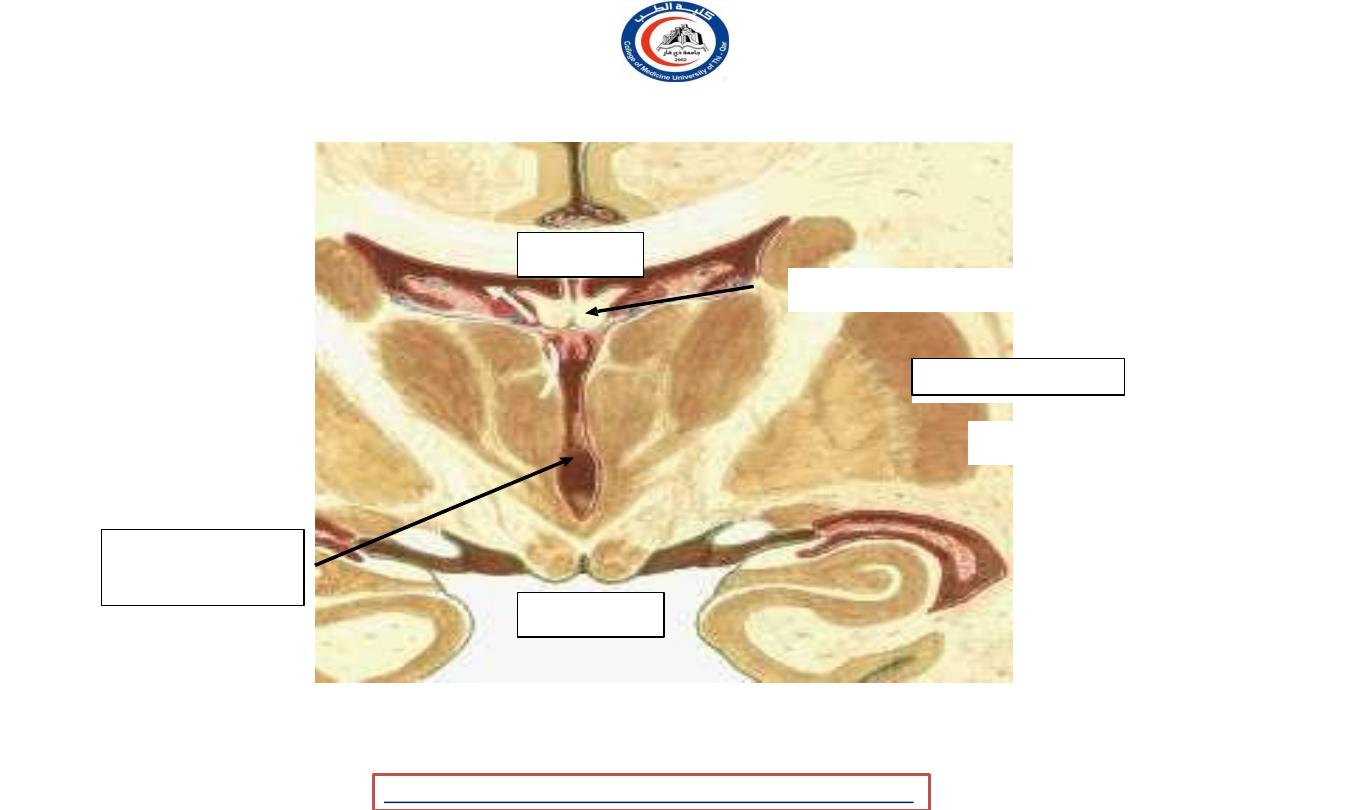
Floor or rhomboid fossa of fourth ventricle :
Formed by ;
1. Posterior
surface of the
pons
2. Cranial ½ of
the medulla
oblongata
Medial
sulcus
(divides the floor
into
symmetrical
halves)
Medial
eminence
Sulcus
limitans
Facial
colliculus
Stria medullaris
(strands of nerve fibers)
Hypoglossal triangle
Vagal triangle
Posterior view of the 4
th
ventricle
University Of Thi-Qar
College Of medicine
Dr.Rafid Remthan AL-Temimi,Clinical Radiology,CAMB, 2020
Anatomy lecture . 2
nd
stage
Dr.Rafid Al-Temimi
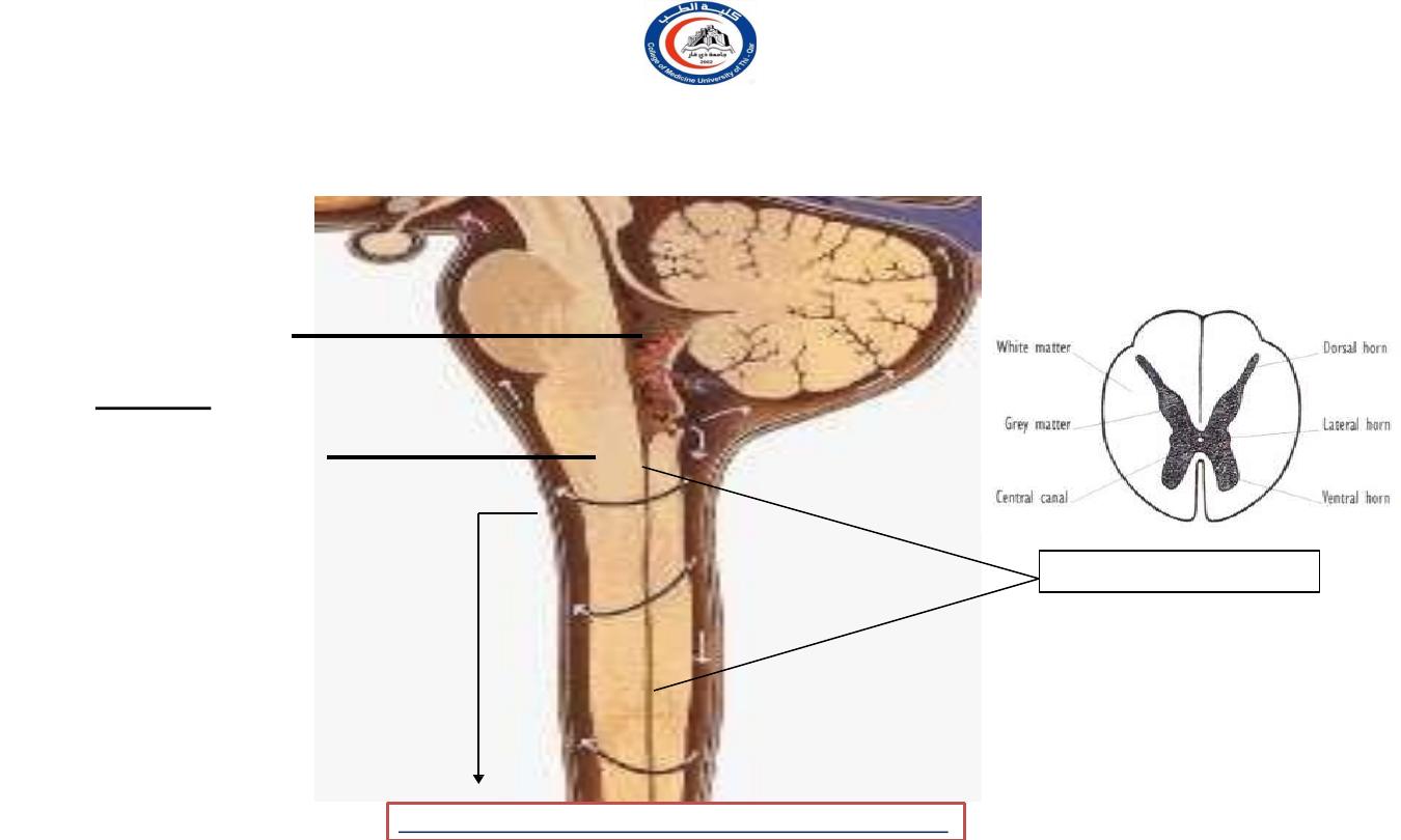
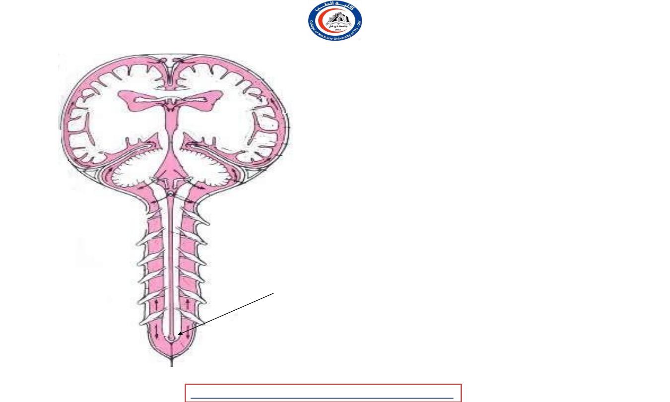
CENTRAL CANAL
Opens superiorly into the fourth ventricle
Entire length
of spinal
cord
Central canal
(Lined with ependyma
but no choroid plexus in
the central canal)
Fourth
ventricle
Extends ;
Inferior ½ of
medulla
oblongata
University Of Thi-Qar
College Of medicine
Dr.Rafid Remthan AL-Temimi,Clinical Radiology,CAMB, 2020
Anatomy lecture . 2
nd
stage
Dr.Rafid Al-Temimi
Frontal lobe
Parietal lobe
Temporal lobe
Occipital lobe
CENTRAL CANAL
University Of Thi-Qar
College Of medicine
Dr.Rafid Remthan AL-Temimi,Clinical Radiology,CAMB, 2020
Anatomy lecture . 2
nd
stage
Dr.Rafid Al-Temimi
Conus medullaris-
Terminal ventricle
University Of Thi-Qar
College Of medicine
Dr.Rafid Remthan AL-Temimi,Clinical Radiology,CAMB, 2020
Anatomy lecture . 2
nd
stage
Dr.Rafid Al-Temimi
University Of Thi-Qar
College Of medicine
Dr.Rafid Remthan AL-Temimi,Clinical Radiology,CAMB, 2020
Anatomy lecture . 2
nd
stage
Dr.Rafid Al-Temimi
University Of Thi-Qar
College Of medicine
Dr.Rafid Remthan AL-Temimi,Clinical Radiology,CAMB, 2020
Anatomy lecture . 2
nd
stage
Dr.Rafid Al-Temimi
University Of Thi-Qar
College Of medicine
Dr.Rafid Remthan AL-Temimi,Clinical Radiology,CAMB, 2020
Anatomy lecture . 2
nd
stage
Dr.Rafid Al-Temimi
Lateral ventricle
Anterior horn
Lateral ventricle
Posterior horn
3
rd
ventricle

University Of Thi-Qar
College Of medicine
Dr.Rafid Remthan AL-Temimi,Clinical Radiology,CAMB, 2020
Anatomy lecture . 2
nd
stage
Dr.Rafid Al-Temimi
Fourth ventricle
University Of Thi-Qar
College Of medicine
Dr.Rafid Remthan AL-Temimi,Clinical Radiology,CAMB, 2020
Anatomy lecture . 2
nd
stage
Dr.Rafid Al-Temimi
intraventricular hemorrhage
Appear hyperdense in CT
(white) like bone
University Of Thi-Qar
College Of medicine
Dr.Rafid Remthan AL-Temimi,Clinical Radiology,CAMB, 2020
Anatomy lecture . 2
nd
stage
Dr.Rafid Al-Temimi
hydrocephalus
Hydrocephalus is a condition in
which an accumulation of
cerebrospinal fluid (CSF) occurs
within the brain.
This typically causes increased
pressure inside the skull.
University Of Thi-Qar
College Of medicine
Anatomy lecture . 2
nd
stage
Dr.Rafid Al-Temimi
Dr.Rafid Remthan AL-Temimi,Clinical Radiology,CAMB, 2020