
Sexually Transmitted Diseases
(STDS)
1

2
STI versus STD----
• STI – Infections acquired through sexual
intercourse (may be symptomatic or
asymptomatic)
• STD – Symptomatic disease acquired through
sexual intercourse
• STI is most commonly used because it applies
to both symptomatic and asymptomatic
infections

Sexually Transmitted disease
presentation:
o Genital ulcers or sores
o Urethral discharge
o Vaginal discharge
o Lower abdominal pain
o Inguinal bubo
o Scrotal swelling
o Rectal or pharyngeal inflammation
o papules
3
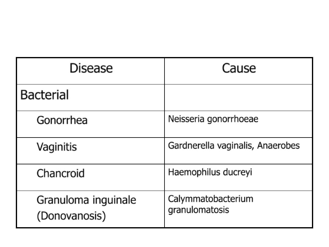
Types and their pathogenic causes
Disease
Cause
Bacterial
Gonorrhea
Neisseria gonorrhoeae
Vaginitis
Gardnerella vaginalis, Anaerobes
Chancroid
Haemophilus ducreyi
Granuloma inguinale
(Donovanosis)
Calymmatobacterium
granulomatosis
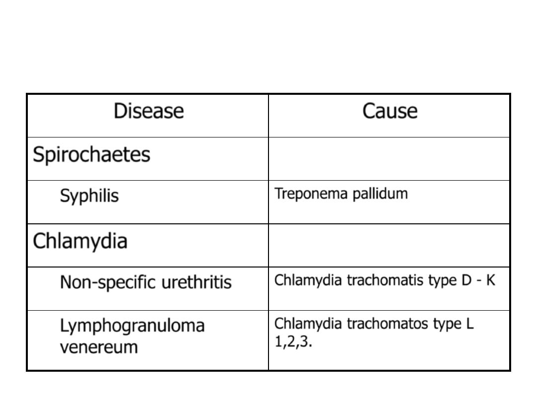
Types and their pathogenic causes
Disease
Cause
Spirochaetes
Syphilis
Treponema pallidum
Chlamydia
Non-specific urethritis
Chlamydia trachomatis type D - K
Lymphogranuloma
venereum
Chlamydia trachomatos type L
1,2,3.
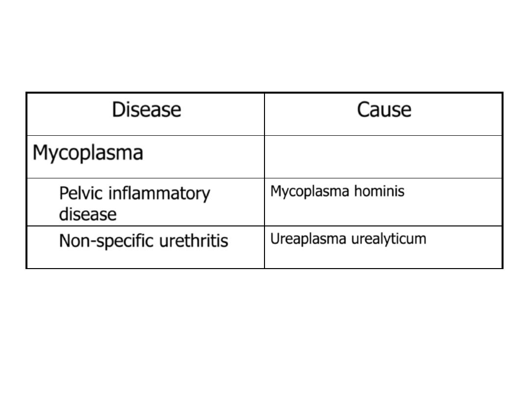
Types and their pathogenic causes
Disease
Cause
Mycoplasma
Pelvic inflammatory
disease
Mycoplasma hominis
Non-specific urethritis
Ureaplasma urealyticum
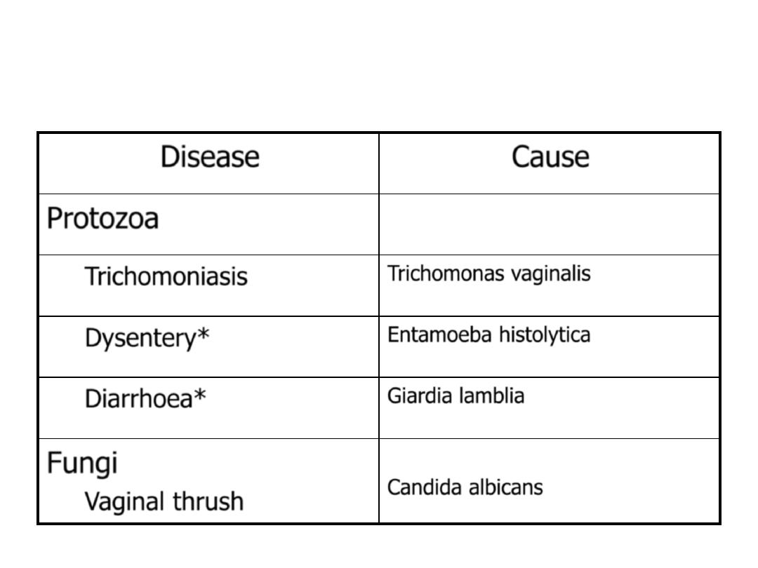
* Not always sexually transmitted
Types and their pathogenic causes
Disease
Cause
Protozoa
Trichomoniasis
Trichomonas vaginalis
Dysentery*
Entamoeba histolytica
Diarrhoea*
Giardia lamblia
Fungi
Vaginal thrush
Candida albicans
Not always sexually transmitted
Types and their pathogenic causes
Disease
Cause
Ectoparasites
Pubic lice
Phthirius pubis
Genital scabies
Sarcoptes scabiei
Viruses
AIDS
HIV
Genital herpes
Herpes simplex, type 2 (and 1)
Warts
Papilloma viruses types 6,11,16 &
18)
Hepatitis*
Hepatitis B

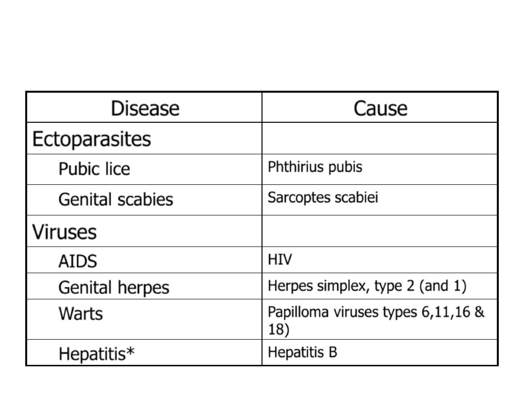
Gonorrhea
• It is bacterial infection caused by Neisseria
gonorrhoeae a gram- negative, infects columnar or
cuboidal epithelium.
• Site of infection: the organism can survive only in
blood and on mucosal surfaces including the urethra,
endocervix, rectum, pharynx, conjunctiva.
Most
common sites:
Cervix (cervicitis) or vagina in the female
Urethra (urethritis) or penis in the male
• Mode of infection: almost always by sexual intercourse.
Greater efficiency of transmission from male to female
• Gonorrhea can also be spread from mother to child
during birth.
9

IN MEN:
➢ Urethritis; Epididymitis
➢ Most infections among men are acute and
symptomatic with purulent discharge &
dysuria (painful urination) after 2-5 day
incubation period
➢ The most common cause of urethritis
among men are Neisseria and Chlamydia.
10

IN WOMEN:
➢ Cervicitis; Vaginitis; Pelvic Inflammatory Disease
(PID); Disseminated Gonococcal Infection (DGI)
➢ Women often asymptomatic ; Often untreated
until PID complications develop.
➢ PID:
• Progressive infection that harms a women’s reproductive
system. Can lead to sterility, ectopic pregnancy and
chronic pain - treated or not.
• Caused by chlamydia and gonorrhea.
• Symptoms - long and painful periods, discharge, low
abdominal pain, fever, chills, nausea, vomiting, pain
during intercourse.
11

IN WOMEN
(cont.)
:
➢ Disseminated Gonococcal Infection (DGI):
• Result of gonococcal bacteremia
• Often skin lesions
• Petechiae (hemorrhagic spots)
• Pustules on extremities
• Arthralgias
• Tenosynovitis
• Septic arthritis
• Occasional complications: Hepatitis; Rarely
endocarditis or meningitis
12

Gonorrhea
Females
1. 50% risk of infection after
single exposure
2. Asymptomatic infections
frequently not diagnosed
3. Genital infection primary
site is cervix (cervicitis),
but vagina, urethra, rectum
can be colonized
4. Ascending infections in 10-
20% including salpingitis,
tubo-ovarian abscesses,
pelvic inflammatory
disease (PID) , chronic
infections can lead to
sterility
Males
1. 20% risk of infection
after single exposure
2. Most initially
symptomatic (95%
acute)
3. Genital infection
generally restricted to
urethra (urethritis) with
purulent discharge and
dysuria
4. Rare complications may
include epididymitis,
prostatitis, and
periurethral abscesses
13

Gonorrhea
Females
5. Disseminated infections
more common, including
septicemia, infection of
skin and joints (1-3%)
6. Can infect infant at
childbirth (conjunctivitis,
ophthalmia neonatorum)
Males
5. Disseminated infections
are very rare
6. More common in
homosexual/bisexual\
heterosexual
14
Extragenital gonorrhea:
oRectal Gonorrhea:
oGonococcal Pharyngitis:
oDisseminated Gonococcal Infection (Arthiritis-Dermatitis
Syndrome):
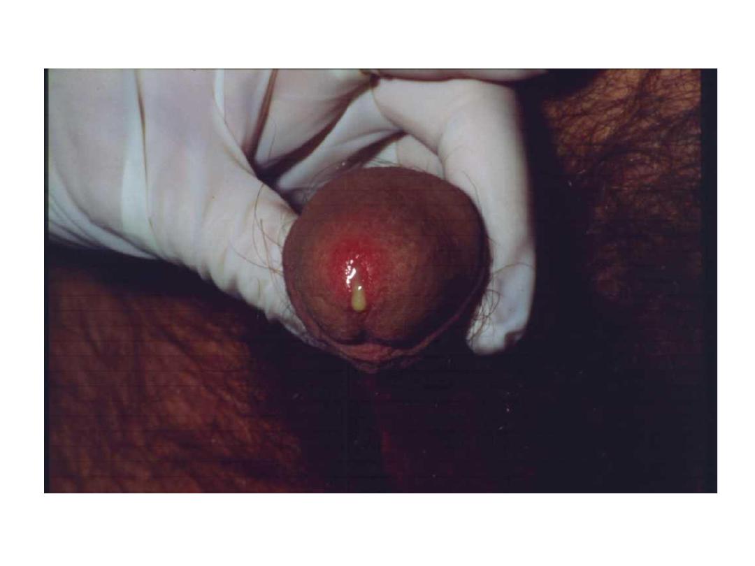
Gonococcal Urethritis

Diagnosis:
• Gram's stain: the presence of intracellular
diplococci within polymorphonuclear
leukocytes→presumptive diagnosis
• Culture →gold slandered for diagnosis
• Nucleic acid amplification tests: have high
sensitivity, and they also test for C.trachomatis.
• serologic test: non-available; all patients should
have a serologic test for syphilis and HIV.
16

Treatment
• the standard therapy recommended in
uncomplicated infections of the urethra,
cervix, rectum, or pharynx in nonpregnant
adults is a single dose of 250mg of ceftriaxone
Plus (if Chlamydia is not Ruled Out ).
• Azithromycin 1 g in a single dose or
• Doxycycline 100 mg twice daily for 7 days
17

Treatment
• Alternative:
• Spectinomycin 2g IM in one dose
• Ciprofloxacin 500mg orally in one dose
• Norfloxacin 800mg orally in one dose
• Cefotaxime 1g in one dose
(To all these: Plus if Chlamydia is not Ruled Out ).
• Azithromycin 1 g in a single dose or
• Doxycycline 100 mg twice daily for 7 days
18

Nongonococcal Urethritis (NGU)
• The diagnosis, as the name implies, used to be one of
exclusion. Any urethral inflammation not caused by
gonorrhea
Organisms:
• Genital chlamydial is responsible for about half of NGU
• Ureaplasma urealyticum and mycoplasma genitalium
cause 10-30% of NGU
• Herpes viruses, T. vaginalis, haemophilus species, and
anaerobic bacteria account less than 10% of cases
• One third of cases, no infectious cause can be found.
19

20
males. NGU begins 7-28 days after sexual contact with
a smarting sensation while urinating and a mucoid
discharge.
females. The sign and symptoms in females are
more nonspecific; may be present mucopurulent
discharge.
Treatment: azithromycin 1gm orally in a single
dose or doxycycline 100mg orally twice a day for 7
days. Alternative: erythromycin 500mg orally four
times a day for 7 days.
Nongonococcal Urethritis

22
Gonococcal
urethritis
NGU
3-5 days
7-28 days
Incubation
period
Abrupt
gradual
Onset
Burning
Smarting feeling
dysuria
Purulent
Mucoid or purulent
discharge
Gram-negative
intracellular
diplococci
Polymorphonuclear
leukocytes
Gram stain

Syphilis
• Also known as lues, is a contagious, sexually-
transmitted disease caused by the spirochete
Treponema Pallidum.
• The spirochete enters through the skin or mucous
membranes, on which the primary
manifestations are seen
• In congenital syphilis the treponema crosses the
placenta and infects the fetus.
• Route of infection: sexual contact (most
important); congenital; acquired by transfusion of
blood; accidental
23

Stages (untreated syphilis)
1.
Primary S: localized infection at site of inoculation
(chancre)
2.
Secondary S: disseminated infection
3.
Latent S: no clinical sign or symptoms (seropositive)
-Early latent S: less than one year duration
-Late latent S: greater than one year duration
4. Syphilis of unknown duration
5. Late (tertiary) S: cutaneous, vascular, neurologic findings
6. Congenital S: acquired in utero
Risk of transmission: during primary, secondary, and early
latent stages of disease. The patient is most infectious
during the first and second year of infection
24

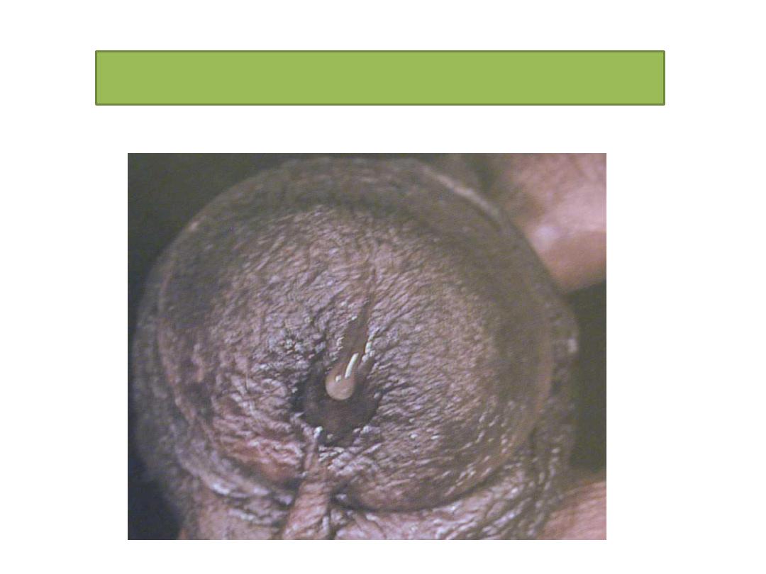
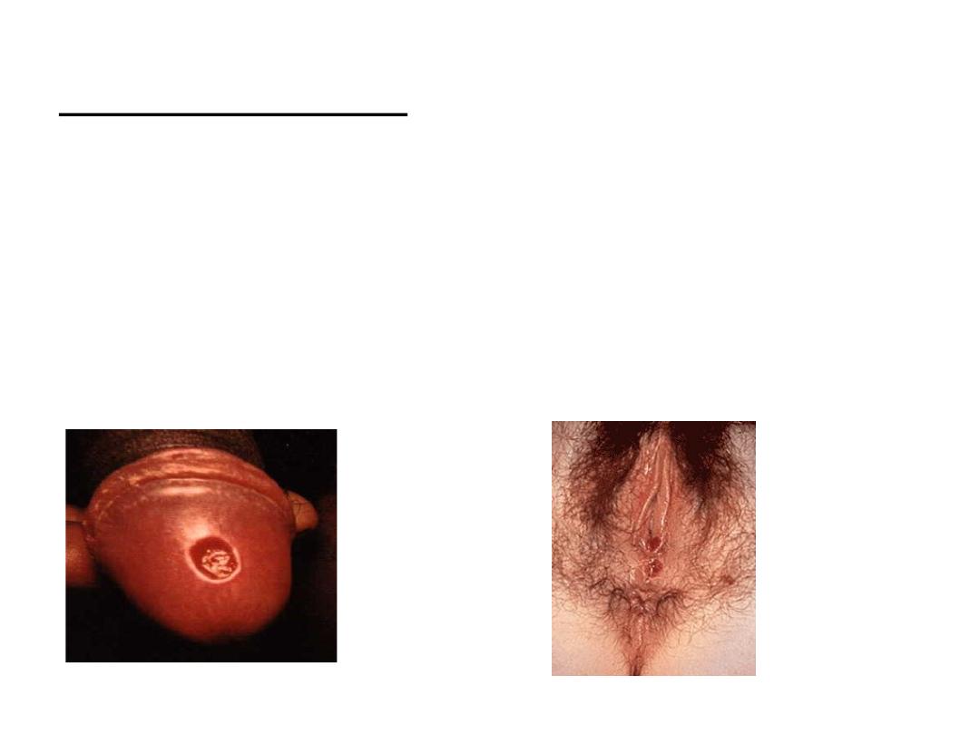
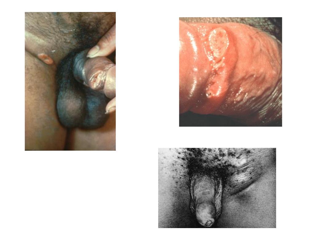
Primary Syphilis; Chancre (primary stage)
• Primary syphilis manifests as a single, painless, clean-
based ulcer . Chancres are usually solitary.
• The lesion usually appears within 3 weeks of
infection (can range from 10-90 days)
• In women the labia and vagina wall are most often
affected, but the cervix may also be involved
• In men: glans of penis, penile shaft, or scrotum
• Can also occur on lips or tongue (infected orally) or in
rectum/anus (infected through anal intercourse)
25
Primary Syphilis; Chancre (primary stage)
• Non tender regional adenopathy.
• On palpation between two fingers, a cartilage-
hard consistency is sensed
26
Glans of penis
labia
27
Course: if left untreated heal spontaneously with scarring
in 3-6 weeks and secondary syphilis appear
Diagnosis: clinical suspicion, conformed by dark filed
examination. Serologically negative.
DD: any genital lesion, primary syphilis should be
considered until ruled out clinically and by specific test

Chancre & Chancroid
chancre
chancroid
Cause
Spirochete in
the serum
Ducrey bacillus in the smear
incubation
3 weeks
4-7 days
Pain
painless
Painful
inflammation
Has no
surrounding
inflammator
y zone
large surrounding
inflammatory zone
Edge
It is not
undermined
It is undermined
Lesions
Usually
single
Multiple
palpation
Cartilage
hard
Soft to the touch
The surface
Has dark,
velvety red
without
membrane
Yellowish red with membrane
adenopathy
Bilateral
usually
Usually unilateral

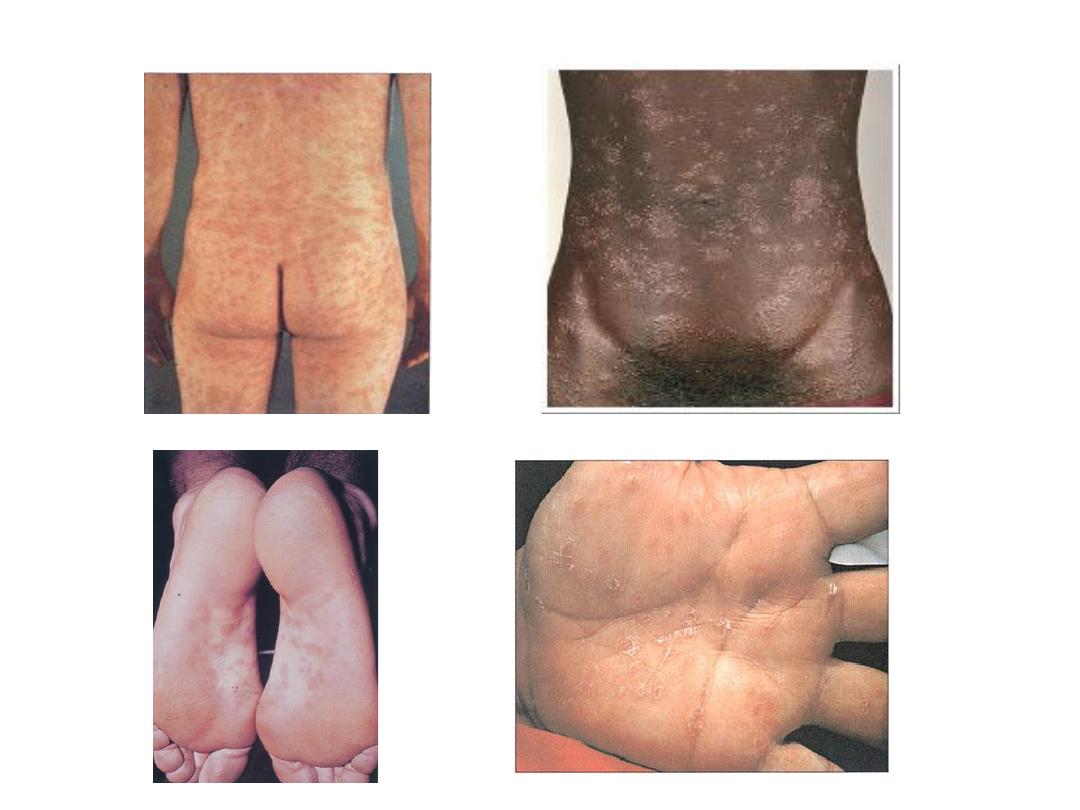
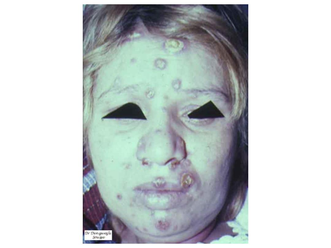
Secondary syphilis
➢ Appears 2-6 months after primary infection and 2-10
weeks after primary chancre.
➢ Lesions: Usually diffuse non-pruritic, indurated rash,
including palms & soles.
➢ the lesions of secondary S have certain characteristics
that differentiate them from other cutaneous diseases:
• There is little or no fever at the onset
• Lesions are noninflammatory, develop slowly, and may
persist for weeks or months
• Pain or itching is minimal or absent
• There is a marked tendency to polymorphism
• Color resembling a "clean-cut ham" or having a
coppery tint
• Lesions have variety of shapes
29

Types of lesions:
• Macular eruption
• Papular eruption
• Papulosquamous syphilids
• Follicular or lichenoid syphilids
• Annular syphilids
30

31
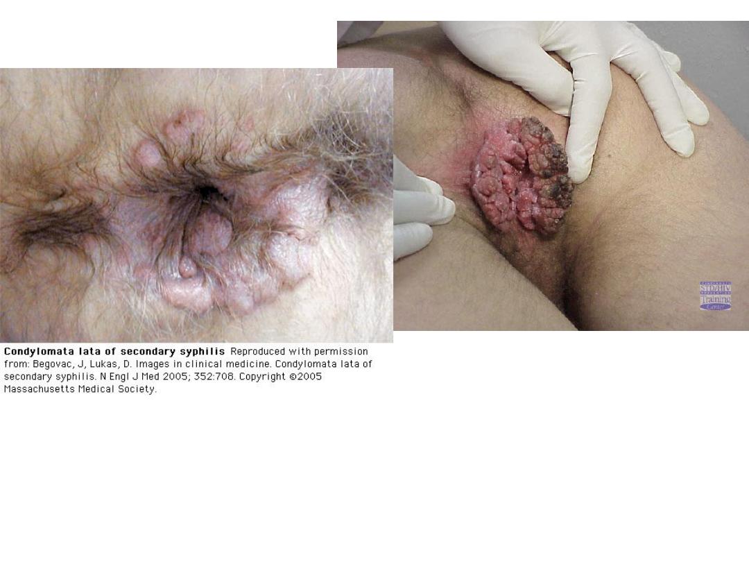
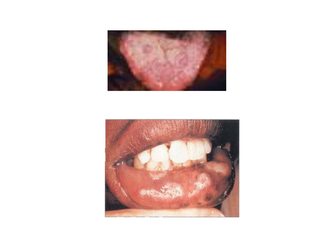
• Condylomata lata: Formed by coalescence of large, pale, flat-
topped papules. Occur in warm, moist areas such as the
perineum. Highly infectious. often mushroom-like mass. They are
not covered by the digitate elevations characteristic of venereal
warts (condylomata acuminata). This later is true verruca,
caused by human papillomavirus.
• Mucosal lesions: ~ 30% of secondary syphilis patients develop
mucous patch
• Note. All cutaneous lesions of secondary syphilis are infectious;
therefore, if you do not know what is, do not touch. Cellular
immune processes are responsible for the cutaneous
manifestations of secondary syphilis.
33
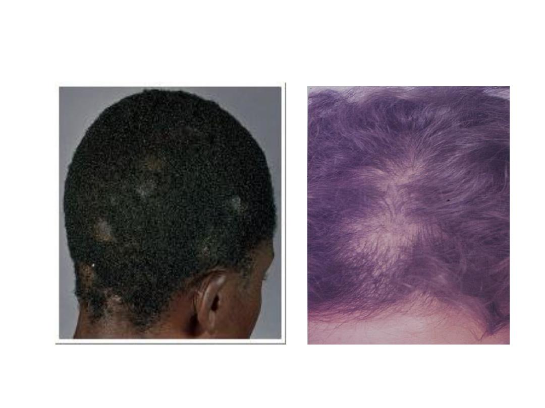
Alopecia areata

36
Diagnosis. Clinical suspicion confirmed by dark-filed
examination and\or serology (STS).
DD. syphilis has long been known as the 'great imitator'
because the various cutaneous manifestation may
simulate almost any cutaneous or systemic disease.
The rash may be confused with:
Pityriasis rosea (usually has a herald patch and lesions
seen along lines of skin cleavage)
Drug eruptions
Acute febrile exanthems
Psoriasis
Lichen planus
Scabies
The mucous patch may be confused with oral thrush

Latent Syphilis
• During this latent period there are no clinical
signs of syphilis, but the serologic tests are
reactive. During the early latent period
infectivity persists: for at least 2 years a
women with early latent S may infect her
unborn child.
• Is divided into early (less than one year
duration) and late (greater than one year
duration).
37

Tertiary Syphilis
• Tertiary S most often occur 3-5 years after infection.
16% of untreated patients will develop tertiary lesions
of the skin, mucous membranes, bone, or joints and
heal with scarring
• Treponema are usually not found by darkfiled
examination. Systemic disease also develop including
cardiovascular disease, CNS lesions.
• Two main types; Nodular syphilid and the Gumma
• Diagnosis: clinical finding, confirmed by STS and
biopsy; darkfiled examination is always negative.
• DD: TB, malignancy, lymphoma.

Congenital Syphilis
• Prenatal syphilis acquired in utero
• Infection through the placenta usually does not occur
before the fourth month, so treatment of the mother
before this time will almost always prevent infection in
the fetus.
• If infection occurs after the fourth month 40% risk of
fetal death
Most neonates with congenital syphilis are normal at
birth.
• Early congenital syphilis; lesions occurring within first
two years of life
• Late congenital syphilis; lesion occur after two years

Early Congenital Syphilis
• Neonates is usually premature, marasmic, fretful,
and dehydrated. The face is pinched and drawn,
resembling that an old man or women.
Multisystem disease is characteristic
• Snuffles, a form of rhinitis, is the most frequent
and often the first specific finding. In persistent
and progressive cases ulceration develop that
may involve the bones and cause perforation of
the septum or development of saddle nose,
which are important stigmata later in the disease
• Cutaneous lesions, resemble those of acquired
secondary S. with exaggeration
41

Late Congenital Syphilis
• Symptoms and signs of late congenital S become more
evident after age 5 years. The most important signs are:
➢ Frontal bosses (bony prominences of the forhead).
➢ Saddle nose
➢ Short maxilla
➢ High arched palate
➢ Mulberry molars (more than four small cusps on a narrow
first lower molar of the second dentition).
➢ Hutchinson's teeth (peg-shaped upper central incisors of
the permanent dentition that appear after age 6 years)
42

Late Congenital Syphilis
➢Higouménaki's sign (unilateral enlargement of
the sternoclavicular portion of the clavicle as
end result of periostitis)
➢Rhagades (linear scars radiating from the
angle of the eyes, nose, mouth, and anus)
➢Hutchinson's triad (Hutchinson's teeth,
interstitial keratitis, and cranial nerve V111
deafness) is considered pathognomonic of late
congenital syphilis.
43

Serologic Tests for Syphilis
There are two types of STS
• Nontreponemal test or classic reaction:
detects antibodies against phospholipids
antigens
• Treponemal test or specific test: detects
antibodies direct against T.Pallidum.
The use of one type is not sufficient for
diagnosis
44

Nontreponemal test
• Correlate with disease activity (reported
quantitatively);
• they are:
• Rapid plasma reagin (RPR).
• Veneral disease research laboratory (VDRL).
45

Treponemal test
• Correlate poorly with disease activity.
• Remains positive lifetime, regardless of
treatment.
• They are:
• Microhemagglutination assay for T.pallidum
(MHA-TP)
• Fluorescent treponemal antibody absorption
(FTA-ABS)
• T-pallidum particle agglutination (TP-PA)
46

Biologic False-Positive Tests Results (BFP)
The term BFP is used to denote a positive STS in
persons with no history or clinical evidence of
syphilis; two types:
• Acute BFP reactions are defined as those that
revert to negative in less than 6 months, may
result in; vaccinations, pregnancy, infections
(hepatitis, measles, typhoid, varicella, influenza,
malaria)
• Chronic BFP reactions positive test persist for
more than than 6 months, seen in: connective
tissue diseases, chronic liver disease, multiple
blood transfusion, and advancing age.
47

Treatment
• Penicillin remains the drug of choice for treatment of
all stages of syphilis.
Patients with primary, secondary, or early latent syphilis
of less than 1 year duration:
recommended treatment: Benzathine penicillin G. 2.4
million units IM in one dose.
alternative treatment in nonpregnant, penicillin allergic:
• Tetracycline 500mg orally four times a day for 2 weeks
• Doxycycline 100mg orally twice a day for 2 weeks.
• Ceftriaxone 1g IM or IV for 8-10 days
• Azithromycin 2g as a single oral dose
48

Treatment
Patients with late latent syphilis of more than one year
duration
• recommended treatment: Benzathine penicillin G. 2.4
MU IM once a week for 3 weeks
alternative treatment in nonpregnant, penicillin allergic:
• Tetracycline 500mg orally four times a day for 30 days
• Doxycycline 100mg orally twice a day for 30 days
Pregnant women with syphilis should be treated with
penicillin in doses appropriate for the stage of
syphilis. Pregnant women who allergic to penicillin
should be skin tested and desensitized if test results
are positive.
49

CHANCROID (Venereal/Soft Sore)
▪ Tropical sexually transmitted disease caused by Haemophillus
ducreyi, a gram negative bacterium.
▪ It is endemic in Africa, Asia and South America
▪ Men outnumber women many fold.
▪ After a one week incubation period a papule develops which
becomes a pustule and then an ulcer, which is
characteristically very painful.
▪ One or more deep or superficial tender ulcer on the genitalia,
and painful adenitis in 50% which may suppurate, are
characteristic of the disease.
▪ 50% of cases have a painful adenopathy with development of
bubos - inflamed lymph nodes with pus and necrosis, fixed to
the skin. There is no systemic component

CHANCROID (Venereal/Soft Sore)
• Diagnosis: the combination of a painful ulcer
with tender inguinal adenopathy is suggestive,
and when accompanied by suppurative
inguinal adenopathy , is almost
pathognomonic.
• In the absence of treatment, the chancroid
lesion can persist for months to years.
• Treatment is with cotrimoxazole or
erythromycin.
51

Granuloma Inguinale
• Granuloma inguinale is a mildly contagious, chronic,
granulomatous, locally destructive disease characterized by
progressive, indolent, serpiginous ulcerations of the groins,
pubes, genitalia, and anus.
• No adenopathy. Inguinal swellings are not lymphadenitis
but represent subcutaneous perilymphatic granulomatous
lesions
• Etiology: Granuloma inguinale is caused by the Gram-
negative bacterium Calymmatobacterium granulomatosis
• The exact mode of transmission of infection is
undetermined. Venereal but also nonvenereal transmition
occurs

Granuloma Inguinale
• Preferred treatment:
– Doxycycline 100 mg twice a day for 3 weeks
• Alternate Treatments:
– Azithromycin 1 gm weekly for 3 weeks
– Ciprofloxacin 750 mg twice a day for 3 weeks
– Erythromycin 500 mg four times a day for 3 weeks

LYMPHO GRANULOMA VENEREUM
is a tropical sexually transmitted disease
caused by Chlamydia trachomatis
• Endemic in Africa, India, SE Asia, South
America and the Caribbean,
• Men affected more commonly than
women, principally between the age 20
to 30 years.

LYMPHO GRANULOMA VENEREUM
• Three stages to the disease:
1) An asymptomatic ulcer which resolves rapidly
2) An inguinal syndrome, between 1 week and 6
months later, with adenopathy (lymph nodes are
painful) and bubo development.
• There is often systemic illness and malaise
3) Proctocolitis regional abscess or fistula,
resulting in regional strictures, e.g. rectal
strictures
• Diagnosis is by serology and intradermal skin test
with LGV antigen - Frei's test.
• Treated with tetracyclines or erythromycin.
55
56
CHANCROID (Venereal/Soft Sore)
Granuloma Inguinale
LYMPHO GRANULOMA VENEREUM