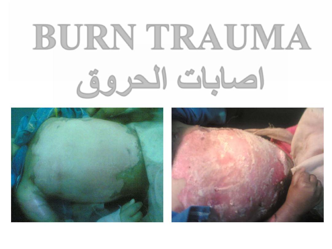
BURN TRAUMA
اصابات الحروق
Definition
Burn:
is a localized tissue injury
caused by exposure to extreme
degrees of temperatures &
associated with multiple
systemic complications.
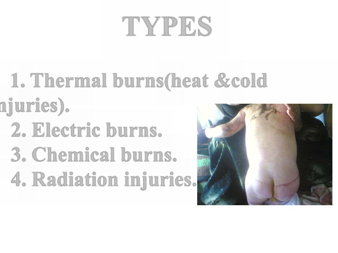
1. Thermal burns(heat &cold
injuries).
2. Electric burns.
3. Chemical burns.
4. Radiation injuries.
TYPES
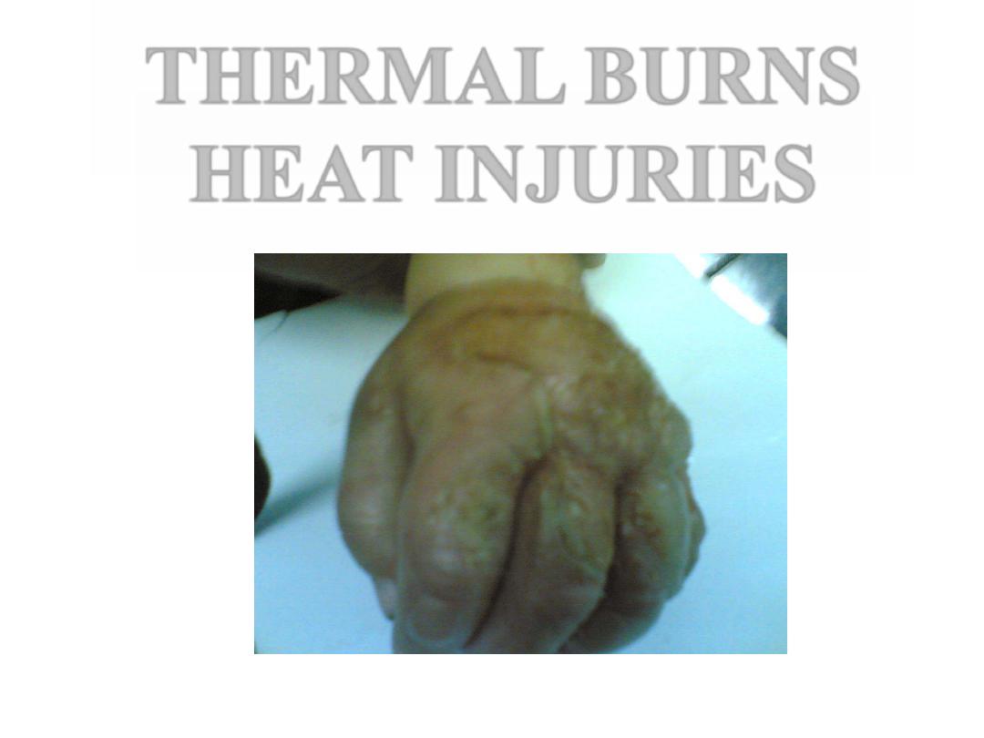
THERMAL BURNS
HEAT INJURIES
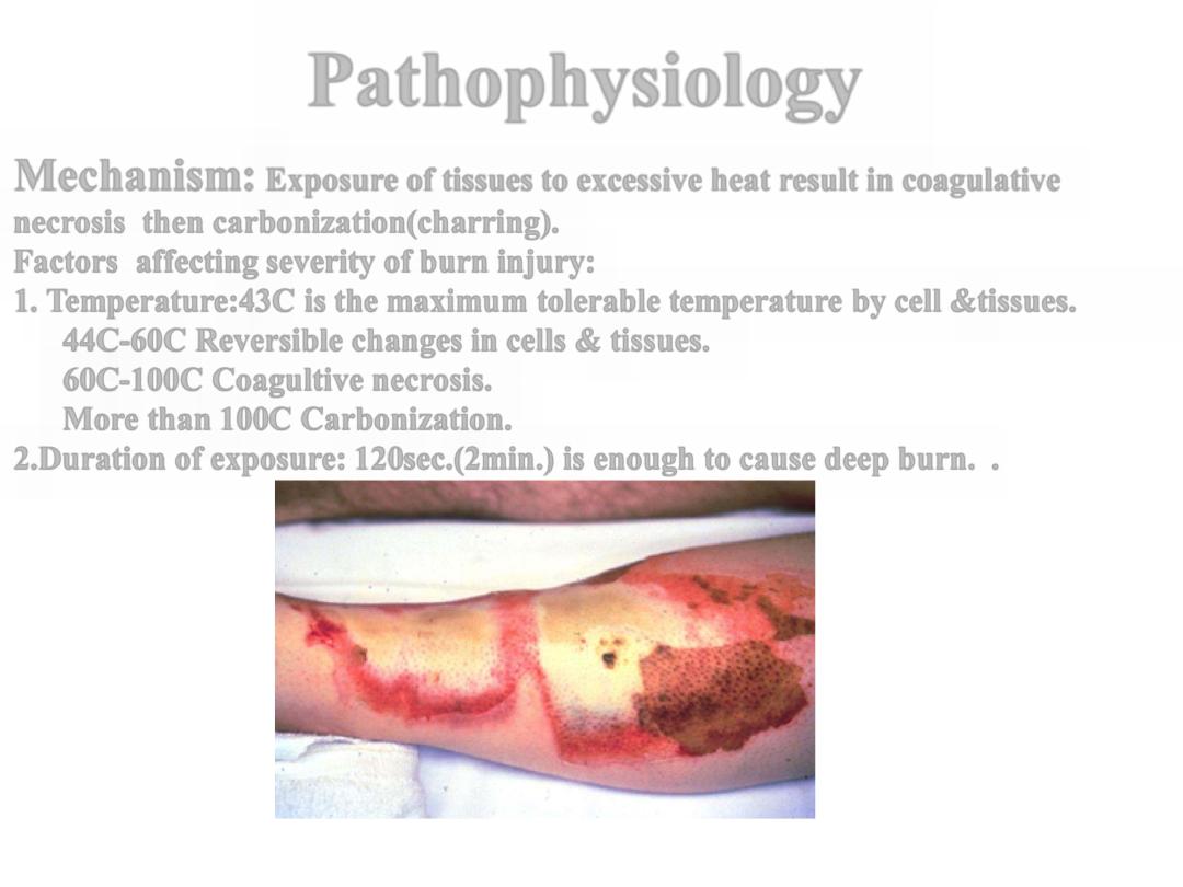
Pathophysiology
Mechanism:
Exposure of tissues to excessive heat result in coagulative
necrosis then carbonization(charring).
Factors affecting severity of burn injury:
1. Temperature:43C is the maximum tolerable temperature by cell &tissues.
44C-60C Reversible changes in cells & tissues.
60C-100C Coagultive necrosis.
More than 100C Carbonization.
.
2.Duration of exposure: 120sec.(2min.) is enough to cause deep burn.
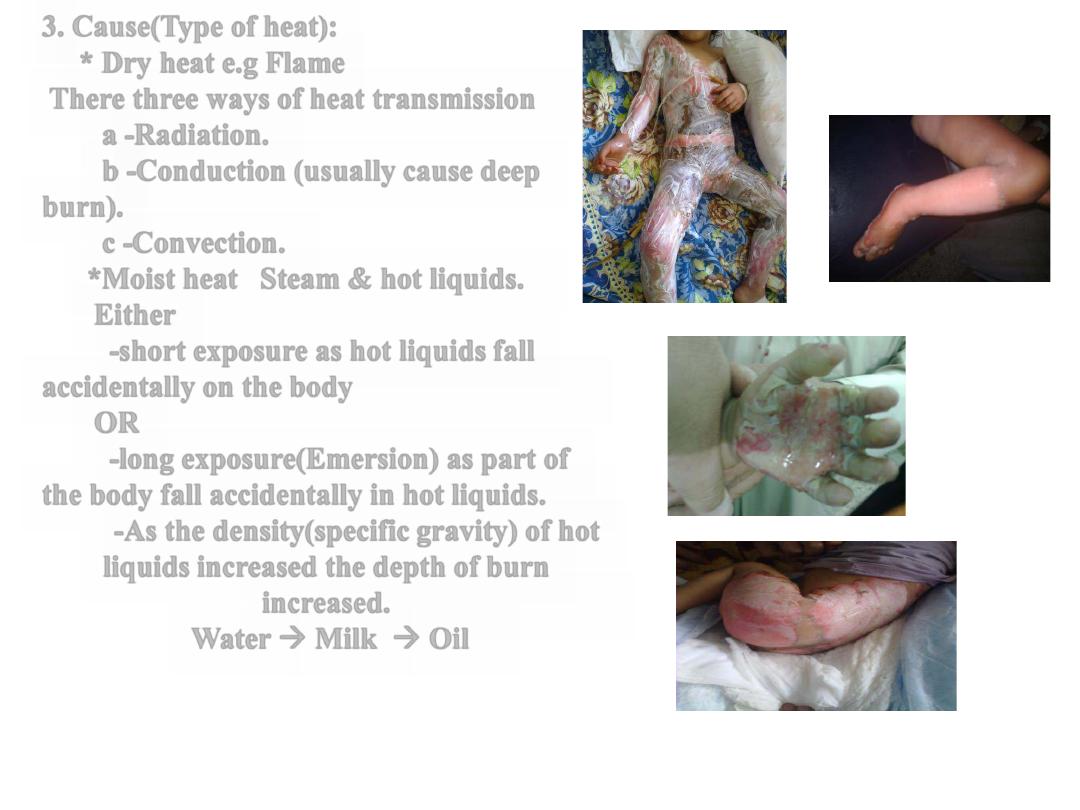
3. Cause(Type of heat):
* Dry heat e.g Flame
There three ways of heat transmission
a -Radiation.
b -Conduction (usually cause deep
burn).
c -Convection.
*Moist heat Steam & hot liquids.
Either
-short exposure as hot liquids fall
accidentally on the body
OR
-long exposure(Emersion) as part of
the body fall accidentally in hot liquids.
-As the density(specific gravity) of hot
liquids increased the depth of burn
increased.
Water → Milk → Oil
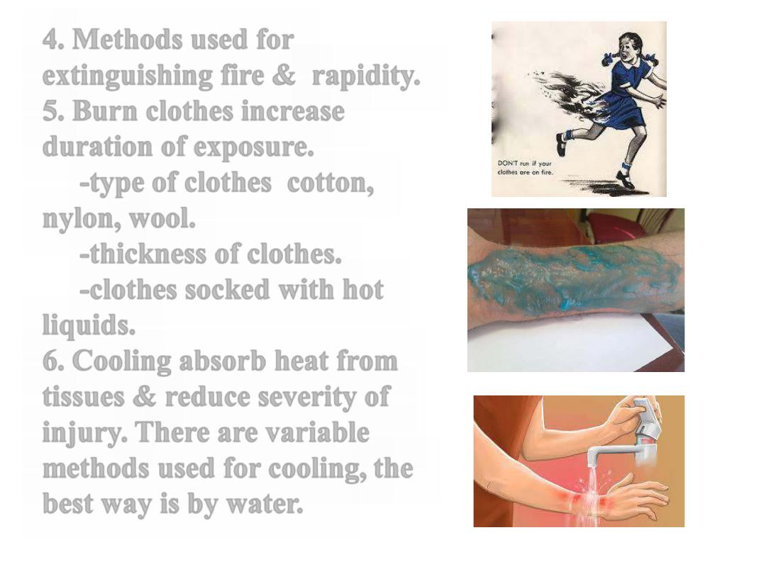
4. Methods used for
extinguishing fire & rapidity.
5. Burn clothes increase
duration of exposure.
-type of clothes cotton,
nylon, wool.
-thickness of clothes.
-clothes socked with hot
liquids.
6. Cooling absorb heat from
tissues & reduce severity of
injury. There are variable
methods used for cooling, the
best way is by water.
Severity
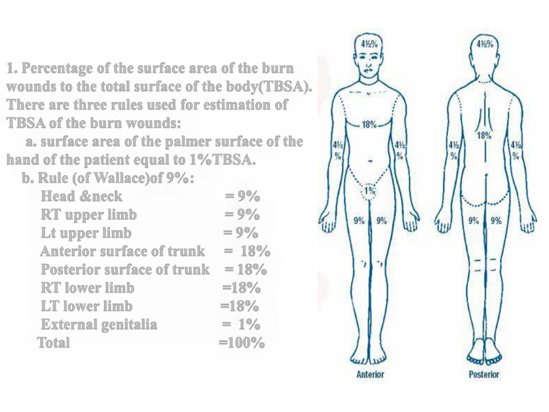
1. Percentage of the surface area of the burn
wounds to the total surface of the body(TBSA).
There are three rules used for estimation of
TBSA of the burn wounds:
a. surface area of the palmer surface of the
hand of the patient equal to 1%TBSA.
b. Rule (of Wallace)of 9%:
Head &neck = 9%
RT upper limb = 9%
Lt upper limb = 9%
Anterior surface of trunk = 18%
Posterior surface of trunk = 18%
RT lower limb =18%
LT lower limb =18%
External genitalia = 1%
Total =100%
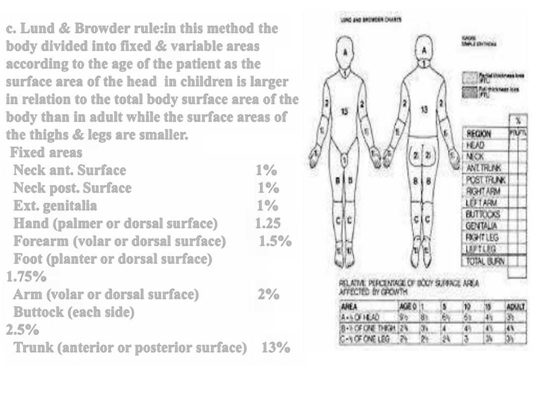
c. Lund & Browder rule:in this method the
body divided into fixed & variable areas
according to the age of the patient as the
surface area of the head in children is larger
in relation to the total body surface area of the
body than in adult while the surface areas of
the thighs & legs are smaller.
Fixed areas
Neck ant. Surface 1%
Neck post. Surface 1%
Ext. genitalia 1%
Hand (palmer or dorsal surface) 1.25
Forearm (volar or dorsal surface) 1.5%
Foot (planter or dorsal surface)
1.75%
Arm (volar or dorsal surface) 2%
Buttock (each side)
2.5%
Trunk (anterior or posterior surface) 13%
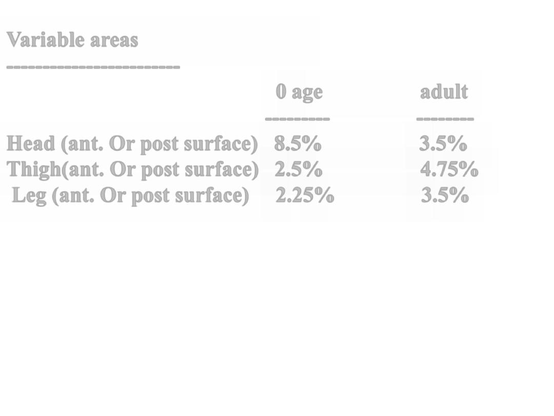
Variable areas
------------------------
0 age adult
---------
--------
Head (ant. Or post surface) 8.5% 3.5%
Thigh(ant. Or post surface) 2.5% 4.75%
Leg (ant. Or post surface) 2.25% 3.5%
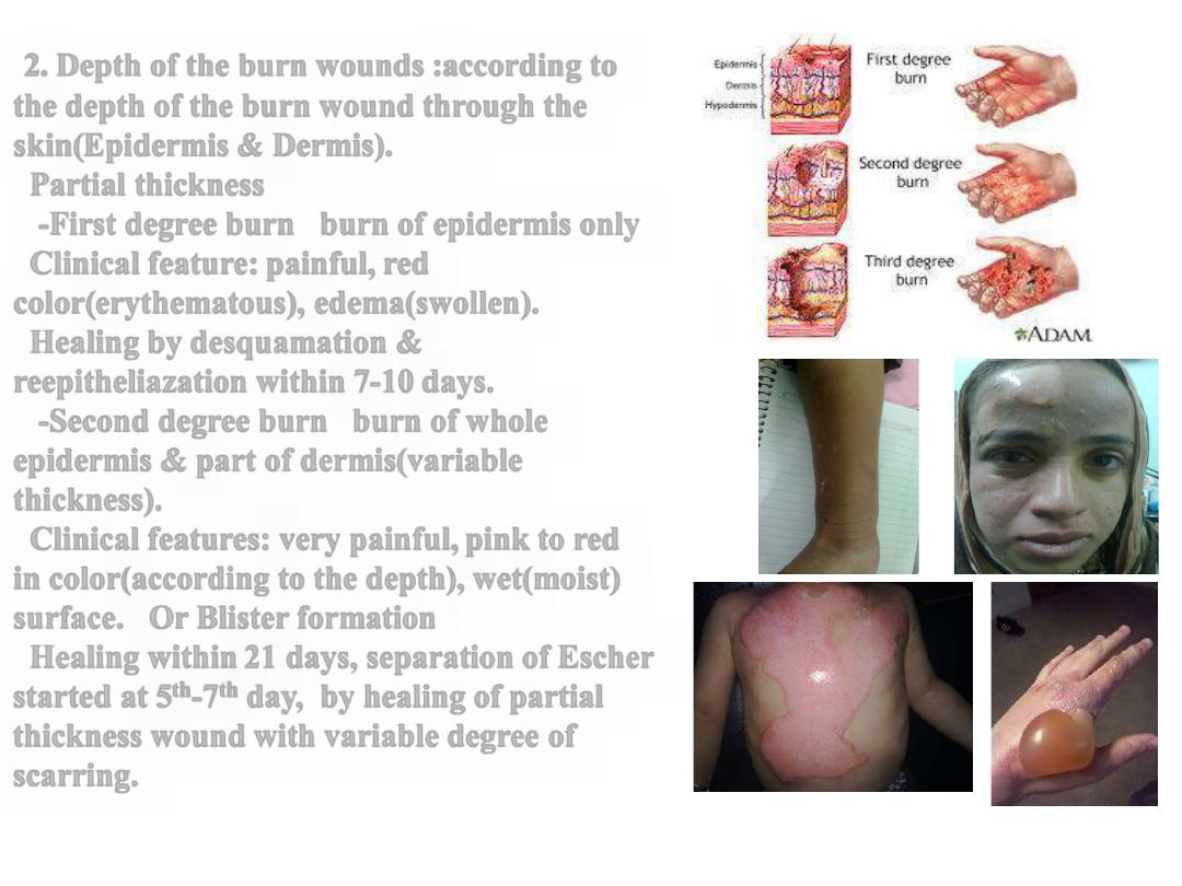
2. Depth of the burn wounds :according to
the depth of the burn wound through the
skin(Epidermis & Dermis).
Partial thickness
-First degree burn burn of epidermis only
Clinical feature: painful, red
color(erythematous), edema(swollen).
Healing by desquamation &
reepitheliazation within 7-10 days.
-Second degree burn burn of whole
epidermis & part of dermis(variable
thickness).
Clinical features: very painful, pink to red
in color(according to the depth), wet(moist)
surface. Or Blister formation
Healing within 21 days, separation of Escher
started at 5
th
-7
th
day, by healing of partial
thickness wound with variable degree of
scarring.
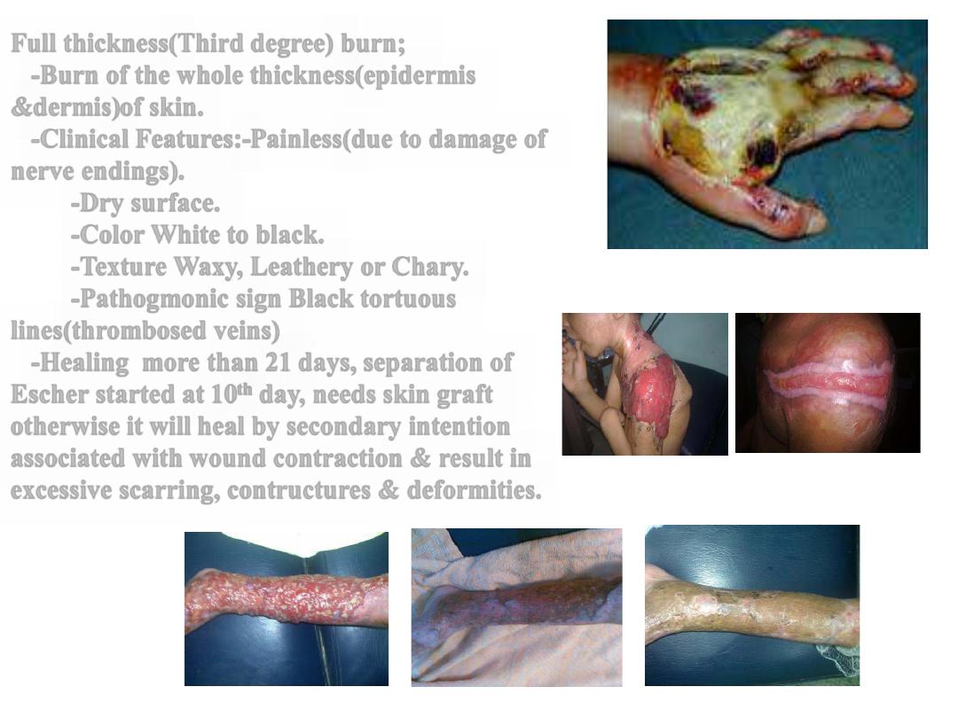
Full thickness(Third degree) burn;
-Burn of the whole thickness(epidermis
&dermis)of skin.
-Clinical Features:-Painless(due to damage of
nerve endings).
-Dry surface.
-Color White to black.
-Texture Waxy, Leathery or Chary.
-Pathogmonic sign Black tortuous
lines(thrombosed veins)
-Healing more than 21 days, separation of
Escher started at 10
th
day, needs skin graft
otherwise it will heal by secondary intention
associated with wound contraction & result in
excessive scarring, contructures & deformities.
PROGNOSTIC FACTORS
1. Age poor prognosis below
2years & over 50years.
2. Percentage more than
30%TBSA is critical|& very
poor above 50%TBSA.
3.Depth of the burn wound.
4.Type of burn injury.
5.Inhalation injury.
6.burn wound infection &
septicemia.
7.Associated trauma.
8.Presence of medical problems
.
INDICATIONS OF ADMISSION OF
BURN PATIENTS
1. 2
nd
&3
rd
degree burn equal or more than 10%TBSA in age below 2
years & above 50 years.
2. 2
nd
&3
rd
degree burn equal or more than 15%TBSA in other age
groups.
3. Third degree burn of 5%TBSA & more.
4. Circumferential burn.
5. Burn of specific areas; face, hands, feet, external genitalia,
perineum & major joints.
6. Inhalation injury.
7. Electric burn.
8. Chemical burn.
9. Associated traumas.
10. Preexisting medical disorders.
11. For learning purposes
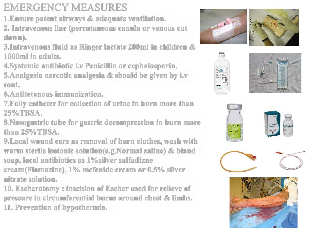
EMERGENCY MEASURES
1.Ensure patent airways & adequate ventilation.
2. Intravenous line (percutaneous canula or venous cut
down).
3.Intravenous fluid as Ringer lactate 200ml in children &
1000ml in adults.
4.Systemic antibiotic i.v Penicillin or cephalosporin.
5.Analgesia narcotic analgesia & should be given by i.v
rout.
6.Antitetanous immunization.
7.Folly catheter for collection of urine in burn more than
25%TBSA.
8.Nasogastric tube for gastric decompression in burn more
than 25%TBSA.
9.Local wound care as removal of burn clothes, wash with
warm sterile isotonic solution(e.g.Normal saline) & bland
soap, local antibiotics as 1%silver sulfadizne
cream(Flamazine), 1% mefenide cream or 0.5% silver
nitrate solution.
10. Escheratomy : inscision of Escher used for relieve of
pressure in circumferential burns around chest & limbs.
11. Prevention of hypothermia.
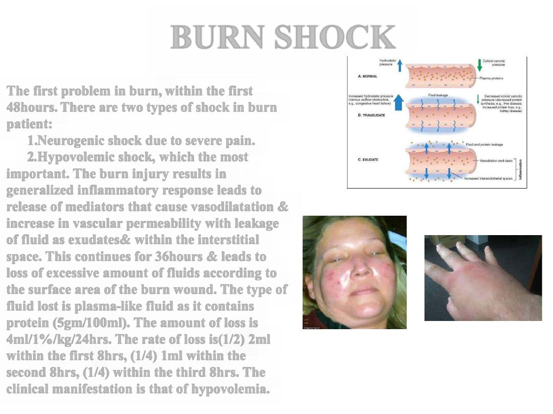
BURN SHOCK
The first problem in burn, within the first
48hours. There are two types of shock in burn
patient:
1.Neurogenic shock due to severe pain.
2.Hypovolemic shock, which the most
important. The burn injury results in
generalized inflammatory response leads to
release of mediators that cause vasodilatation &
increase in vascular permeability with leakage
of fluid as exudates& within the interstitial
space. This continues for 36hours & leads to
loss of excessive amount of fluids according to
the surface area of the burn wound. The type of
fluid lost is plasma-like fluid as it contains
protein (5gm/100ml). The amount of loss is
4ml/1%/kg/24hrs. The rate of loss is(1/2) 2ml
within the first 8hrs, (1/4) 1ml within the
second 8hrs, (1/4) within the third 8hrs. The
clinical manifestation is that of hypovolemia.
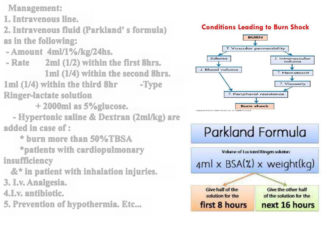
Management:
1. Intravenous line.
2. Intravenous fluid (Parkland' s formula)
as in the following:
- Amount 4ml/1%/kg/24hs.
- Rate 2ml (1/2) within the first 8hrs.
1ml (1/4) within the second 8hrs.
1ml (1/4) within the third 8hr -Type
Ringer-lactate solution
+ 2000ml as 5%glucose.
- Hypertonic saline & Dextran (2ml/kg) are
added in case of :
* burn more than 50%TBSA
*patients with cardiopulmonary
insufficiency
&* in patient with inhalation injuries.
3. I.v. Analgesia.
4.I.v. antibiotic.
5. Prevention of hypothermia. Etc...
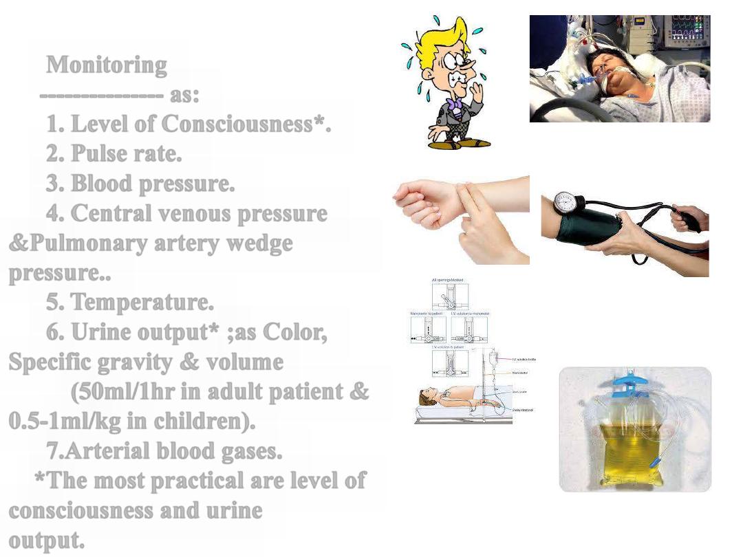
Monitoring
--------------- as:
1. Level of Consciousness*.
2. Pulse rate.
3. Blood pressure.
4. Central venous pressure
&Pulmonary artery wedge
pressure..
5. Temperature.
6. Urine output* ;as Color,
Specific gravity & volume
(50ml/1hr in adult patient &
0.5-1ml/kg in children).
7.Arterial blood gases.
*The most practical are level of
consciousness and urine
output.