Occupational Physical Hazards (Cont.)Ionizing and Non Ionizing Radiation
ByDr. Ashraf Hussain
MBChB, PhD/Com. Med
Ionizing Radiation
Ionising RadiationIonising radiation can be described as
the transfer of energy in the form of particles (such as alpha, beta and neutronic particles) or electromagnetic waves (such as X-rays and gamma rays) capable of producing ions directly or indirectly.
Ionizing radiation with enough energy can remove tightly bound electrons from the orbit of an atom, causing the atom to become charged or ionized.
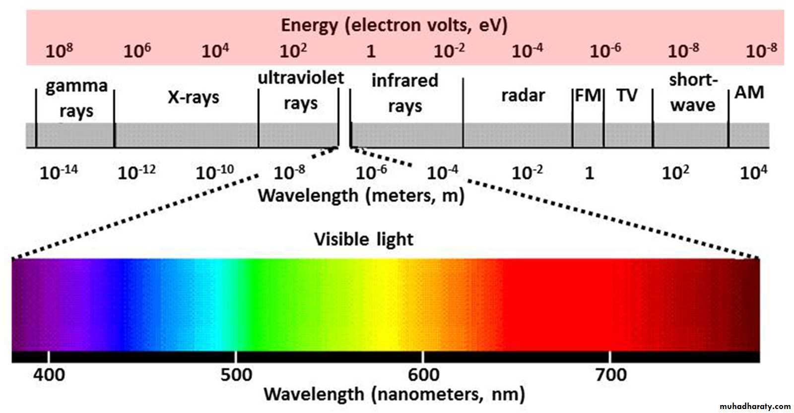
Longer wave length, lower frequency waves (heat and radio) have less energy than shorter wave length, higher frequency waves (X and gamma rays). Not all electromagnetic (EM) radiation is ionizing. Only the high frequency portion of the electromagnetic spectrum which includes X rays and gamma rays is ionizing.
Radiation Sources
Natural sources: including more than 60 naturally-occurring radioactive materials found in soil, water and air.Radon, a naturally-occurring gas, emanates from rock and soil and is the main source of natural radiation.
People are also exposed to natural radiation from cosmic rays, particularly at high altitude (Aircrew).
Man-made sources ranging from nuclear power generation to medical uses of radiation for diagnosis or treatment.
Today, the most common human-made sources of ionizing radiation are medical devices, including X-ray machines
Workers exposed to ionizing radiation
• Healthcare and veterinary workers who are:Caring for patients who have been treated with Iodine-131 or other nuclear medicine radioactive materials
Assisting with fluoroscopy procedures
Working around portable X-ray machines in medical, dental, or veterinary offices.
• Certain industrial and laboratory workers.
• Aircrew (flight attendants and pilots)
Biological Effects
The effect on body tissues will depend on:• The type of radiation,
• The amount of radiation generated and the dose being absorbed
• Duration of exposure
• The sensitivity of exposed different tissues and organs.
• Whether the source is internal or external to the body.
• Distance from the source
• Availability and effectiveness of shields.
Deterministic OR Acute effect of radiation
acute effects cause cell damage and death
Occurs when certain threshold been exceeded.
They may include skin redness and skin damage, hair loss, radiation burns, or acute radiation syndrome.
The effective dose is used to measure ionizing radiation in terms of its potential to cause harm.
The sievert (Sv) or More concisely Milli Sievert (mSv) is the unit of effective dose that takes into account the type of radiation and sensitivity of tissues and organs.
For instance, the dose threshold for acute radiation syndrome is about 70 rad (700 mSv) and for skin damage is 3000 mSv.
Stochastic effects or "long-term“ Health effects
If the radiation dose is low and/or delivered over a long period of time (low dose rate), the risk of cell damage and death is substantially reduced.if the damaged cell survives, its DNA can be affected so the cell mutate and reproduce.
They occur by statistical chance. The probability of the effect occurring in a population increases with the dose received, and the severity of the effect does not depend on the dose.
Cancer is the main stochastic effect that can result often many years following the exposure.
Stochastic health effects are assumed not to have a threshold dose below which they do not occur.
This is the reason that no level of radiation dose is considered to be completely "safe" and why doses should always be kept as low as reasonably achievable (ALARA).
Although it may not accurately describe all stochastic health effects, they are sometimes described as "long-term" health effects.
Epidemiological studies on populations exposed to radiation, such as atomic bomb survivors or radiotherapy patients, showed a significant increase of cancer risk at doses above 100 mSv.
More recently, some epidemiological studies in individuals exposed to medical exposures during childhood (paediatric CT) suggested that cancer risk may increase even at lower doses (between 50-100 mSv).
Acute Radiation Syndrome
Acute Radiation Syndrome (ARS), or radiation sickness, occurs when all or most of the body receives a very high dose—around 70 rad (700mSv) or higher—of penetrating radiation in a short period of time
ARS is a collection of symptoms attributable to damage to the bone marrow and the gastrointestinal, cardiovascular, and central nervous systems resulting from such a dose
These symptoms include loss of appetite, fatigue, fever, nausea, vomiting, diarrhea, and possibly even seizures, coma and can sometimes result in death over the following days or weeks..
This seriously ill stage may last from a few hours up to several months.
TYPES OF IONIZING RADIATIONS
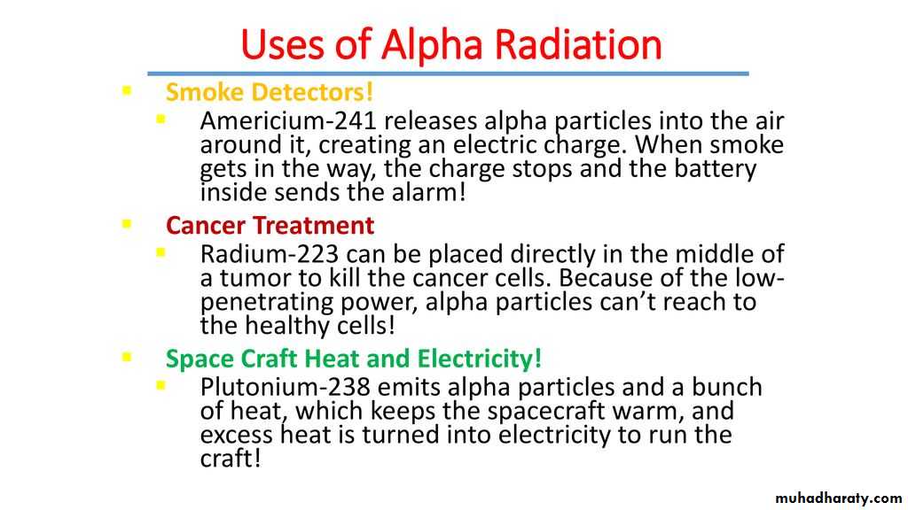
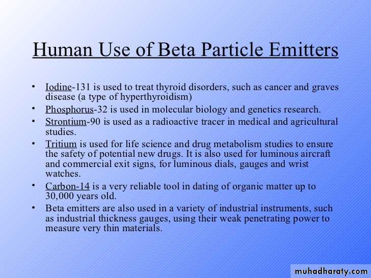
Particulate vs. Electromagnetic Radiations:Particulate Radiations are sub-atomic particles with mass (e.g., alpha and Beta particles, electrons, neutrons).
EM Radiations (X-rays and gamma rays) have no mass and no charge.
Alpha is the least penetrating, while gamma is the most penetrating.
Dose Limit
The most important dose limit is the annual dose limit of 20 mSv. It means that a worker can receive a dose of 20 mSv per year from ionising sources they are working with.Natural background radiation due to radioactivity in soil, water, air, food, on average the annual dose is around 2 mSv
So a worker using ionising radiation sources can receive ten times the dose of the natural background at the workplace.
Examples of suspected radiation doses in different radioimaging applications
1.Computed Tomography (CT)–Abdomen and Pelvis
7mS
2.
Computed Tomography (CT)–Head
2 mSv
3.
Extremity (hand, foot, etc.) X-ray
0.001 mSv
4.
Spine X-ray
• 1.5 mSv
5.
Chest X-ray
0.1 mSv
6.
Dental X-ray
0.005 mSv
7.
Mammography
0.4 mSv
Pregnant and Nursing Mother Workers (Special Precautions)
For women, there are special limitations during pregnancy or breast feeding.
Pregnant woman can work in a radiation area but the dose to the foetus must be below 1 mSv during pregnancy.
Breast feeding woman can work in a radiation area when only exposure to external radiation is possible (X-ray devices or encapsulated radioactivity sources). In that case, the limit of 20 mSv per year applies.
A breastfeeding mother is not allowed to work in an area where contamination and intake of radioactivity is possible.
Protection
To keep radiation doses low, three options are used: time, distance and shielding.1. The dose is proportional to the time of exposure
The more time one is exposed to ionising radiation, the larger the dose that will be received and the more harmful the radiation will be.
2. The radiation reduces with the distance from the source.
Protection: Cont.
Shielding: There are activities that require workers to be close to the source and in a high radiation field.In that case, we minimise the doses by using shielding and protective clothing.
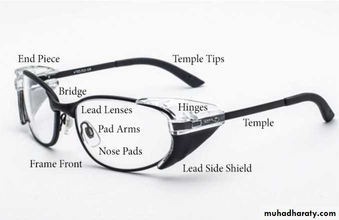
When working with X-ray devices in medicine, the most common personal protective clothing is lead aprons. Led aprons made of 0.25 mm thick lead attenuate X-rays more than 100 times.
In some cases when eyes are exposed, spectacles made of lead glass are used as protection. Also, lead gloves can be used, however such gloves are quite thick and not appropriate for detailed work.


https://www.who.int/news-room/fact-sheets/detail/ionizing-radiation-health-effects-and-protective-measures
https://www.osha.gov/SLTC/radiationionizing/healtheffects.html.
https://www.cdc.gov/niosh/topics/repro/ionizingradiation.html
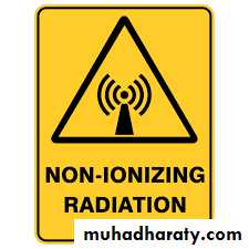
Non Ionizing Radiation
Non-ionizing radiation is a series of energy waves composed of oscillating electric and magnetic fields traveling at the speed of light.
They includes the spectrum of ultraviolet (UV), visible light, infrared (IR), microwave (MW), radio frequency (RF), and extremely low frequency (ELF).
Lasers commonly operate in the UV, visible, and IR frequencies.
Non-ionizing radiation is found in a wide range of occupational settings and can pose a considerable health risk to potentially exposed workers if not properly controlled.
Ultraviolet light
Generated by black light, sunlight
Visible light
Generated by sunlight, LEDs, light bulbs, lasers
Infrared light
Generated by sunlight, thermal radiation, incandescent light bulbs, lasers, remote controls
Microwave radiation
Generated by microwaves, cellphones, and data transmission
Radio frequency radiation
Generated by AM and FM radio signals, cellphone and data communications, and by radio frequency heaters used to bond vinyls and plastics
Extremely low frequency radiation
Generated by transmission lines, old CRT computer monitors
1. Extremely Low Frequency (ELF) radiation
Extremely Low Frequency (ELF) radiation at 60 HZ is produced by power lines, electrical wiring, and electrical equipment.Common sources of intense exposure include ELF induction furnaces and high-voltage power lines.
No data really support the presence of adverse health effects
Sources of RF and MW radiation include radio emitters and cell phones.
power supplies may create microwave/RF radiation as a byproduct of their operation.Less likely are leaking ovens and heaters
In general, most biological effects of exposure to microwave/RF radiation are related to the direct heating of tissues (thermal effects) or the flow of current through tissue (induced current effects).
2. Radiofrequency and Microwave Radiation
Occupations with RF/MW exposures
Television transmitter stationInduction and dielectric heaters
Military exposure
surface-to-air missile system radars.
Aircraft and helicopters.
Communication devices,
Airport radars.
Radio navigation systems
Non-thermal effects resulting in carcinogenesis, teratogenesis, etc. have been demonstrated in animals but have not been proven by epidemiological studies on humans.
The following biological effects have been demonstrated in humans:
• Cataract formation (from eye exposure).
• RF (induction) burns.
• Burns from contact with metal implants, spectacles, etc
3. Non-Coherent Light Source Safety
Ultraviolet radiationSunburn
Skin cancer
Welder's "arc-eye"
Cataracts
Infrared radiation
Skin burns
Cataracts
Retinal burns
Many devices (or sources) produce either broadband or discrete wavelength radiation between 100 nm and 1 mm. Under certain conditions, these sources may present a health hazard.
Welders arc eye
The cornea is like the skin in that it can be “sunburned” by exposure to too much UV radiation.
This is called keratoconjunctivitis (snow blindness or welders flash), a condition where the corneal (epithelial) cells are damaged or destroyed.
This condition usually does not present until 6 to 12 hours following the UV exposure.
Although very painful (often described as having sand in the eyes) this condition is usually temporary (a few days) because the corneal cells will grow back.
In very severe cases, the cornea may become clouded and corneal transplants may be needed to restore vision.
Exposure to the UV-C and UV-B present the greatest risk to the cornea.
a pair of polycarbonate safety glasses or a polycarbonate face shield always should be worn whenever there is a potential for ongoing UV radiation exposure.
References
https://www.osha.gov/SLTC/radiation_nonionizing/index.htmlhttps://www.cdc.gov/nceh/radiation/nonionizing_radiation.html
https://ehs.berkeley.edu/laser-safety/non-ionizing-radiation-safety-manual#introduction.