
PLEURAL EFFUSION
Dr.Ahmed Hussein Jasim F.I.B.M.S (resp)
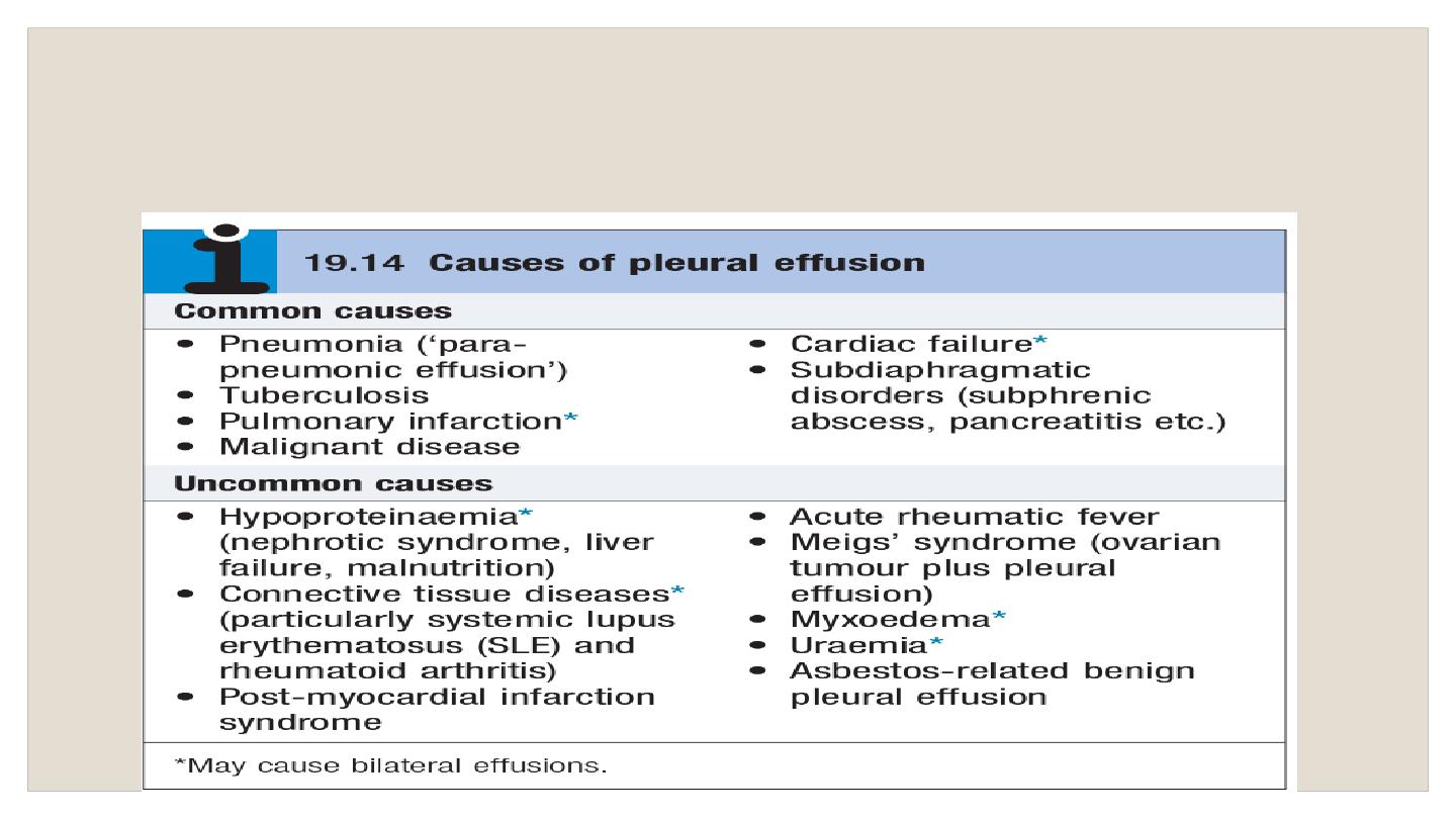
A pleural effusion results from the accumulation of abnormal volumes
(>10–20mL) of fluid in the pleural space.
Commonest causes in the UK and US (in order):
cardiac failure,
pneumonia, malignancy, PE.

Clinical features
May be asymptomatic or associated with breathlessness, dry cough,
pleuritic chest pain (suggesting pleural inflammation), chest
‘heaviness’, and sometimes pain referred to the shoulder or abdomen
signs on examination include reduced chest expansion, reduced
tactile vocal fremitus, a stony dull percussion note, quiet breath
sounds, and sometimes a patch of bronchial breathing above the
fluid level. a friction rub may be heard with pleural inflammation.
Imaging CXR
• Sequential blunting of posterior, lateral, and then anterior costophrenic
angles are seen on radiographs as effusions increase in size
•
PA CXr will usually detect effusion volumes of 200mL or more; lateral CXr
is more sensitive and may detect as little as 50mL pleural fluid
• Classical CXr appearance is of basal opacity obscuring hemidiaphragm,
with concave upper border.

US has a much higher sensitivity than CXr at detecting and localizing
pleural fluid and is useful for distinguishing pleural fluid from pleural
masses or thickening.
CT chest with pleural contrast is useful in distinguishing benign and
malignant pleural disease:
nodular, mediastinal, or circumferential
pleural thickening and parietal pleural thickening >1cm are all highly
specific for malignant disease.
scans are best performed prior to
complete drainage of fluid
Role of MRI is unclear; it may have increasing role in distinguishing
benign from malignant pleural disease.

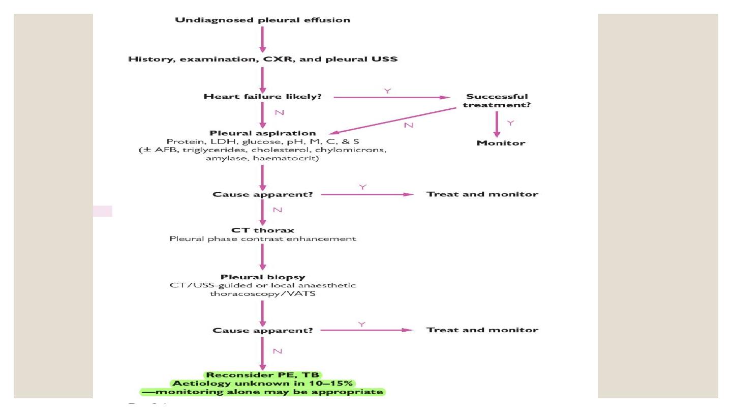
Thoracentesis
(= ‘pleural tap’ or pleural fluid aspiration) may be
diagnostic and/or therapeutic, depending on the volume of
fluid
removed.
Following diagnostic tap: • note pleural fluid appearance
• Send sample to biochemistry for measurement of glucose, protein,
and lactate dehydrogenase (LDH)
• Send a fresh 20mL sample in sterile pot to cytology for examination
for malignant cells (yield 60% in malignancy) and differential cell count
• Send samples in sterile pot to microbiology for Gram stain and
microscopy, culture. For suspected pleural infection, also send pleural
fluid in blood culture bottles. Low threshold for AFB stain and tB culture
• Process non-purulent, heparinized samples in ABG analyser for pH
• Consider measurement of cholesterol, triglycerides, chylomicrons,
haematocrit, adenosine deaminase, and amylase, depending on the
clinical circumstances.

Is the pleural effusion a transudate or an exudate? Helpful in narrowing
the differential diagnosis. In patients with a normal serum protein,
pleural fluid protein <30g/L = transudate, and protein >30g/L = exudate.
In borderline cases (protein 25–35g/L) or in patients with abnormal
serum protein,
apply Light’s criteria—effusion is exudative if it meets one
of following criteria.
• Pleural fluid protein/serum protein ratio >0.5
• Pleural fluid LDH/serum LDH ratio >0.6
• Pleural fluid LDH > two-thirds the upper limit of normal serum LDH
Pleural tissue biopsy for histology and tB culture using image-guided or
thoracoscopic biopsies.

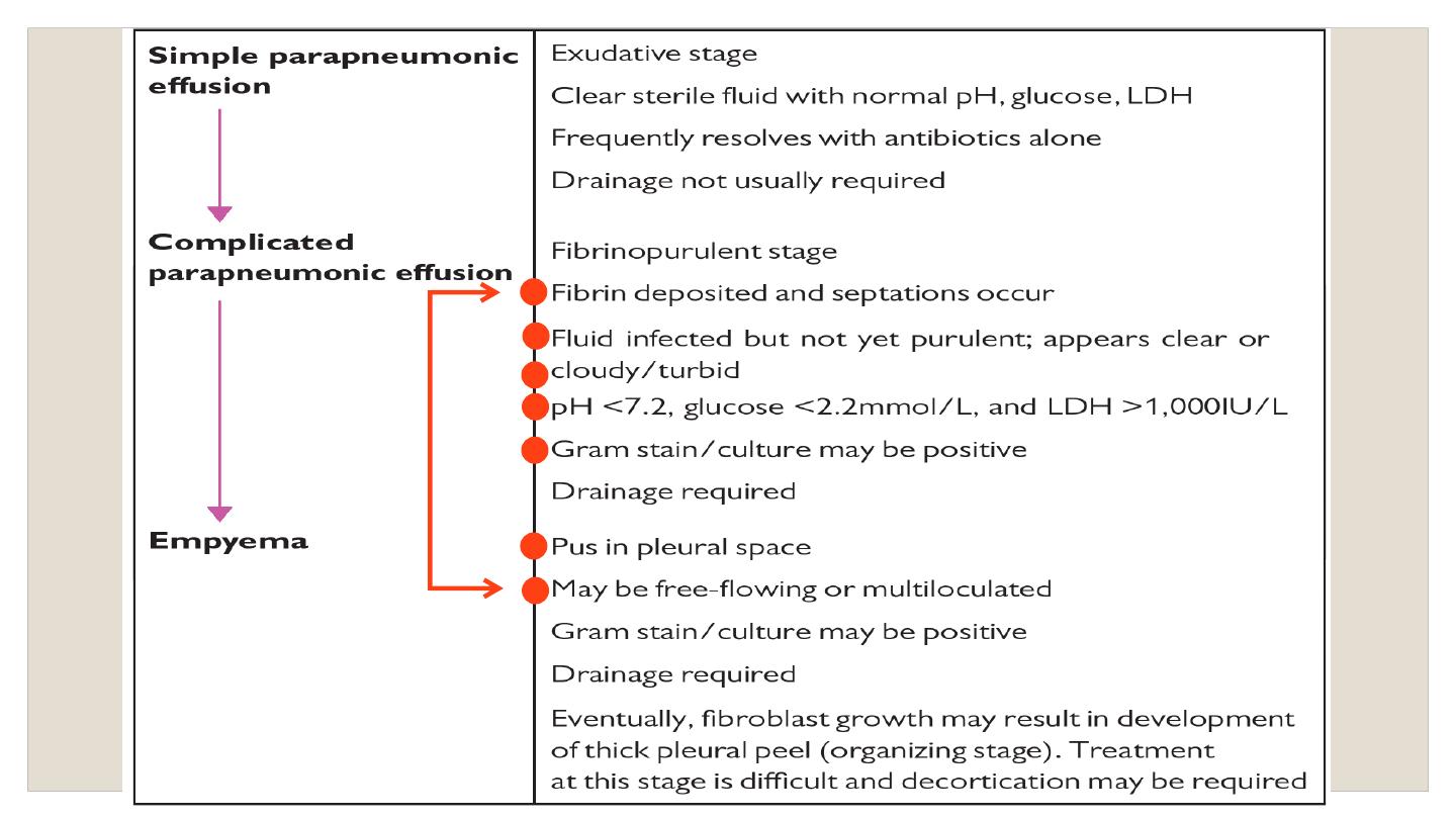
Parapneumonic effusion and empyema
Definition and pathophysiology pleural effusions occur in up to 57% of
patients with pneumonia.
an initial sterile exudate (simple
parapneumonic effusion) may, in some cases, progress to a
complicated parapneumonic effusion and eventually empyema
pleural infection may also occur in the absence of
a preceding
pneumonic illness (‘ primary empyema’).
Clinical features
Consider the diagnosis particularly in cases of
‘slow-to-respond’
pneumonia (e.g. failure of CRP to fall ≥50% in first 3 days), pleural
effusion with fever, or high-risk groups with non-specific symptoms such
as weight loss ,
anaerobic empyema may present less acutely, often
with weight loss and without fever.

Risk factors for developing empyema include
diabetes, alcohol abuse,
gastro-oesophageal reflux, and IV drug abuse
. anaerobic infection is
associated particularly with aspiration or poor dental hygiene.
clinical variables associated with development of pleural infection in
those with pneumonia:
albumin <30g/L, CRP >100mg/L, platelets >400 ×
10
9
/l, sodium <130mmol/L, IVDU, and chronic alcohol use.
Bacteriology
Community-acquired infection (% of cases): •Streptococcus ‘milleri’
group (30%) •anaerobes (5–30%) •Streptococcus pneumoniae (15%)
•Staphylococcus aureus (10%)
Hospital-acquired infection (% of cases): •MRSA (25-30%)
•Staphylococcus aureus (10–20%) • enterobacteriaceae (20%)
pleural infection is frequently polymicrobial.
Indications
for drainage