LEC 1:
ا.م.د.ﺣﺴﻦ اﻟﺠﻤﻴﻠﻲ
Liver and biliary tract disease
Introduction:
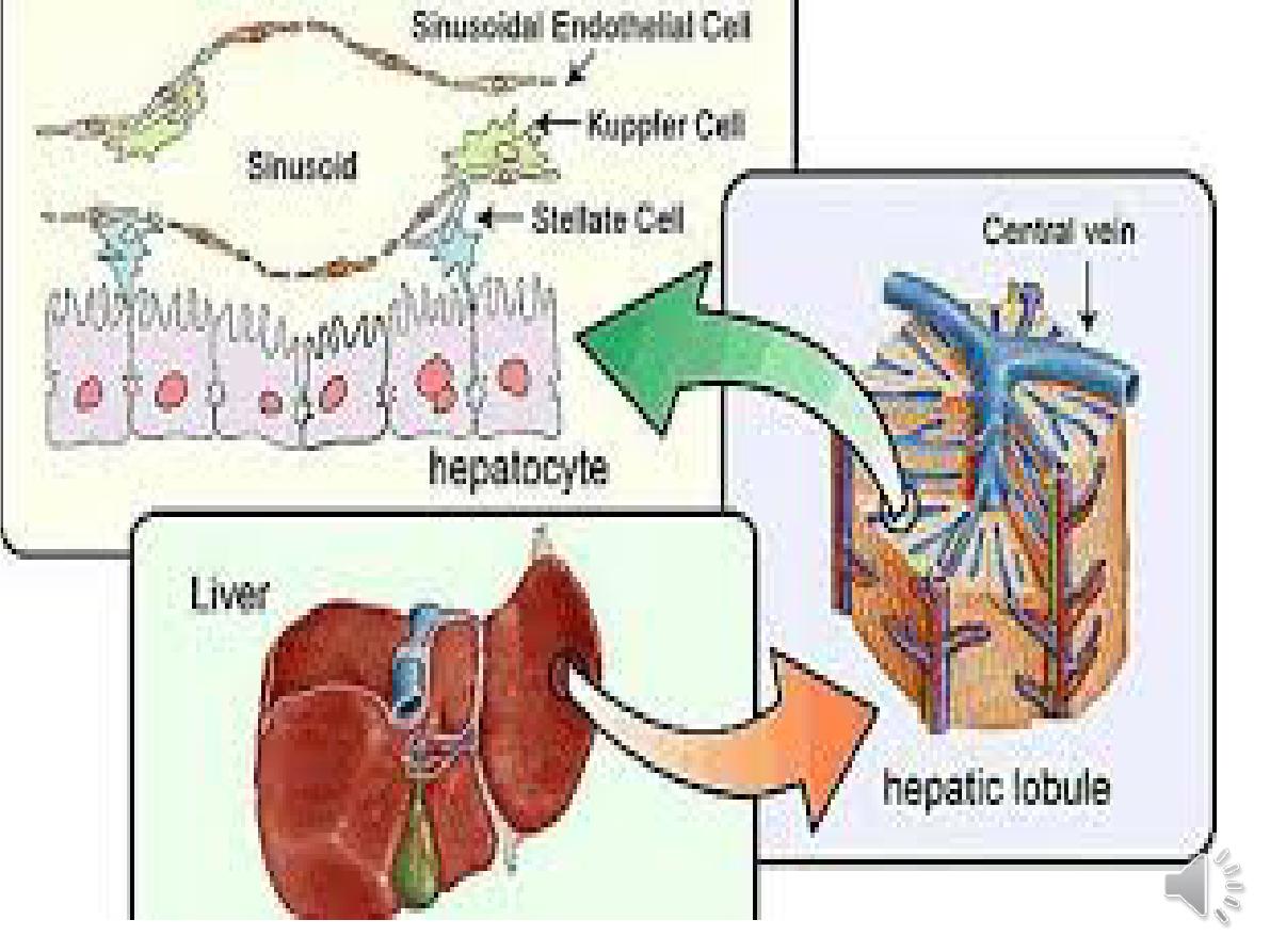
Liver cells
Hepatocytes comprise 80% of liver cells.
The remaining 20% are the endothelial
cells lining the sinusoids ,epithelial cells
lining the intrahepatic bile ducts, cells of
the immune system (including
macrophages (Kupffer
cells)
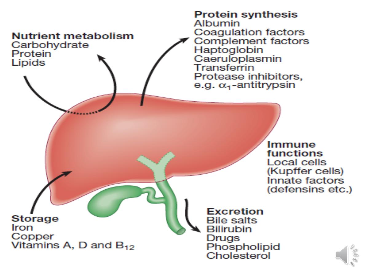
Hepatic function
1-Carbohydrate, amino acid and lipid metabolism
•Amino acids from dietary proteins are used for synthesis of
plasma proteins, including albumin.The liver produces 8–14 g
of albumin per day..
• Following a meal, more than half of the glucose absorbed is
taken up by the liver and stored as glycogen --preventing
hyperglycaemia. During fasting, glycogen is broken down to
release glucose (gluconeogenesis), thereby preventing
hypoglycaemia
• The liver plays a central role in lipid metabolism, producing
VLDL and further metabolising LDL &HDL .Dysregulation of
lipid metabolism is thought to have a critical role in the
pathogenesis of NAFLD.

2-Clotting factors
The liver produces key proteins that are
involved in the coagulation cascade. Reduced
clotting factor synthesis is an important and
easily accessible biomarker of liver function
in the setting of liver injury.
Prothrombin time
(PT or INR) is therefore one
of the most important clinical tools available
for the assessment of hepatocyte function.
.

3-Storage of vitamins and minerals
Vitamins A, D and B12
are stored by the liver in large
amounts, while others, such
as vitamin K and folate
,
are stored in smaller amounts and disappear rapidly
if dietary intake is reduced.
The liver is also able to metabolise vitamins to more
active compounds, e.g. 7-dehydrocholesterol to
25(OH) vitamin D.
Vitamin K is a fat-soluble vitamin
and so the inability
to absorb fat soluble vitamins, as occurs in biliary
obstruction, results in a coagulopathy.
The liver stores minerals such as
iron, in ferritin and
haemosiderin, and copper,
which is excreted in bile.

4 Immune regulation
Approximately 9% of the normal liver is
composed of immune cells .
Kupffer
cells
derived from blood monocytes, the
liver macrophages and natural killer
(
NK
)cells, as well as ‘classical’ B and T
cells
.
They remove aged and damaged red
blood cells, bacteria, viruses, antigen–
antibody complexes and endotoxin
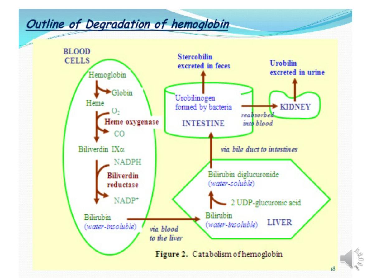
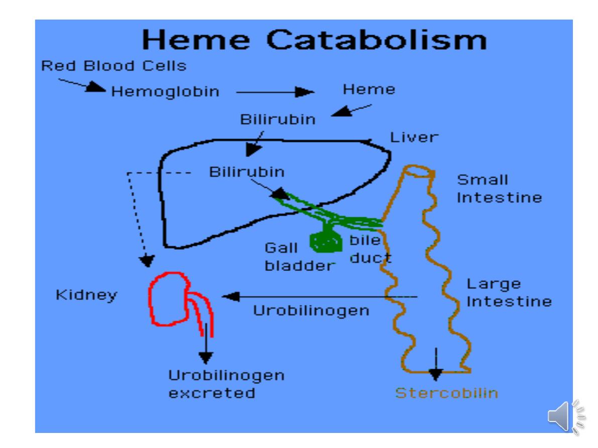
5-billrubin and bile metabolism:
Bile contains bile acids ,phospholipids ,
bilirubin and cholesterol
Investigations
1- Bilirubin and albumin
The degree of elevation of bilirubin can reflect the degree of
liver damage. A raised bilirubin often occurs earlier in the
natural history of biliary disease (e.g. primary biliary
cirrhosis) than in disease of the liver parenchyma (e.g.
cirrhosis) where the hepatocytes are primarily
involved.
Serum albumin levels are often low in patients with liver
disease.. Since the plasma half-life of albumin is about 2
weeks, albumin levels may be normal in acute liver failure
but are almost always reduced in chronic liver failure.
2-Alanine aminotransferase (ALT) and aspartate
aminotransferase (AST) :
are located in the cytoplasm of the hepatocyte;
Although both transaminase enzymes are widely
distributed, expression of ALT outside the liver is
relatively low and this enzyme is therefore
considered more specific for hepatocellular damage.
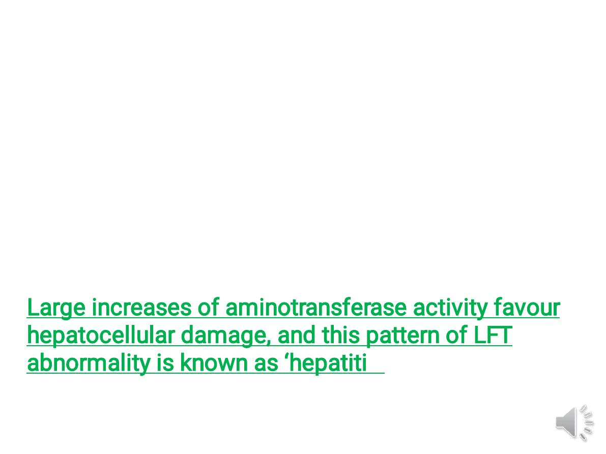
Large increases of aminotransferase activity favour
hepatocellular damage, and this pattern of LFT
abnormality is known as ‘hepatiti
c’
3-Alkaline phosphatase and gamma-glutamyl transferase
:
(
ALP)
is widely distributed in the body, but the main sites of
production are the
liver, gastrointestinal tract, bone, placenta and
kidney
, levels rise with intrahepatic and extrahepatic biliary obstruction
and in infiltrative liver disease.
(
GGT
) is a microsomal enzyme found in many cells and tissues of the
body. The highest concentrations are located in the liver
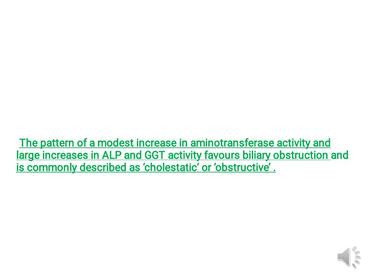
-The pattern of a modest increase in aminotransferase activity and
large increases in ALP and GGT activity favours biliary obstruction and
is commonly described as ‘cholestatic’ or ‘obstructive’ .
Isolated elevation of the serum GGT is relatively common, and may
occur during ingestion of microsomal enzyme-inducing drugs,
including alcohol ,but also in NAFLD.

4-Other biochemical tests
•
Hyponatraemia
occurs in severe liver disease
due to increased production of ADH
•
Serum urea
may be reduced in hepatic failure,
whereas levels of urea may be increased
following
GI haemorrhage.
• Significantly elevated
ferritin
suggests
haemochromatosis. Modest elevations can be
seen in inflammatory disease and alcohol
excess.
5-Haematological tests
•
A normochromic normocytic anaemia
-gastrointestinal
haemorrhage,
chronic blood loss is characterised by a hypochromic
microcytic anaemia secondary to iron deficiency.
-(macrocytosis) is associated with alcohol misuse,
•
Leucopenia
may complicate portal hypertension and
hypersplenism, whereas leucocytosis may occur with
cholangitis, alcoholic hepatitis and hepatic abscesses.
. •
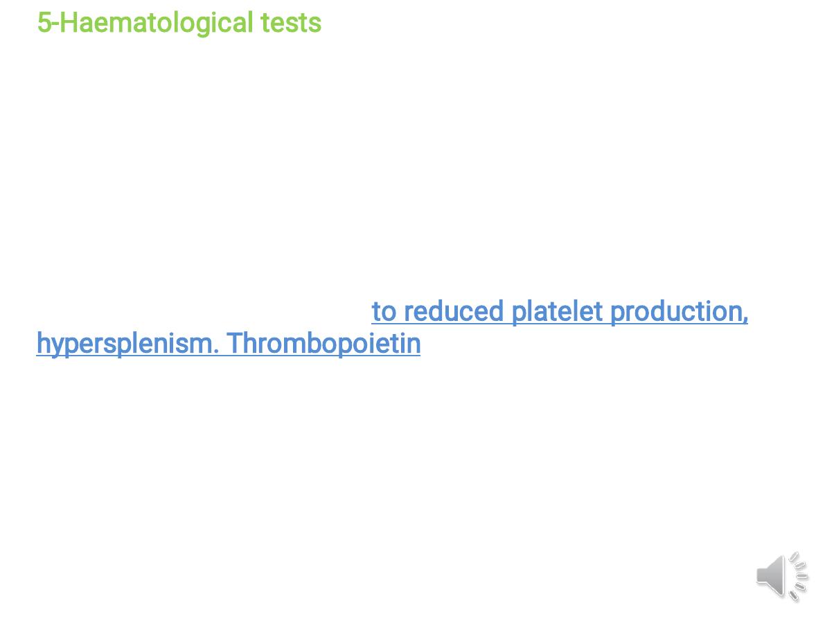
Thrombocytopenia
--due to reduced platelet production,
hypersplenism. Thrombopoietin, required for platelet
production, is produced in the liver and levels fall with
worsening liver function. A low platelet count is often an
indicator of chronic liver disease,
Thrombocytosis
is unusual in patients with liver disease but
may occur in those with active GI haemorrhage and, rarely, in
hepatocellular carcinoma
.
6-Coagulation tests
vitamin K-dependent coagulation factors
(1972)in the blood are short (5–72 hours) and
so
changes in
the prothrombin time
occur
relatively quickly following liver damage. An
increased PT is evidence of severe liver
damage in chronic liver disease.
Vitamin K does not reverse this deficiency if it
is due to liver disease, but will correct the PT
if the cause is vitamin K deficiency, as may
occur with biliary obstruction due to non-
absorption of fat-soluble vitamins.
7-Immunological tests
- Elevation in overall serum
immunoglobulin levels can also be
suggestive of autoimmunity
(immunoglobulin (Ig)G and IgM).
-Elevated serum IgA can be seen,
often in more advanced
alcoholic liver disease and NAFLD

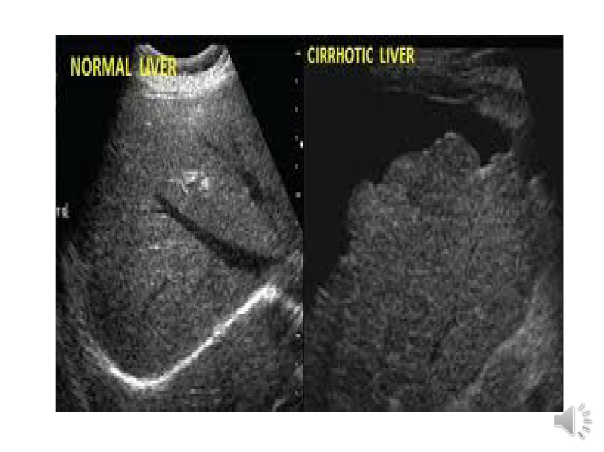
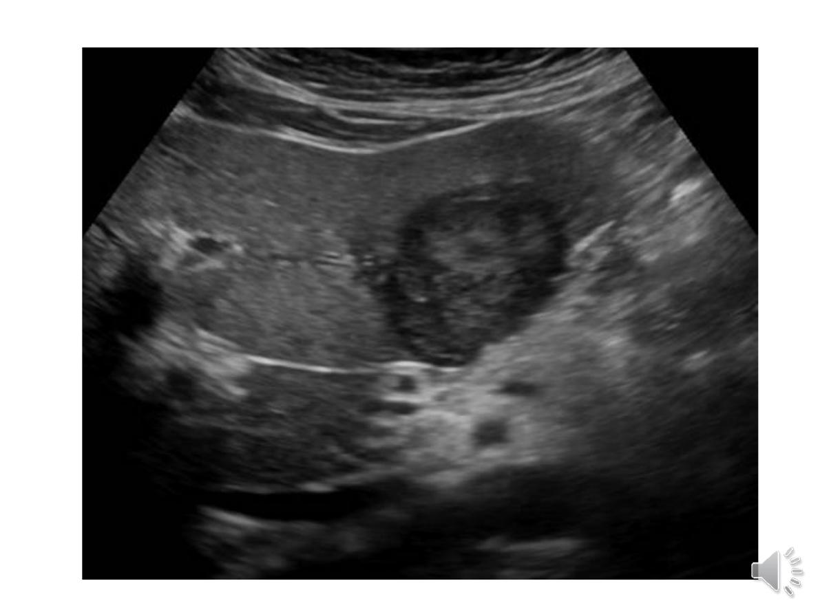
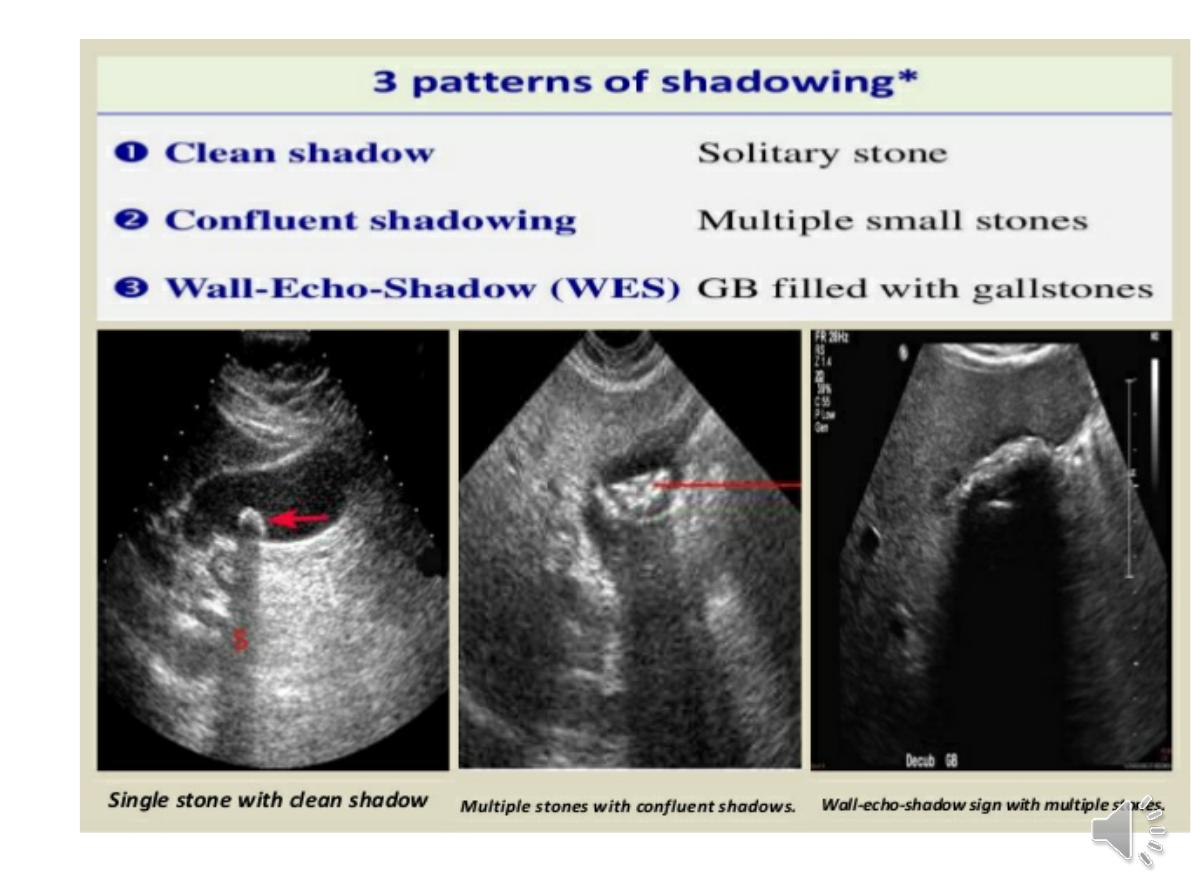
8 Ultrasound
a ‘first-line’ test to identify gallstones, biliary
obstruction or thrombosis in the hepatic
vasculature.
Focal lesions, such as tumours, may not be
detected if they are below 2 cm in diameter
and have echogenic characteristics similar to
normal liver tissue.
. Doppler ultrasound allows blood flow in the
hepatic artery, portal vein and hepatic veins to
be investigated
.
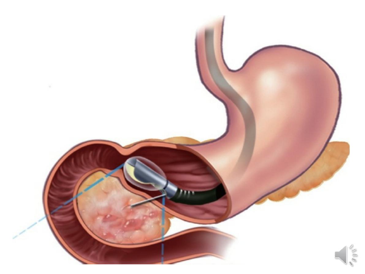
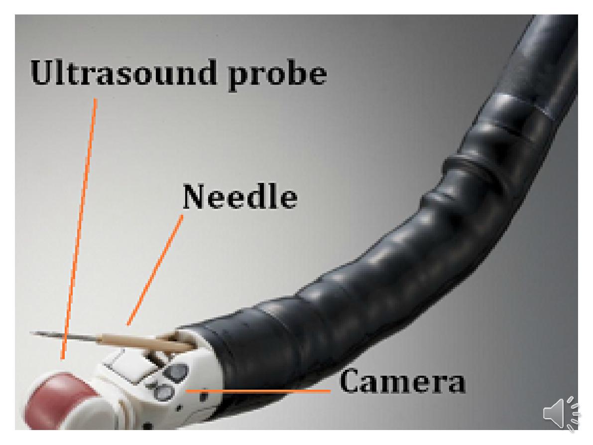
Endoscopic ultrasound provides high-
resolution images of the pancreas, biliarytree
and liver

9-
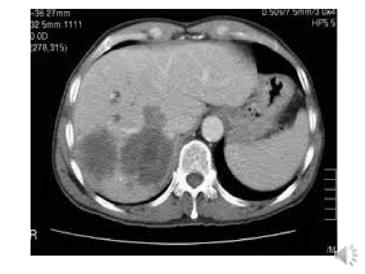
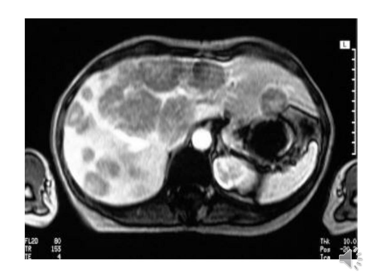
(CT)
detects smaller focal lesions in
the liver, especially when combined
with contrast injection .
(MRI
) can also be used to localise
and confirm the aetiology of focal
liver lesions, particularly primary and
secondary tumours

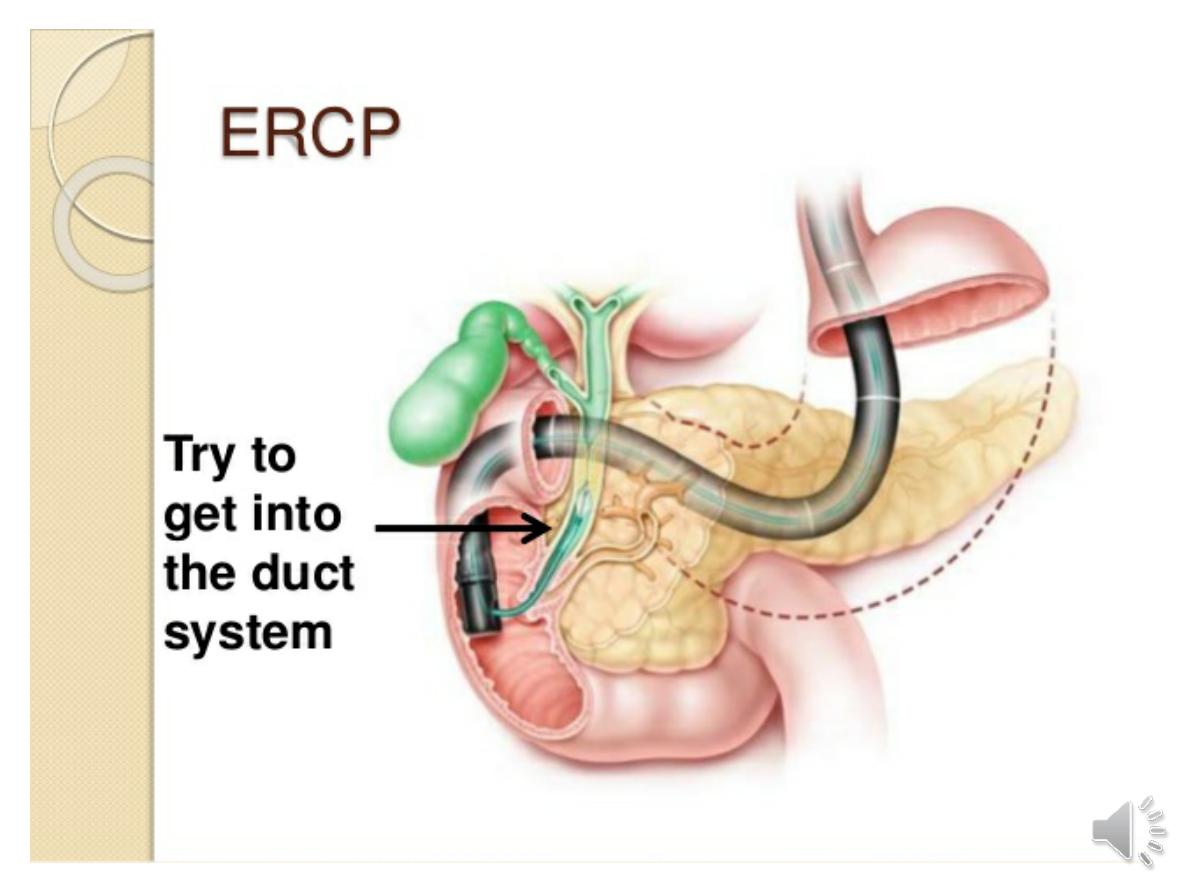
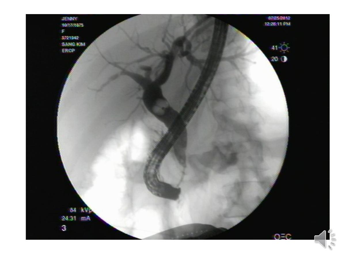
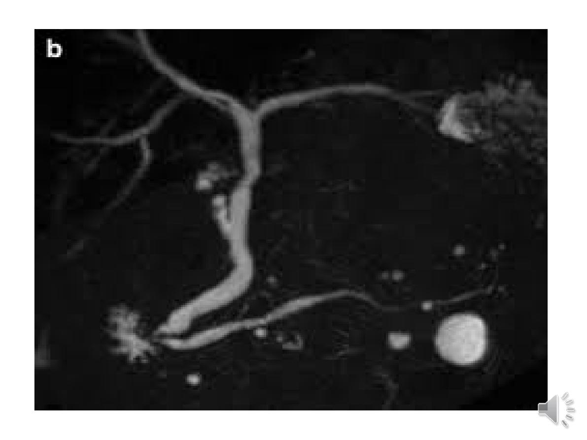
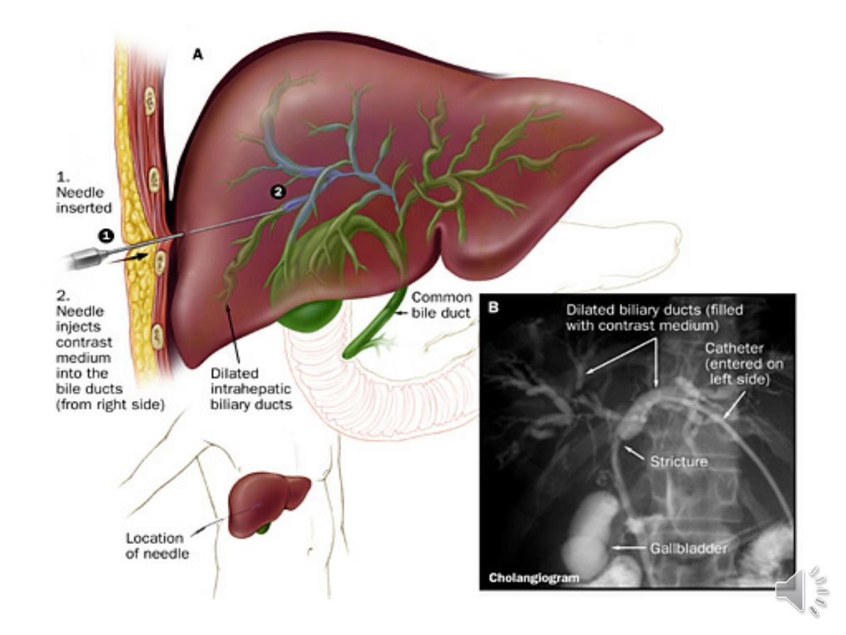
10-Cholangiography
(MRCP)& ERCP
, or the
percutaneous
approach (percutaneous
transhepatic cholangiography,
PTC
). The latter does not allow
the ampulla of Vater or
pancreatic duct to be visualised
.
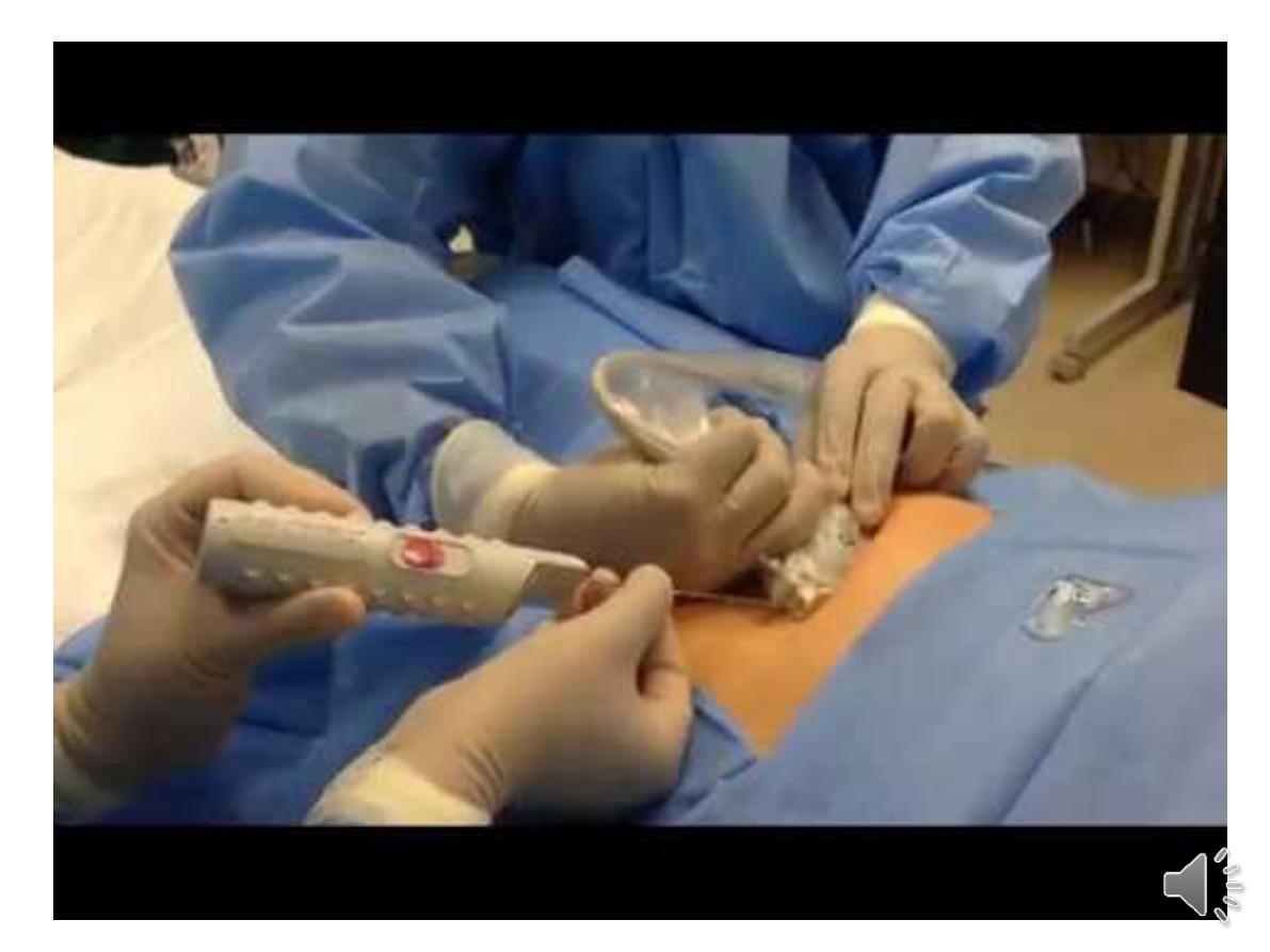
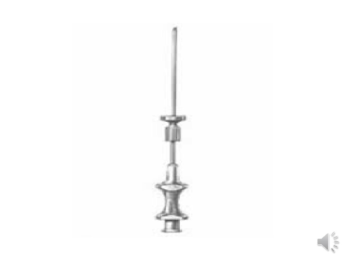
11-liver biopsy
can confirm the severity of liver
damage and provide aetiological information.
It is performed percutaneously with a Trucut or
Menghini
needle, usually through an intercostal space
under local anaesthesia, Percutaneous liver biopsy is
a relatively safe procedure.
mortality of about 0.01%. The main complications
are abdominal and/or shoulder pain, bleeding and
biliary peritonitis.
liver disorders can be broadly classified histologically
into fatty liver (steatosis), hepatitis (inflammation,
‘grade’) and cirrhosis (fibrosis, ‘stage’).
12-Non-invasive markers of hepatic fibrosis
***Serological markers of hepatic fibrosis, such
as α2-
macroglobulin, haptoglobin.
The ELF®(Enhanced Liver Fibrosis) serological assay uses
a combination
of hyaluronic acid, procollagen peptide III
(PIIINP) and tissue inhibitor of metalloproteinase 1 (TIMP1)
.
These tests are good at differentiating severe fibrosis
from mild scarring, but are limited in their ability to detect
subtle changes.
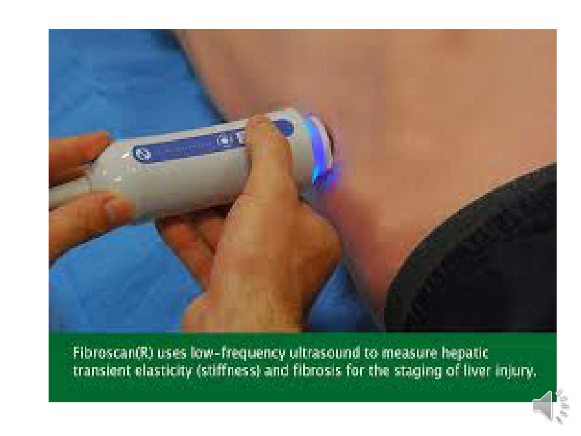
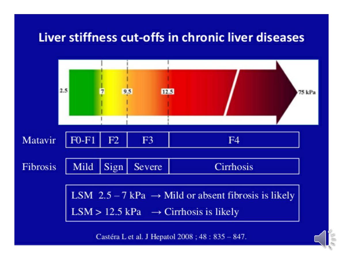
-***fibroscan.:
elastography in which ultrasound-based shock waves are
sent through the liver to measure liver stiffness as a
surrogate for hepatic fibrosis.