Haemolytic anaemia
Genetic disorders of hemoglobin
Dr. Zainab W. Al-Maaroof
•
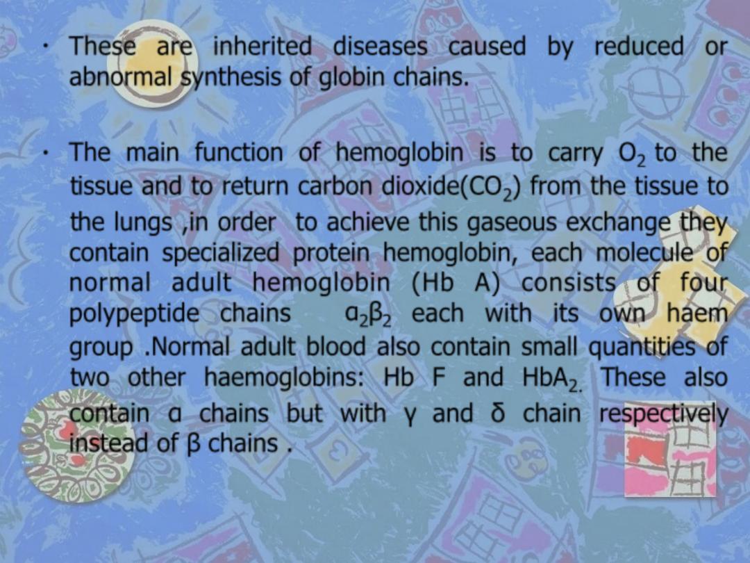
These are inherited diseases caused by reduced or
abnormal synthesis of globin chains.
•
The main function of hemoglobin is to carry O
2
to the
tissue and to return carbon dioxide(CO
2
) from the tissue to
the lungs ,in order to achieve this gaseous exchange they
contain specialized protein hemoglobin, each molecule of
normal adult hemoglobin (Hb A) consists of four
polypeptide chains α
2
β
2
each with its own haem
group .Normal adult blood also contain small quantities of
two other haemoglobins: Hb F and HbA
2.
These also
contain α chains but with γ and δ chain respectively
instead of β chains .
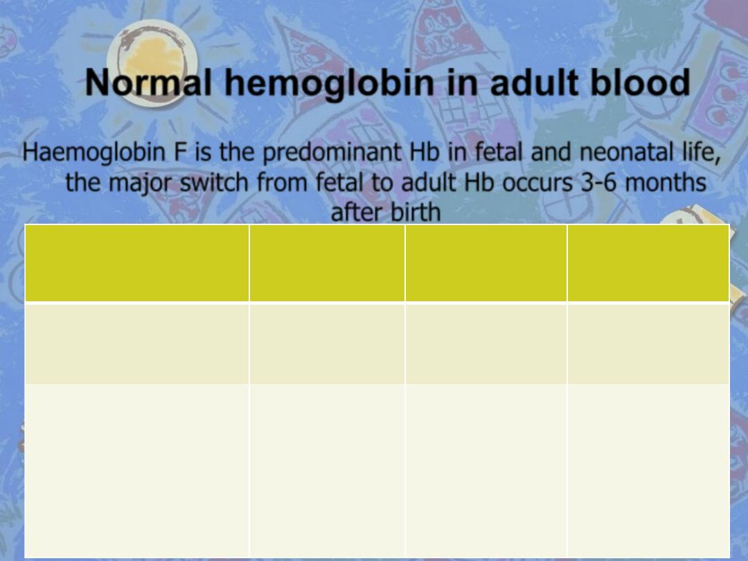
Normal hemoglobin in adult blood
Haemoglobin F is the predominant Hb in fetal and neonatal life,
the major switch from fetal to adult Hb occurs 3-6 months
after birth
Hb A
Hb F
Hb A2
Structure (globin chains)
α
2
β
2
α
2
γ
2
α
2
δ
2
Normal (%)
96-98
0.5-0.8
1.5-3.2
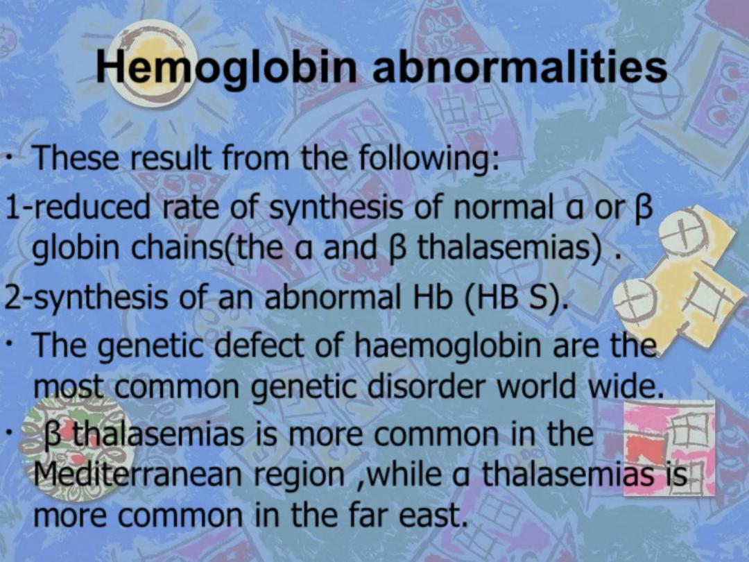
Hemoglobin abnormalities
•
These result from the following:
1-reduced rate of synthesis of normal α or β
globin chains(the α and β thalasemias) .
2-synthesis of an abnormal Hb (HB S).
•
The genetic defect of haemoglobin are the
most common genetic disorder world wide.
•
β thalasemias is more common in the
Mediterranean region ,while α thalasemias is
more common in the far east.
Thalassemias
•
These are a heterogeneous group of genetic disorders
that result from a reduced rate of synthesis of α and β
chains.
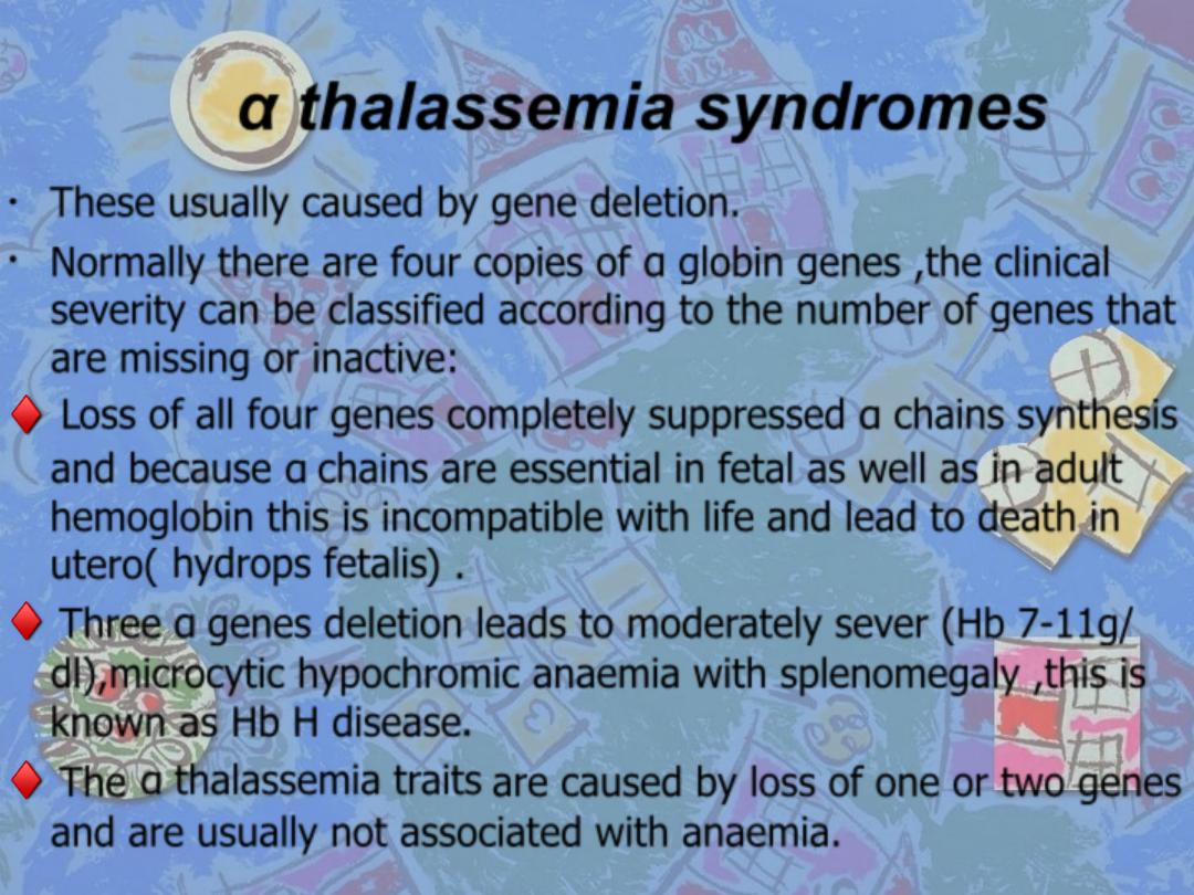
α thalassemia syndromes
•
These usually caused by gene deletion.
•
Normally there are four copies of α globin genes ,the clinical
severity can be classified according to the number of genes that
are missing or inactive:
♦
Loss of all four genes completely suppressed α chains synthesis
and because α chains are essential in fetal as well as in adult
hemoglobin this is incompatible with life and lead to death in
utero(
hydrops fetalis
) .
♦
Three α genes deletion leads to moderately sever (Hb 7-11g/
dl),microcytic hypochromic anaemia with splenomegaly ,this is
known as
Hb H disease
.
♦
The
α thalassemia traits
are caused by loss of one or two genes
and are usually not associated with anaemia.
β thalassemia syndromes
β thalassemia major
•
This condition occurs in one of four offspring if both
parents are carrier of the β thalassemia trait.
•
Either no β chains or small amounts are synthesized.
•
Excess α chains precipitate in erythroblasts and in
mature red cells causing sever ineffective erythropoisis
and haemolysis that are typical of this disease .
•
β thalassemia unlike α thalassemia ,the majority of
genetic lesions are point mutation rather than gene
deletions.
Clinical features
1-Sever anaemia
becomes apparent at 3-6 months after birth
when the switch from γ to β chains production should take
place.
2-Enlargment of the liver and spleen
occurs as the result of
excessive red cells destruction ,extramedullary haemopoisis,
and because of iron overload.
3-Expantion of the bones
caused by intense marrow
hyperplasia leads to
thalassemic facies
and to thinning of the
cortex of many bones with a tendency to fracture and
bossing of the skull with a
(hair-on-end
)appearance on x-
ray.
Clinical features
4-The patient can be sustained by blood transfusion but
iron
overload
caused by repeated transfusion is inevitable unless
chelating therapy is given.
•
Iron damages the liver and endocrine organs
with failure of the
growth delayed or absent puberty ,diabetes mellitus,
hypothyroidisms and hypoparayhyroidism.
•
Skin pigmentations
as a result of excess melanin and
haemosidrin gives a gray appearance at an early stages of iron
overload.
•
Most importantly iron damages the heart and in the absence of
intensive iron chelating thereby, death occurs in the second or
third decade usually from
congestive heart failure or cardiac
arrhythmia.
Clinical features
5-infection
can occur in thalassemic patient for a variety of
reasons:
●In infancy anaemic child is prone to bacterial infection.
●If splenectomy has been carried out and prophylactic penicillin
is not given ,pneumococcal ,haemophilus and meningococcal
infection are likely to occur.
●Yersinia entercolitica occur in iron-loded patient being treated
with deferoxamine and it may cause sever gasteroenteritis.
●Transmission of viruses(such HIV virus) by blood transfusion,
may happened in some patients.
6- osteoporosis
may occur ,it is more common in diabetic
patient with endocrine abnormalities.
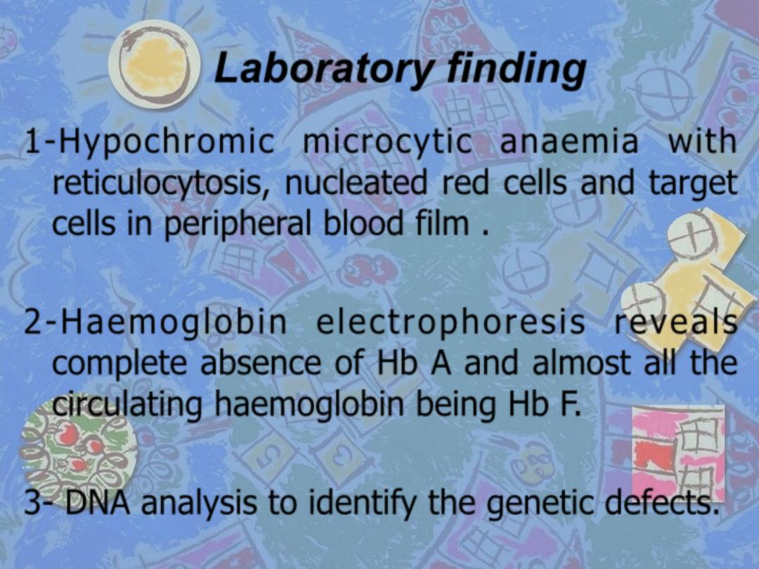
Laboratory finding
1-Hypochromic microcytic anaemia with
reticulocytosis, nucleated red cells and target
cells in peripheral blood film .
2-Haemoglobin electrophoresis reveals
complete absence of Hb A and almost all the
circulating haemoglobin being Hb F.
3- DNA analysis to identify the genetic defects.
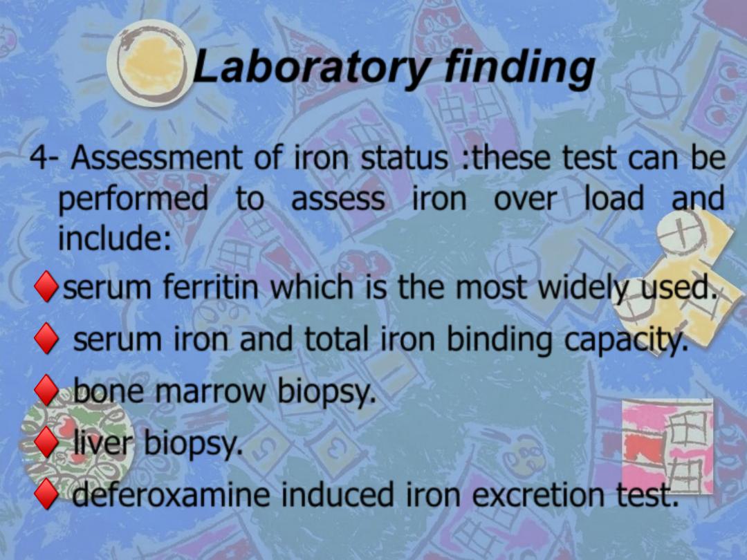
Laboratory finding
4- Assessment of iron status :these test can be
performed to assess iron over load and
include:
♦
serum ferritin which is the most widely used.
♦
serum iron and total iron binding capacity.
♦
bone marrow biopsy.
♦
liver biopsy.
♦
deferoxamine induced iron excretion test.
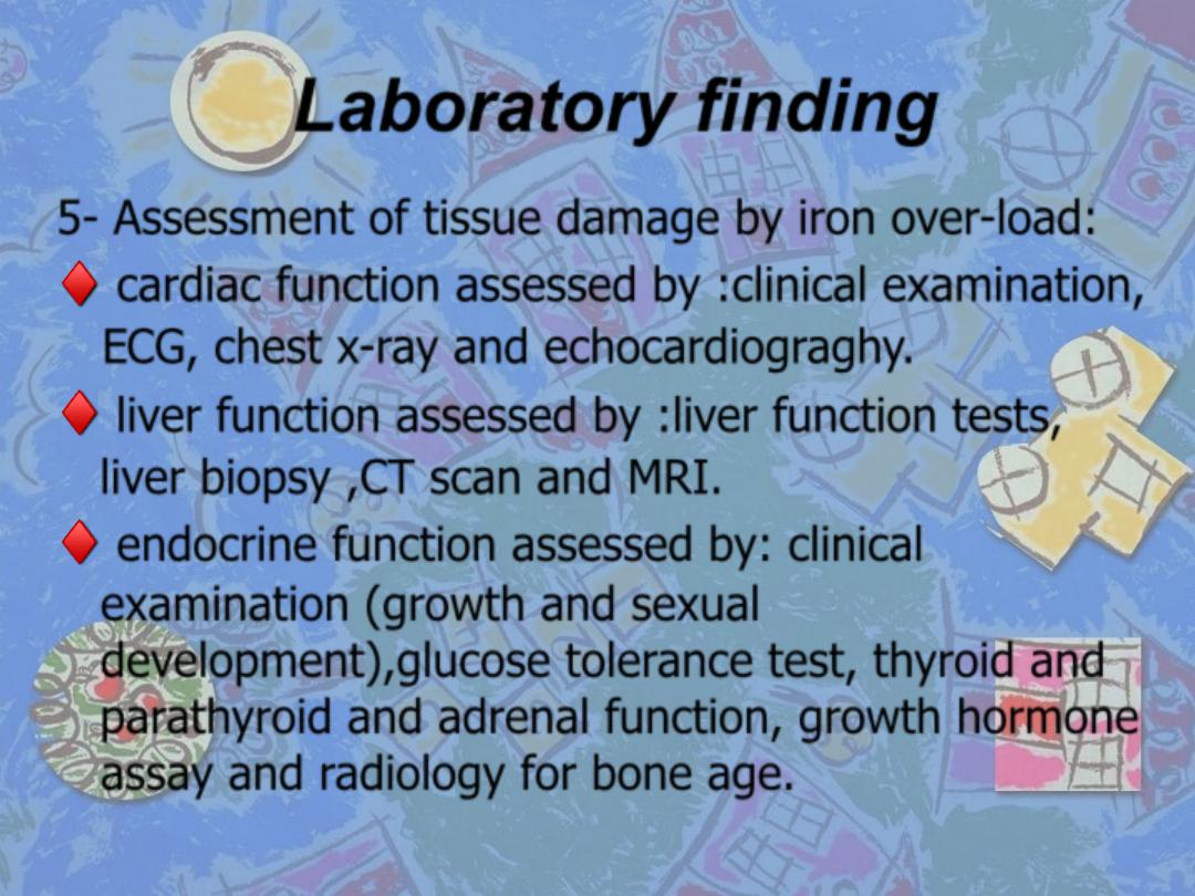
Laboratory finding
5- Assessment of tissue damage by iron over-load:
♦
cardiac function assessed by :clinical examination,
ECG, chest x-ray and echocardiograghy.
♦
liver function assessed by :liver function tests,
liver biopsy ,CT scan and MRI.
♦
endocrine function assessed by: clinical
examination (growth and sexual
development),glucose tolerance test, thyroid and
parathyroid and adrenal function, growth hormone
assay and radiology for bone age.
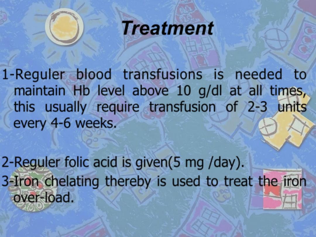
Treatment
1-Reguler blood transfusions is needed to
maintain Hb level above 10 g/dl at all times,
this usually require transfusion of 2-3 units
every 4-6 weeks.
2-Reguler folic acid is given(5 mg /day).
3-Iron chelating thereby is used to treat the iron
over-load.
Treatment
4-
Vitamin C
( 200 mg/ day), increases excretion of iron
produced by deferoxamine.
5-
Splenectomy
, may be needed to reduce blood
requirement which should be delayed until the patient
is over 6 years old because of the high risk of
dangerous infections post splenectomy.
6-
Endocrine therapy
: is given either as replacement
because of end organ failure or to stimulate the
pituitary if puberty is delayed. Diabetes will require
insulin therapy.
7-
Immunization
against hepatitis B.
8-
Allogeneic Bone Marrow transplantation.
β thalassemia minor
•
This is a common usually a symptomatic
characterized by hypochomic microcytic
blood picture and mild anemia, a raised Hb
A2 ( > 3.5%) confirms the diagnosis
Thalassemia Intermedia
•
Are cases of thalassemia of moderate
s e v e r i t y w h o d o n ’ t n e e d r e g u l a r
transfusions and this is a clinical syndrome
which may be caused by a variety of
genetic defects. The patients may show
bone deformity, hepatoslenomegaly,
extramedullary haemopoiesis and features
of iron over load caused by increased iron
absorption.
Sickle cell anaemia
❖
Sickle cell disease is a group of haemoglobin disorder in
which the sickle β globin gene is inherited.
❖
Sickle β globin gene result from single base substitution
at position number 6,this leads to an amino acids
changes from glutamic acid to valine in the β chain.
❖
Hb S (Hb α
2
β
2
s
) is insoluble and form crystals when
exposed to low oxygen tension, deoxygenated sickle Hb
polymerized into long fibers and red cells become
sickle-like which may block different areas of
microcirculations or large vessels causing infarct of
various organs.
•
Clinical features
•
Sever haemolytic anemia associated with crises.
•
The symptoms of anemia are usually mild in relation
to the severity of anemia, this because HbS gives up
oxygen to the tissue easily compared with Hb A.
•
The crises may be vaso-occlusive, visceral, aplastic or
haemolytic.
Clinical features
1-Painful vaso-occlusive crises:
•
These are the most frequent and precipitated by many factors such
as infection, dehydration, acidosis and deoxygenated states like
high altitude, operation ,exposure to cold and obstetric delivery.
•
Infarct can occur in a variety of organs including the bones, the
lungs and the spleen, the most serious vaso-occlusive crises is of
the brain which cause a stroke. Hand-foot syndrome (painful
dactylitis caused by infarcts in small bones) is frequently the first
presentation of the disease and may lead to digits of varying
lengths.
Clinical features
2- Visceral sequestration crises:
❑
These are caused by sickling within organs and pooling of the
blood, often with a sever exacerbation of anemia.
❑
The acute chest syndrome is serious complication and the most
common cause of death after puberty ,it present with dyspnea
and chest pain ,treatment by analgesia, oxygen and exchange
transfusion .
❑
Splenic sequestration present with splenomegaly, falling
Hemoglobin and abdominal pain ,it is typically seen in infants
and treatment is with blood transfusion ,attacks may be
recurrent and splenectomy is often needed.
Clinical features
3-Aplastic crises:
➢
These occur as a result of infection with parvovirus or from
folate deficiency and characterized by fall in hemoglobin as well
as reticulocytes.
4- Hemolytic crisis:
➢
characterized by an increased rate of haemolysis with a fall in
hemoglobin and a rise in reticulocytes.
Clinical features
5-Other clinical features:
•
Ulcer of lower legs as a result of vascular stasis and local
ischemia.
•
Enlarged spleen in infancy and early childhood but later is often
reduced in size as a result of infarction ( Autosplenectomy).
•
Pulmonary hypertension.
•
Proliferative retinopathy.
•
Chronic damage to the liver due to micro infarct.
•
Pigment gall stones.
•
Osteomylitis.

Laboratory finding
•
The hemoglobin is usually 6-9 g/ dl, low in comparison to
symptoms of anemia.
•
Sickle cells and target cells . Features of splenic atrophy ( Howell-
Jolly bodies).
•
Screening tests for sickling are positive when the blood is
deoxygenated.
•
Hemoglobin electrophoresis: in HbSS, no Hb A is detected, Hb F is
usually 5-15%.
Treatment
1- Prophylactic: avoid factors known to precipitate crisis, especially
dehydration, anoxia, infections, stasis of circulation and cooling of
the skin surface.
2- Folic acid, (5 mg once weekly).
3- Good general nutrition and hygiene.
4- Pneumococcal, Haemophilus and Meningococcal vaccination.
Hepatitis B vaccination is also given as transfusion may be
needed.
5- Regular oral penicillin should start at diagnosis and continue at
least until puberty.
Treatment
6-
Crisis treatment
: rest, warmth, rehydration and antibiotics if
infection is present. Analgesia ( paracetamol, non-steroidal anti-
inflammatory agent and opiate). Blood transfusion if there is sever
anemia with symptoms. Exchange transfusion may be needed if
there is neurological damage, visceral sequestration crisis or
repeated painful crisis. This is aimed to achieve an Hb S
percentage of less than 30 in sever cases and after a stroke is
continued for at least 2 years.
7- Careful anesthetic and recovery technique to avoid hypoxemia or
acidosis.
8- Routine transfusions throughout pregnancy are given to those
with a poor obstetric history or a history of frequent crises .
Treatment
9-
Transfusions
: sometimes given repeatedly as prophylaxis to
patients having frequent crises or who have had major organ
damage (e.g. of the brain). The aim is to suppress Hb S
production over a period of several months or even years. Iron
overload which may need chelation therapy is common problem.
10-
Hydroxyurea
( 15-20 mg/ kg) can increase Hb F and improve
clinical course of patients who are having three or painful crises
each year. It should not be used during pregnancy.

Treatment
11-
Stem cell transfusion
can cure the disease.
12-
Butyrates
which enhance Hb F synthesis and
increase solubility of Hb S.
13-
Gene- therapy
.
Sickle cell trait
•
This is a benign condition in which there is
inheritance of normal hemoglobin and sickle
hemoglobin .
•
There is no anemia and normal appearance of
red cells in blood film.
•
Haematuria is the common symptom and
caused by minor infarcts to the renal papillae.