
Management of
labour

Learning objectives:
1- to distinguish between normal and
abnormal labour
2- to learn the clinical approach and
dealing with a woman with labour, from
the time of diagnosis to the end of the 3rd
stage of labour

When a pregnant woman started
labour or when she has
spontaneous rupture of membranes
at term she should be admitted and
full assessment of her condition is
accomplished.

FULL HISTORY ON ADMISSION
contractions
vaginal discharge or bleeding
LMP, GA , ANC
past obstetrical history, mode of
deliveries, any history of delivering big
baby? C/S
recent activity of the fetus

PROCEED FOR EXAMINATION
General examination, vital signs
abdominal examination:
previous scars
Leopold's maneuvers
Palpate the abdomen for assessment of the
uterine contractions for at least ten minutes
FHR: pinard stethoscope
or sonicaid
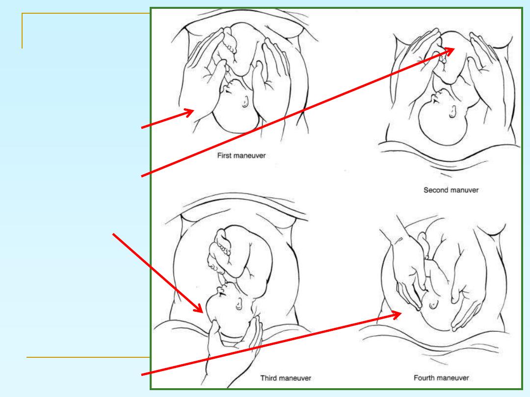
Leopold's
maneuvers
1- lateral grip
2- fundal grip
3- pawlick
4- pelvic grip

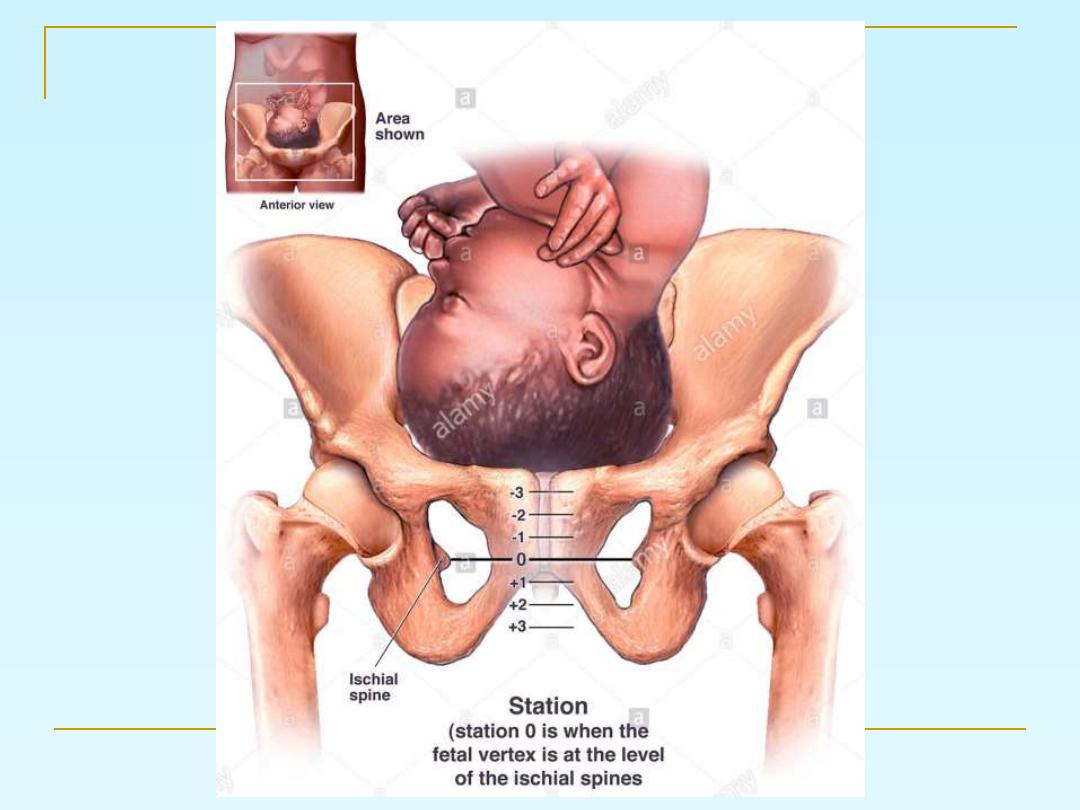
Vaginal examination to assess
cervix and station of PP
Bishop’s score:
It include:
1- dilatation
2- effacement
3- station
4- position of the cervix
5- consistency

ST
1
MANAGEMENT OF THE
STAGE
Woman in the latent phase:
Encouraged mobilization,
Adequate analgesia, and support
Light foods and drinks
Urine testing (for protein and glucose),
CBC.
Blood sampling to be available for cross-
match
If she is low risk she can go home and come
back when contractions increased

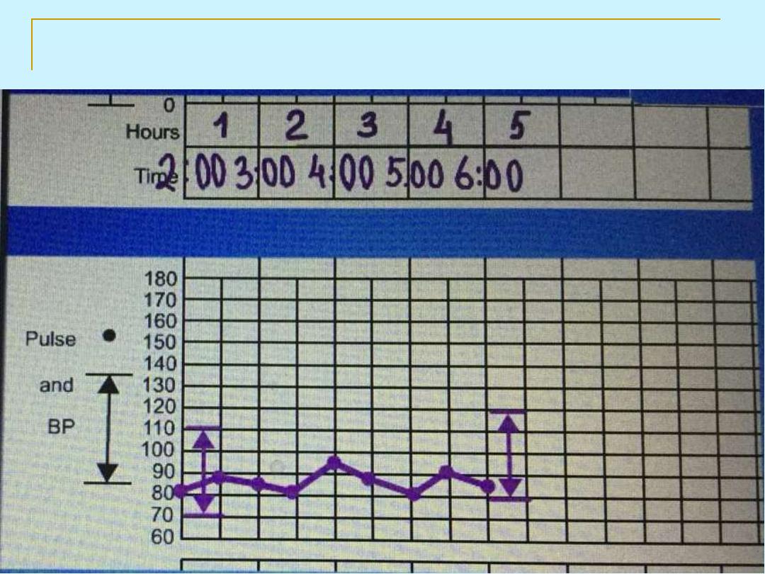
Maternal blood pressure (BP) and temperature
recorded every 4 hours,
pulse should be recorded every hour during the first
stage of labor and every 10 minutes during the
second stage of labor.
Vaginal examination in early labour is infrequently
performed (4 hourly is the standard) and the
frequency may be increased accordingly to assess
dilatation and descent of the presenting part, and
every 1 hour in the 2
nd
stage
No need to do ARM if the labor is progressing well.
STAGE
ST
1
MANAGEMENT OF THE
active phase:

Adequate monitoring of both the maternal and
fetal conditions
giving her antacid, adequate analgesia and may
be urinary catheter if labor is prolonged and
abnormal, or if she has epidural analgesia
evacuate the rectum ( may be done by enema) in
the 1st stage.
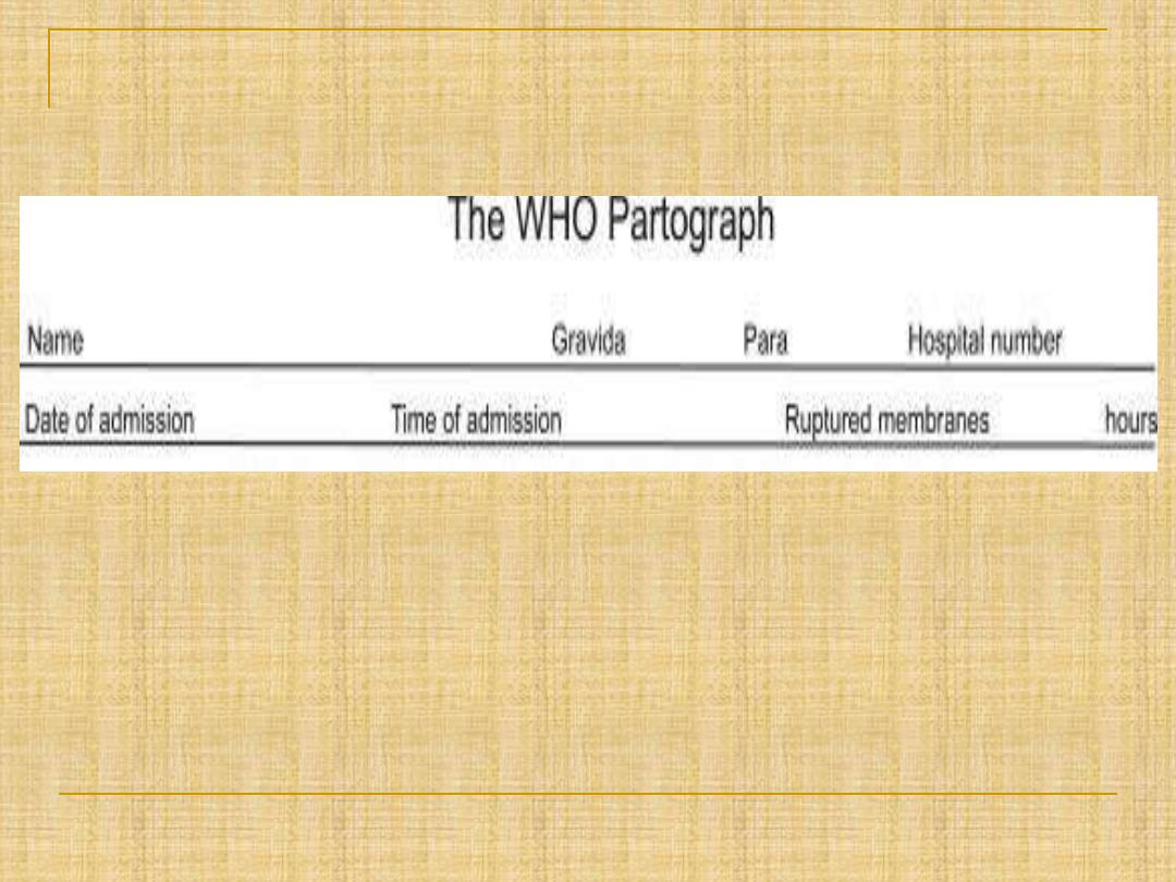
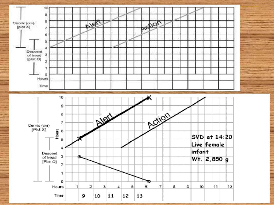
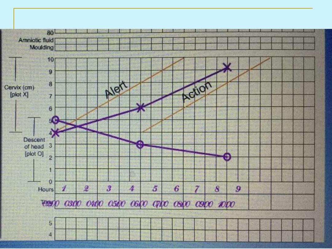
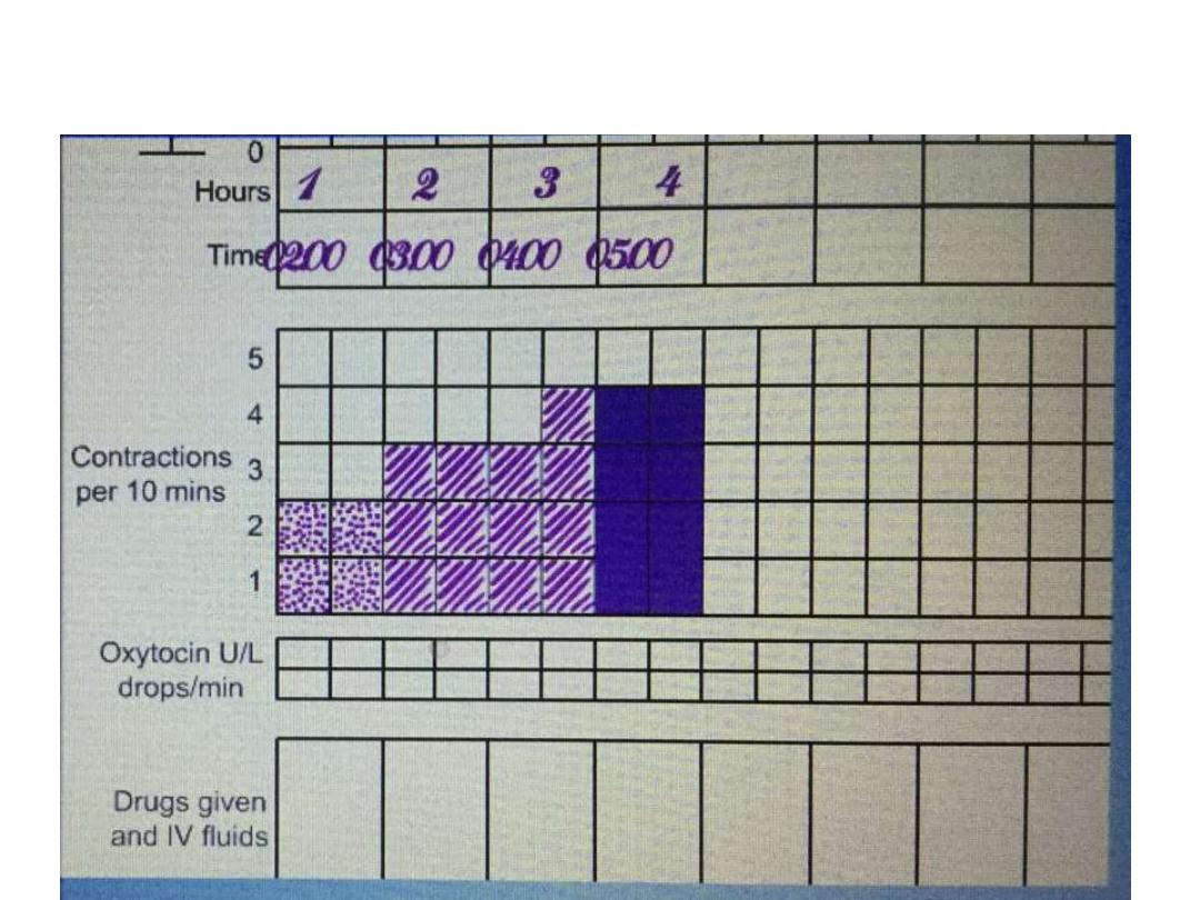
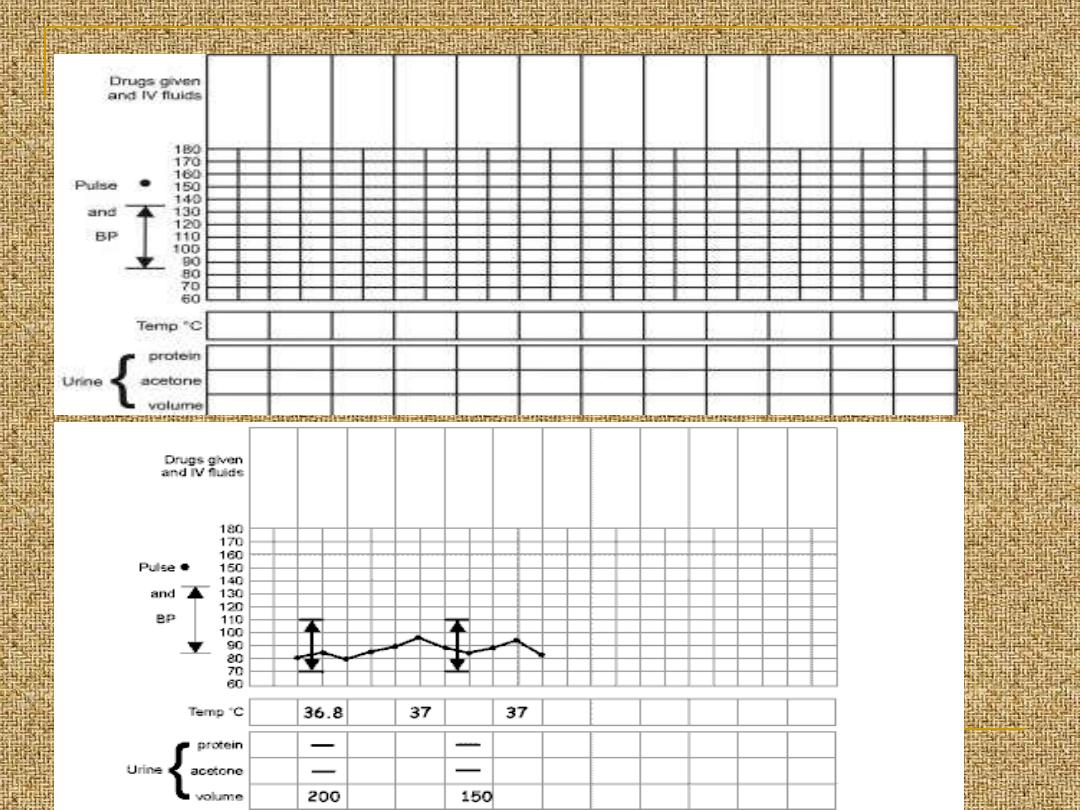
All of the data obtained since the admission to
the labour world should be recorded on a
partogram
ST STAGE
1
MANAGEMENT OF THE
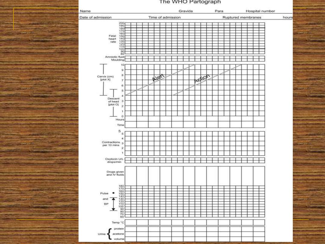
P
A
R
T
O
G
R
A
P
H
WHO PARTOGRAPH 2010
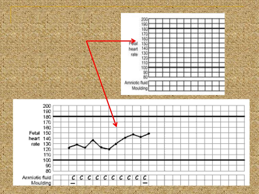
Fetal condition
fetal heart
recording
AF
moulding
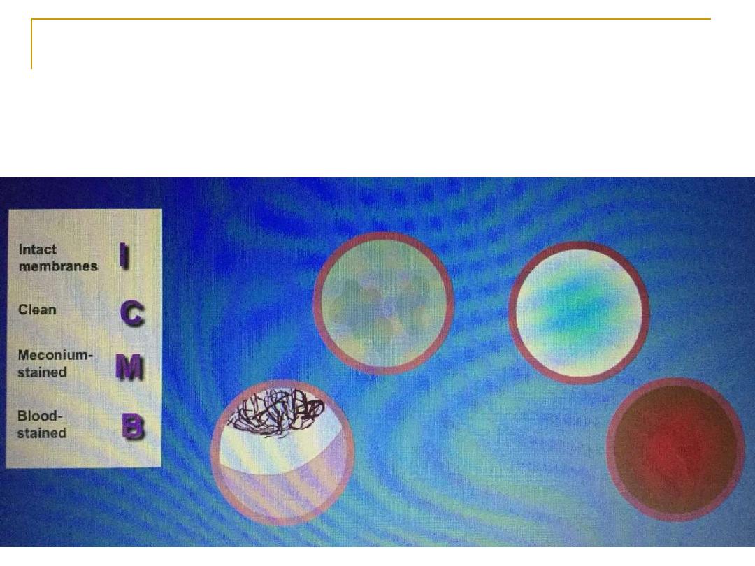
Amniotic fluid
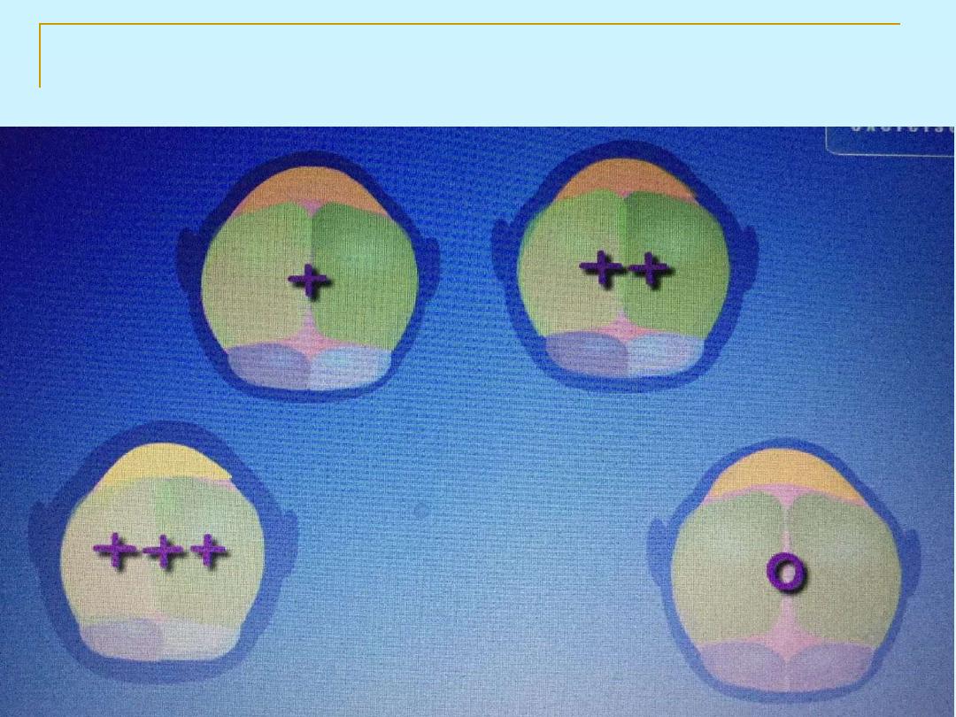
moulding
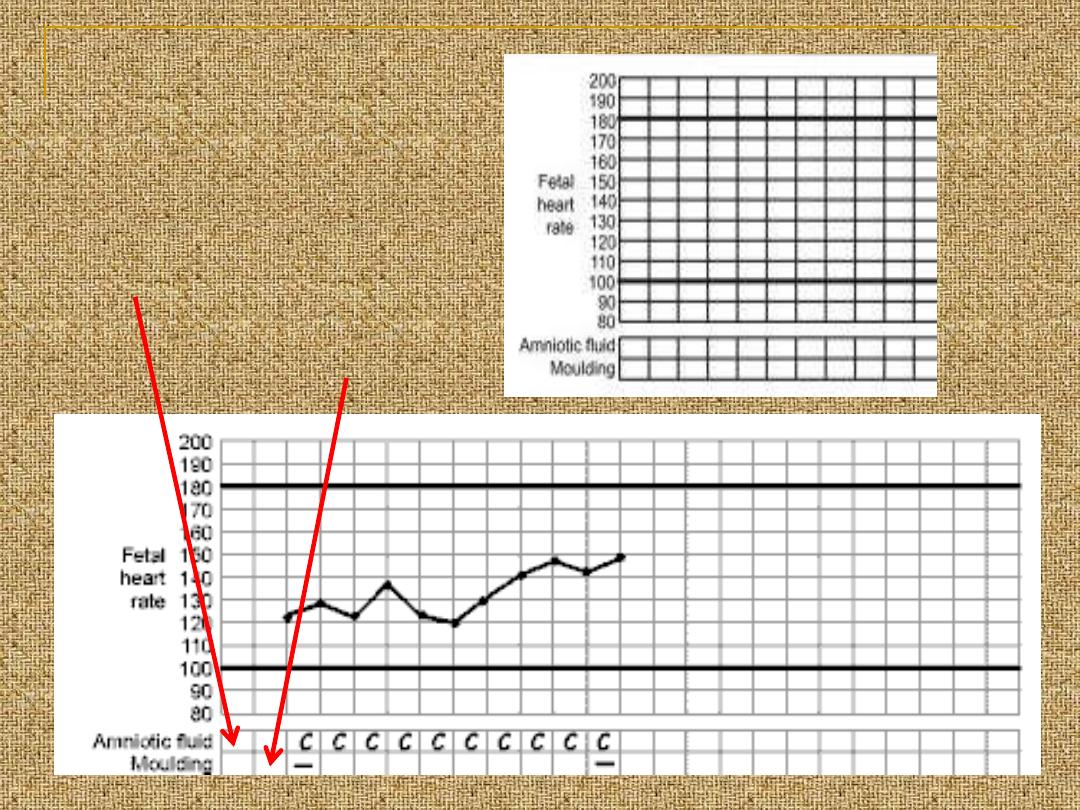
Fetal condition
fetal heart
recording
AF
moulding

contractions
Ut contractions
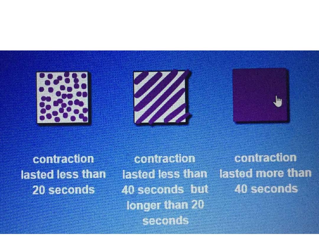
Uterine contractions
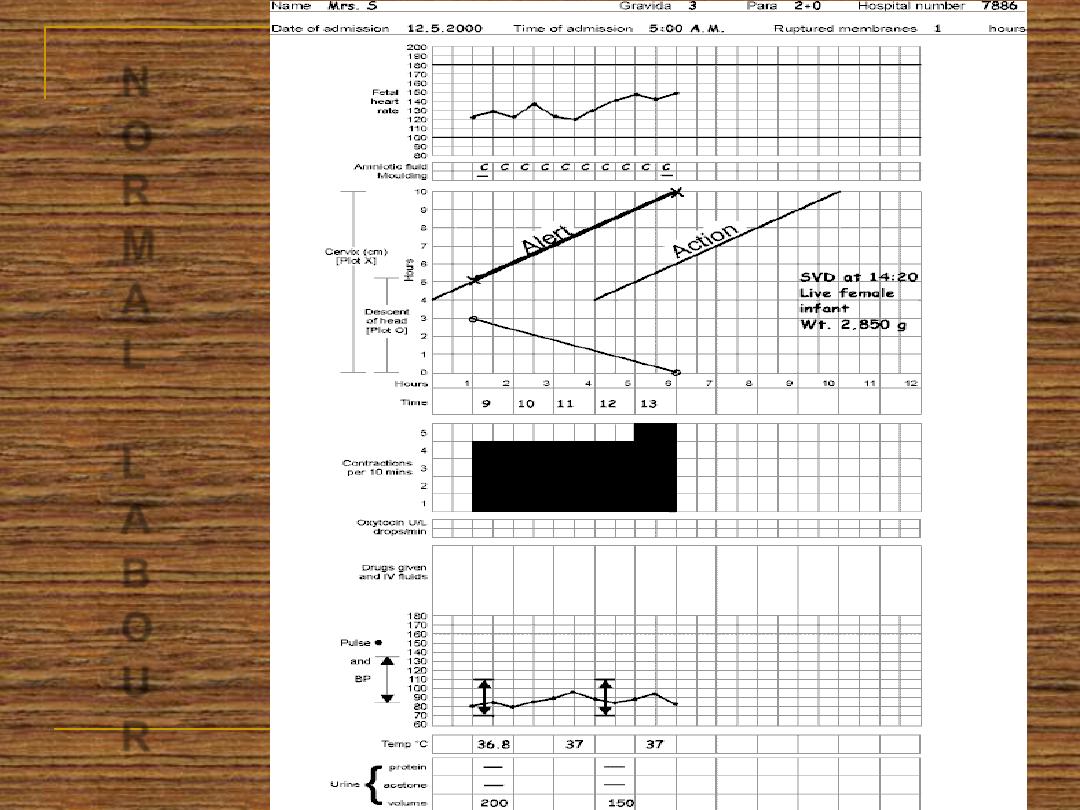
N
O
R
M
A
L
L
A
B
O
U
R