Disorders of the lymphatic system
ByDr.Ahmed Abdul-Ameer Daffar
( Cardio-Thoracic &Vascular Surgeon )
Classification
Primary lymphedemaLymphedema congenita
The onset is before the first year of life.
Lymphedema praecox
The onset is between the ages of 1 and 35 years. It is the most common form of primary lymphedema, accounting for more than 80% of the cases.Lymphedema tarda
The onset is after the age of 35 years. Approximately half of these are associated with an inciting event, such as infection and injury.Secondary Lymphedema
Infiltration of regional nodes by tumor
Surgical excision of regional nodes in treatment for malignancy
Fibrosis after infectious or inflammatory processes or radiation
Infestation by filaria (the most common being Wuchereria bancrofti) is the most frequent cause of secondary lymphedema in tropical countries.
Tuberculous lymphangitis.
Clinical features:-
Patients commonly complain of heaviness and fatigue in the affected extremity.
The limb size increases throughout the day and decreases over the course of the night when the patient is in bed.
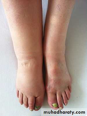
In the lower extremity, the swelling involves the dorsum of the foot, and the toes have a squared-off appearance.
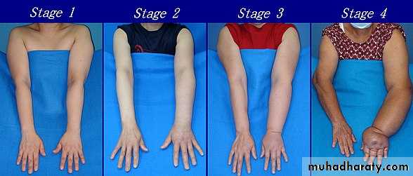
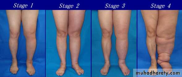
Initially, this swelling pits easily with pressure. This early edema is often responsive to elevation and may decrease or disappear entirely with elevation of the limb.
Later, the edema becomes nonpitting & limb elevation and compression with elastic garments then are much less successful at reducing the extremity volume.
The skin becomes thickened, hypertrophic, and hyperkeratotic. Recurrent spontaneous attacks of cellulitis & lymphangitis are common.
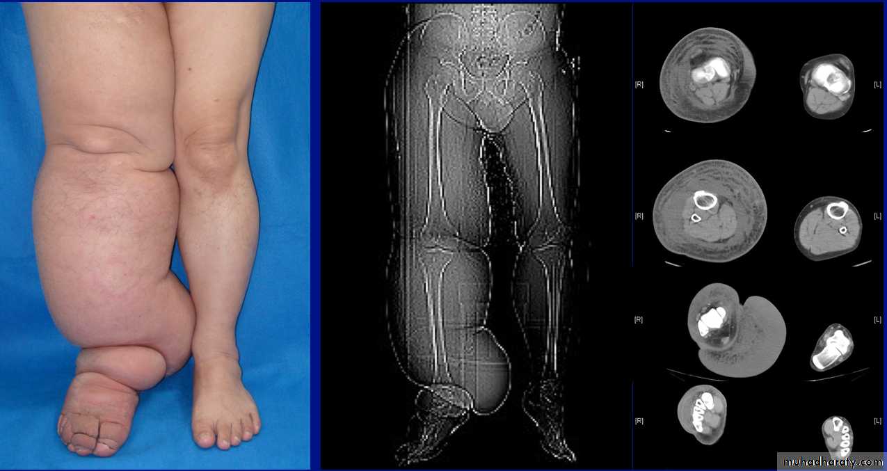
Lymphoedema of right leg
Investigations
Duplex UltrasoundWhen a patient is evaluated for edema, it is often difficult to distinguish the early stages of lymphedema from venous insufficiency. Duplex ultrasound of the venous system can determine if there is concomitant venous thrombosis or venous reflux.
Lymphoscintigraphy
It requires only subcutaneous injection of technetium labeled colloid and serial gamma camera imaging of the extremity.Lymphangiography
Contrast medium is slowly infused, and the transit of dye is observed by serial roentgenograms.MRI
TREATMENT
Nonoperative TherapyMeticulous skin hygiene
Bed rest and Leg elevation
External compression with elastic support garments.
Lymphatic massage:- The strategy is to massage the excess interstitial fluid from areas with deficient lymphatic drainage to areas with more normal lymphatic function.
Avoidance of local injury as well as walking barefoot.
Care should be taken to keep the interdigital spaces dry, and frequent use of an antifungal powder may be helpful.
Aggressive antibiotic therapy is recommended at the earliest signs or symptoms of cellulitis & lymphangitis.
Use of benzopyrones to reduce the formation of high-protein edema.
Treatment of the underlying disorder in the secondary lymphedemas, such as using diethylcarbamazine for filariasis or appropriate antibiotics for tuberculosis or lymphogranuloma venereum.
Surgical Therapy
It's rarely indicated.