1
بسم هللا الرحمن الرحيم
Lecture -3- Medical Physiology (GIT system)
2
nd
stage Dr. Noor Jawad
Ingestion of food
Objectives of our lecture:
1. What is the mechanisms of food ingestion?
2. Stages of swallowing?
The amount of food that a person ingests is determined principally
by an intrinsic desire for food called hunger. The type of food that a
person preferentially seeks is determined by appetite. These
mechanisms are extremely important for maintaining an adequate
nutritional supply for the body and in relation to nutrition of the
body.
Mechanisms of food ingestion:
1. Mastication ( chewing)
The teeth are admirably designed for chewing. The anterior teeth
(incisors) provide a strong cutting action, and the posterior teeth
(molars) provide a grinding action. All the jaw muscles working
together can close the teeth with a force as great as 55 pounds on the
incisors and 200 pounds on the molars.
2
Most of the muscles of chewing are innervated by the motor branch
of the fifth cranial nerve, and the chewing process is controlled by
nuclei in the brain stem. Stimulation of specific reticular areas in the
brain stem taste centers will cause rhythmical chewing movements.
In addition, stimulation of areas in the hypothalamus, amygdala, and
even the cerebral cortex near the sensory areas for taste and smell
can cause chewing.
Much of the chewing process is caused by a chewing reflex. The
presence of a bolus of food in the mouth at first initiates reflex
inhibition of the muscles of mastication, which allows the lower jaw
to drop. This action automatically raises the jaw to cause closure of
the teeth, but it also compresses the bolus again against the linings
of the mouth, which inhibits the jaw muscles once again, allowing

3
the jaw to drop and rebound another time; this process is repeated
again and again.
Chewing is important for digestion of all foods, but it is especially
important for most fruits and raw vegetables because they have
indigestible cellulose membranes around their nutrient portions that
must be broken before the food can be digested. Furthermore,
chewing aids the digestion of food for another simple reason:
Digestive enzymes act only on the surfaces of food particles;
therefore, the rate of digestion is dependent on the total surface area
exposed to the digestive secretions.
2. Swallowing (Deglutition)
Swallowing is a complicated mechanism, principally because the
pharynx subserves respiration and swallowing. The pharynx is
converted for only a few seconds at a time into a tract for propulsion
of food. It is especially important that respiration not be
compromised because of swallowing.
In general, swallowing can be divided into (1) a voluntary stage,
which initiates the swallowing process; (2) a pharyngeal stage,
which is involuntary and constitutes passage of food through the
pharynx into the esophagus; and (3) an esophageal stage, another
involuntary phase that transports food from the pharynx to the
stomach.
4
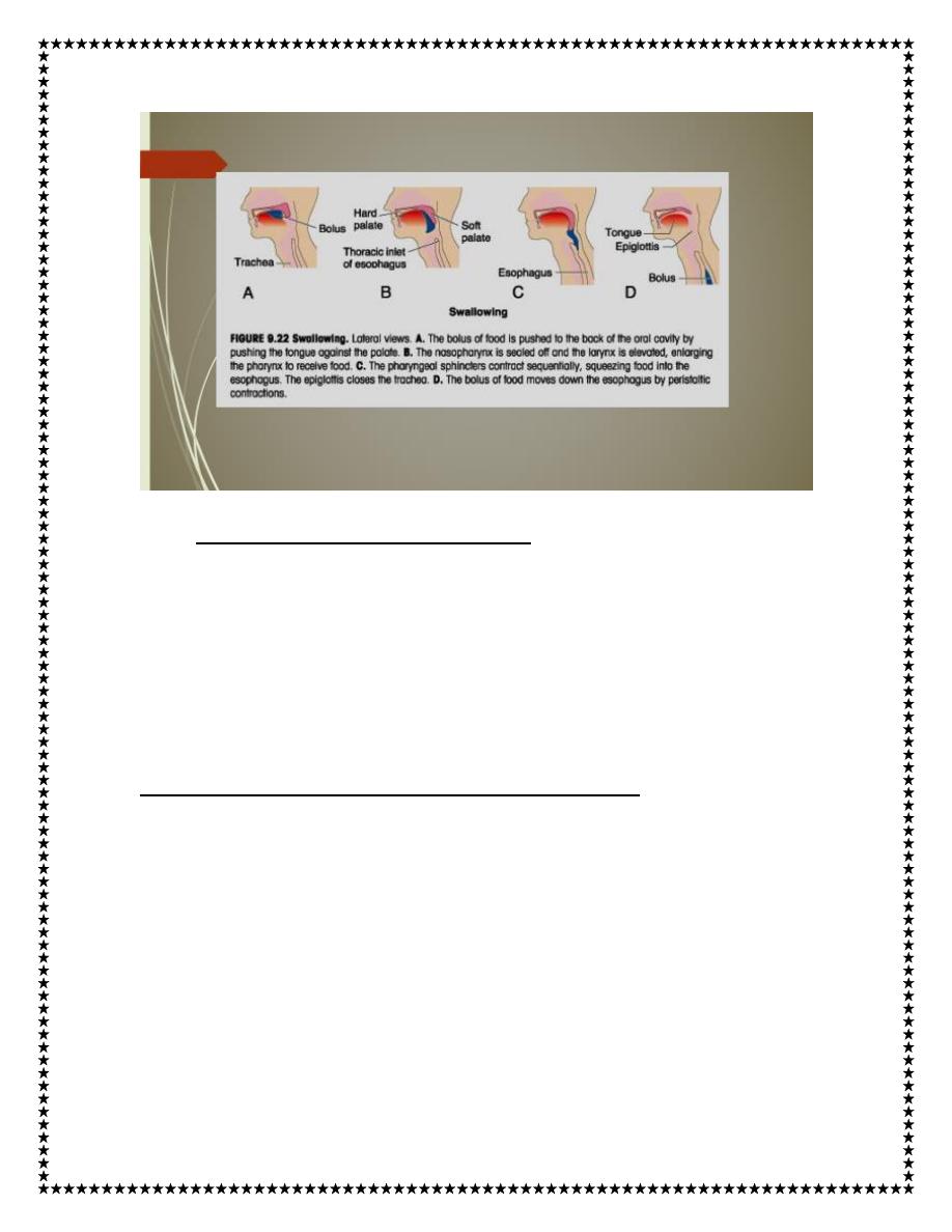
1. Voluntary Stage of Swallowing.
When the food is ready for swallowing, it is “voluntarily” squeezed
or rolled posteriorly into the pharynx by pressure of the tongue
upward and backward against the palate, as shown in Figure .
2. Involuntary Pharyngeal Stage of Swallowing
As the bolus of food enters the posterior mouth and pharynx, it
stimulates epithelial swallowing receptor areas all around the
opening of the pharynx, especially on the tonsillar pillars, and
impulses from these areas pass to the brain stem to initiate a series
of automatic pharyngeal muscle contractions as follows:
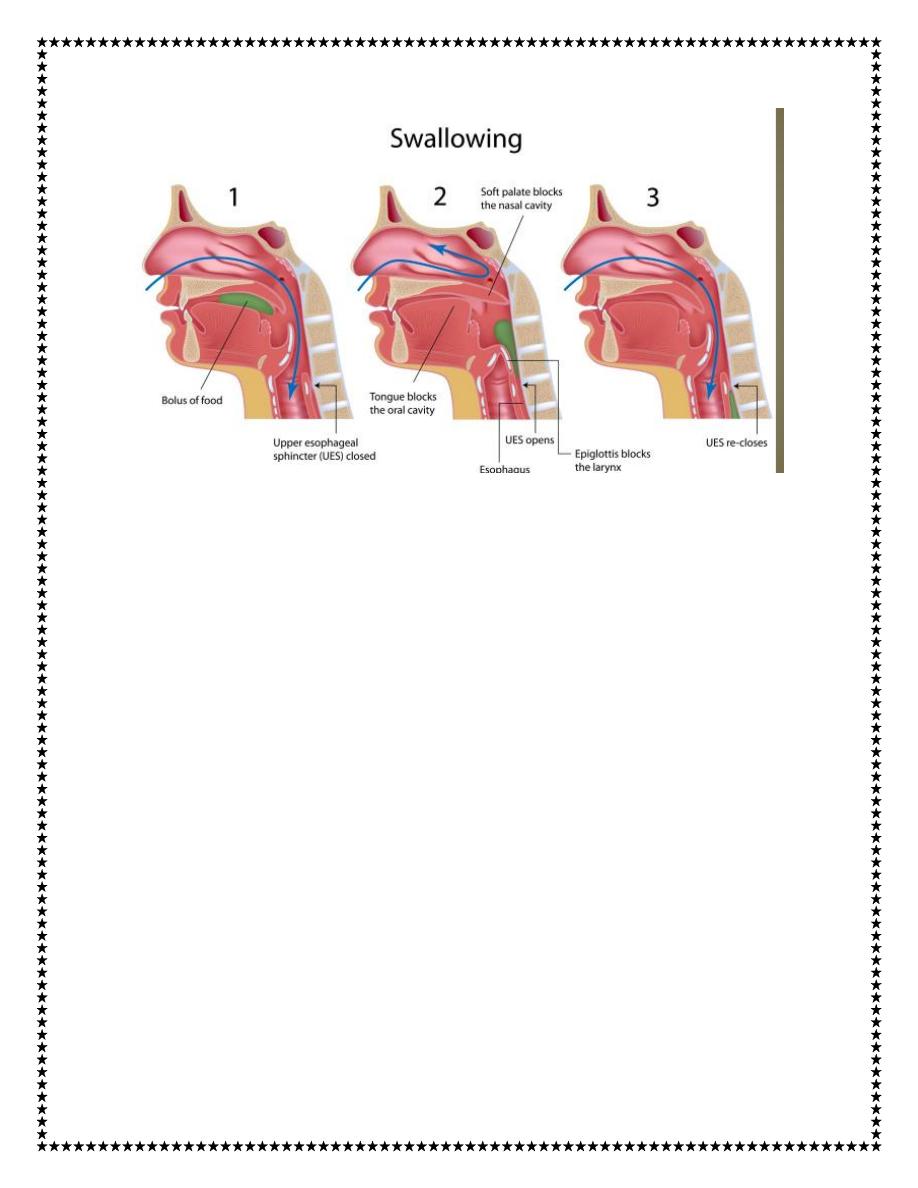
5
1. The soft palate is pulled upward to close the posterior nares to
prevent reflux of food into the nasal cavities.
2. The palatopharyngeal folds on each side of the pharynx are pulled
medially to approximate each other. In this way, these folds form a
sagittal slit through which the food must pass into the posterior
pharynx. This slit performs a selective action, allowing food that has
been masticated sufficiently to pass with ease. Because this stage of
swallowing lasts less than 1 second, any large object is usually
impeded too much to pass into the esophagus.
3. The vocal cords of the larynx are strongly approximated, and the
larynx is pulled upward and anteriorly by the neck muscles. These
actions, combined with the presence of ligaments that prevent
upward movement of the epiglottis, cause the epiglottis to swing
6
backward over the opening of the larynx. All these effects acting
together prevent passage of food into the nose and trachea.
4. The upward movement of the larynx also lifts the glottis out of
the main stream of food flow, so the food mainly passes on each side
of the epiglottis rather than over its surface; this action adds still
another protection against entry of food into the trachea.
5. Once the larynx is raised and the pharyngoesophageal sphincter
becomes relaxed, the entire muscular wall of the pharynx contracts,
beginning in the superior part of the pharynx, then spreading
downward over the middle and inferior pharyngeal areas, which
propels the food by peristalsis into the esophagus.
To summarize the mechanics of the pharyngeal stage of swallowing:
The trachea is closed, the esophagus is opened, and a fast peristaltic
wave initiated by the nervous system of the pharynx forces the bolus
of food into the upper esophagus, with the entire process occurring
in less than 2 seconds.
3. The Esophageal Stage of Swallowing Involves Two Types
of Peristalsis.
The esophagus functions primarily to conduct food rapidly from the
pharynx to the stomach, and its movements are organized
specifically for this function. The esophagus normally exhibits two

7
types of peristaltic movements: primary peristalsis and secondary
peristalsis.
Primary peristalsis is simply continuation of the peristaltic wave
that begins in the pharynx and spreads into the esophagus during the
pharyngeal stage of swallowing. This wave passes all the way from
the pharynx to the stomach in about 8 to 10 seconds.
Food swallowed by a person who is in the upright position is usually
transmitted to the lower end of the esophagus even more rapidly than
the peristaltic wave itself, in about 5 to 8 seconds, because of the
additional effect of gravity pulling the food downward.
If the primary peristaltic wave fails to move all the food that has
entered the esophagus into the stomach, secondary peristaltic waves
result from distention of the esophagus itself by the retained food;
these waves continue until all the food has emptied into the stomach.
The secondary peristaltic waves are initiated partly by intrinsic
neural circuits in the myenteric nervous system and partly by
reflexes that begin in the pharynx and are then transmitted upward
through vagal afferent fibers to the medulla and back again to the
esophagus through glossopharyngeal and vagal efferent nerve
fibers.
The musculature of the pharyngeal wall and upper third of the
esophagus is striated muscle. Therefore, the peristaltic waves in
8
these regions are controlled by skeletal nerve impulses from the
glossopharyngeal and vagus nerves. In the lower two thirds of the
esophagus, the musculature is smooth muscle, but this portion of the
esophagus is also strongly controlled by the vagus nerves that act
through connections with the esophageal myenteric nervous system.
When the vagus nerves to the esophagus are cut, the myenteric nerve
plexus of the esophagus becomes excitable enough after several
days to cause strong secondary peristaltic waves even without
support from the vagal reflexes. Therefore, even after paralysis of
the brain stem swallowing reflex, food fed by tube or in some other
way into the esophagus still passes readily into the stomach.
Function of the Lower Esophageal Sphincter (Gastroesophageal
Sphincter).
At the lower end of the esophagus, extending upward about 3
centimeters above its juncture with the stomach, the esophageal
circular muscle functions as a broad lower esophageal sphincter,
also called the gastroesophageal sphincter. This sphincter
normally remains tonically constricted with an intraluminal pressure
at this point in the esophagus of about 30 mm Hg, in contrast to the
midportion of the esophagus, which normally remains relaxed.
When a peristaltic swallowing wave passes down the esophagus,
“receptive relaxation” of the lower esophageal sphincter occurs

9
ahead of the peristaltic wave, which allows easy propulsion of the
swallowed food into the stomach. Rarely, the sphincter does not
relax satisfactorily, resulting in a condition called achalasia.
The stomach secretions are highly acidic and contain many
proteolytic enzymes, and esophageal mucosa, except in the lower
one eighth of the esophagus, is not capable of resisting the digestive
action of gastric secretions for long. Fortunately, the tonic
constriction of the lower esophageal sphincter helps prevent
significant reflux of stomach contents into the esophagus except
under abnormal conditions
Additional Prevention of Esophageal Reflux by Valvelike
Closure of the Distal End of the Esophagus.
Another factor that helps prevent reflux is a valvelike mechanism of
a short portion of the esophagus that extends slightly into the
stomach. Increased intraabdominal pressure caves the esophagus
inward at this point. Thus, this valvelike closure of the lower
esophagus helps to prevent high intra-abdominal pressure from
forcing stomach contents backward into the esophagus. Otherwise,
every time we walked, coughed, or breathed hard, we might expel
stomach acid into the esophagus.
Thank you
10
References : Guyton and Hall textbook of medical physiology,
thirteen edition.