.أ
م
.
د
.
احمد
عبداالمير دفار
(
اختصاصي جراحة الصدر و القلب و االوعية الدموية
)
1
TETRALOGY OF FALLOT
Objective :
To show the definition, criteria and management of TOF (
Tetralogy
of Fallot )
It’s the most common cyanotic congenital heart disease &0
consists of four major
defects:
- Pulmonary stenosis
- Ventricular septal defect
- Aortic overriding
- Right ventricular Hypertrophy
Clinical
…
.
features
- The severity of obstruction of the right ventricular outflow tract determines the
degree of right-to-left shunting, which usually determines the degree of
cyanosis
and
the age of presentation.
- Complications of TOF include
polycythemia
leading to cerebrovascular thrombosis,
hemoptysis
secondary to enlarged bronchial arteries,
paradoxical embolism
( leading
to stroke or end organ failure ),
subacute bacterial endocarditis
,
or brain abscess
.
-
Squatting
is a classical behavioral adaptation of older children with TOF whereby
systemic vascular resistance is increased, producing more pulmonary blood flow.
- Hypercyanotic episodes (“tet spells”)
are characterized by intense cyanosis that may
last minutes to hours, during which time oxygen delivery may be so compromised as
to cause loss of consciousness or impairment of myocardial function. Dehydration,
viral respiratory infection, and the injudicious administration of medications that lead
to peripheral vasodilation all may cause a hypercyanotic episode in a patient who has
been previously stable.
Physical Examination
- Cyanosis
- Ejection systolic murmur heard loudest in the second left intercostal space.
- Digital clubbing.
Investigations
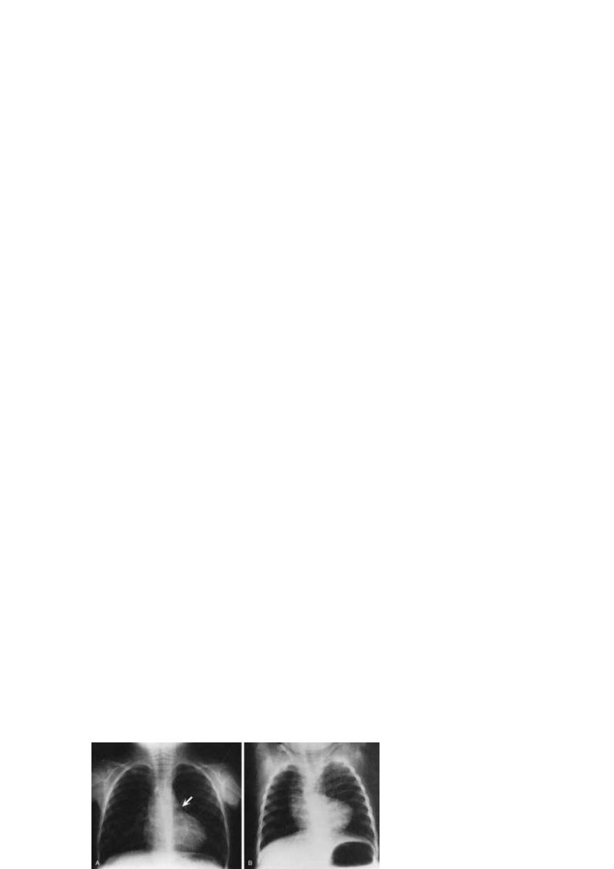
Chest X-Ray
- Boot-shaped heart due to RVH + small pulmonary artery.
- The lung fields usually appear oligemic
- A right aortic arch occurs in approximately 25% of cases.
Chest film of TOF

.أ
م
.
د
.
احمد
عبداالمير دفار
(
اختصاصي جراحة الصدر و القلب و االوعية الدموية
)
2
Electrocardiogram
It demonstrates right ventricular hypertrophy.
Laboratory Studies
Polycythemia
Echocardiography
It's diagnostic
Cardiac Catheterization
For further evaluation
MEDICAL MANAGEMENT
Outpatient Management
The goals of medical management are to allow growth and development of the
child until surgical repair is undertaken while hypercyanotic episodes or
complications arising from the condition are prevented.
The preoperative management includes:
- Maintaining these infants in a well-hydrated state.
- Adequate feeding regimen.
- Every effort should be made to protect these infants from respiratory viruses and
dehydration accompanying diarrheal illnesses
- Β-Blockers such as propranolol (Inderal) due to their negative chronotropic effects
with increased ventricular filling at slower heart rates & relief of RVOT spasm.
-
Diuretics are contraindicated in cyanotic tetralogy.
Management of Hypercyanotic Episodes
Basic treatment principles include:
Administration of intravenous fluids, morphine or other intravenous sedatives,
and oxygen.
Placing the infant in a knee-chest position can elevate systemic vascular
resistance with a resultant increase in pulmonary blood flow.
Intravenous β-blockers such as esmolol and α-agonists such as phenylephrine
may also be used to temporize.
Intubation and positive pressure ventilation may also be used to attempt to
increase oxygenation.

.أ
م
.
د
.
احمد
عبداالمير دفار
(
اختصاصي جراحة الصدر و القلب و االوعية الدموية
)
3
SURGICAL TECHNIQUES
Palliative Procedures
- Blalock-Taussig shunt
In performance of a subclavian-pulmonary anastomosis, the incision is generally
made on the side opposite that on which the aorta descends. Ideally, the
subclavian branch of the innominate artery is used for the anastomosis.
- Modified Blalock-Taussig Shunt
It requires interposition of a segment graft material between the subclavian
artery and the pulmonary artery.
- Central Aortopulmonary Shunt
This includes interposition of a graft material between the ascending aorta and
main (or right) pulmonary artery.
-
Waterston shunt
It’s between the ascending aorta to right pulmonary artery.
- Potts shunt
It’s between the descending aorta to left pulmonary artery .
- RV Outflow Tract Patch.
Is to relieve the pulmonary stenosis but leave the ventricular septal defect
open.
- Balloon Angioplasty of RV Outflow Tract.
Total Correction
The goals of Correction
1-To close the ventricular septal defect
2-To relieve right ventricular outflow obstruction
3-To
repair
any
stenoses
in
the
pulmonary
arteries.

.أ
م
.
د
.
احمد
عبداالمير دفار
(
اختصاصي جراحة الصدر و القلب و االوعية الدموية
)
4