
The ear
BY FIRAS AL-HAMEED
THI-QAR MEDICAL SCHOOL
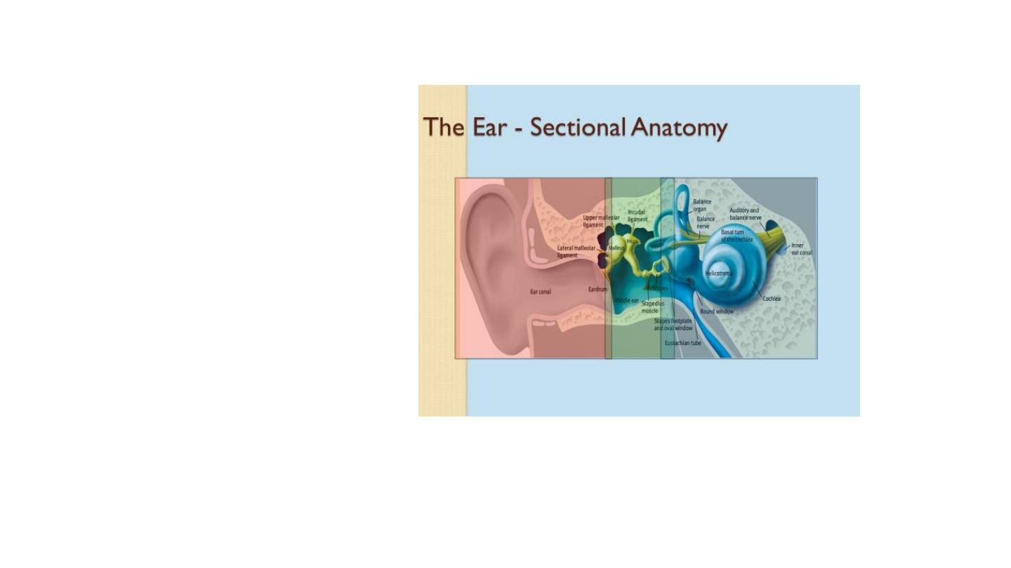
• External, middle and inner
External ear
• Composed of : the auricle (or pinna), and the
external acoustic meatus – which ends at the
tympanic membrane.
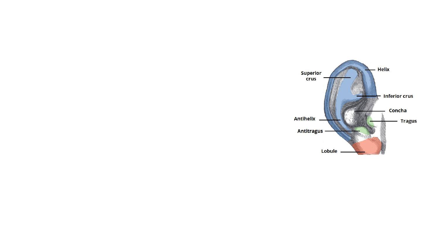
• Auricle
• Capture and direct sound waves towards the external
acoustic meatus.
• Single piece of yellow elastic cartilage covered with skin
closely adherent to the perichondrium on lateral surface
than the medial
• Earlobe: tough areolar and adipose tissue
• Curvatures: helix, antihelix
• Concha
• Tragus and antitragus

External Acoustic Meatus
• The outer one-third of the canal is cartilage and the medial two-thirds
bony
• The two parts meet at an angle; hence the need for traction on the
pinna to straighten the canal at otoscopy.
• 2.5 cm long, anterior wall is longer than posterior
• Wax glands and hair follicles only in the outer third
• The fissures of Santorini (in the floor of cartilaginous portion), are
notorious routes for spread of infection to the parotid and skull base
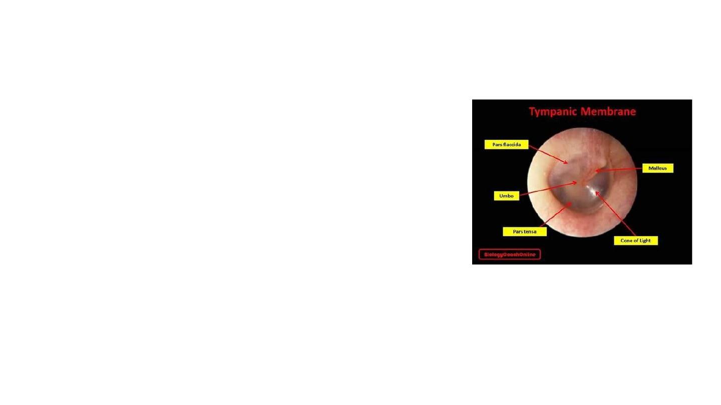
Tympanic membrane
• Diameters
:
1 cm and its area 85 mm2, of which only 55 mm2 is
physiologically effective.
• Colour: pearly grey membrane
• Pars tensa and pars flaccida
• Three layers: keratinizing squamous epithelium, the lamina
propria, shows radiating and circular fibres, the inner layer is
mucous epithelium of the middle ear.
• Superiorly, the middle layer is deficient (but not absent), forming
the pars flaccida
• Tympanic annulus: fibrocartilaginous ring that connects TM to the
surrounding temporal bone.
• Umbo: point of attachment of the malleus.
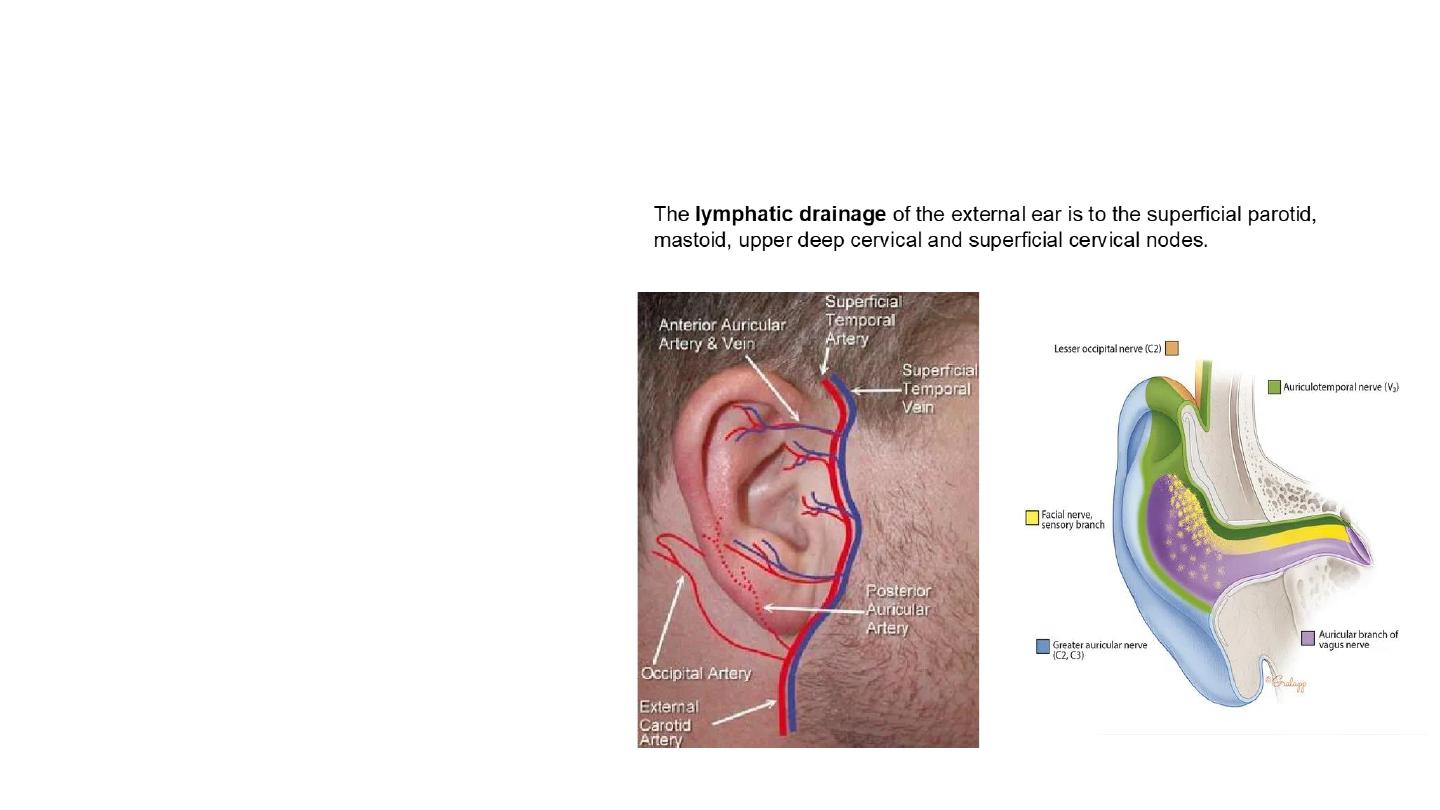
Vasculature
The external ear is supplied by branches of the external carotid artery:
•
Posterior auricular artery
•
Superficial temporal artery
•
Occipital artery
•
Maxillary artery (deep auricular branch)
– supplies the deep
aspect of the external acoustic meatus and tympanic membrane
only.
Venous drainage is via veins following the arteries listed above.
Innervation
The sensory innervation to the skin of the auricle comes from numerous
nerves:
•
Greater auricular nerve (branch of the cervical plexus)
–
innervates the skin of the auricle
•
Lesser occipital nerve (branch of the cervical plexus)
–
innervates the skin of the auricle
•
Auriculotemporal nerve (branch of the mandibular nerve)
–
innervates the skin of the auricle and external auditory meatus.
•
Branches of the facial and vagus nerves
– innervates the
deeper aspect of the auricle and external auditory meatus
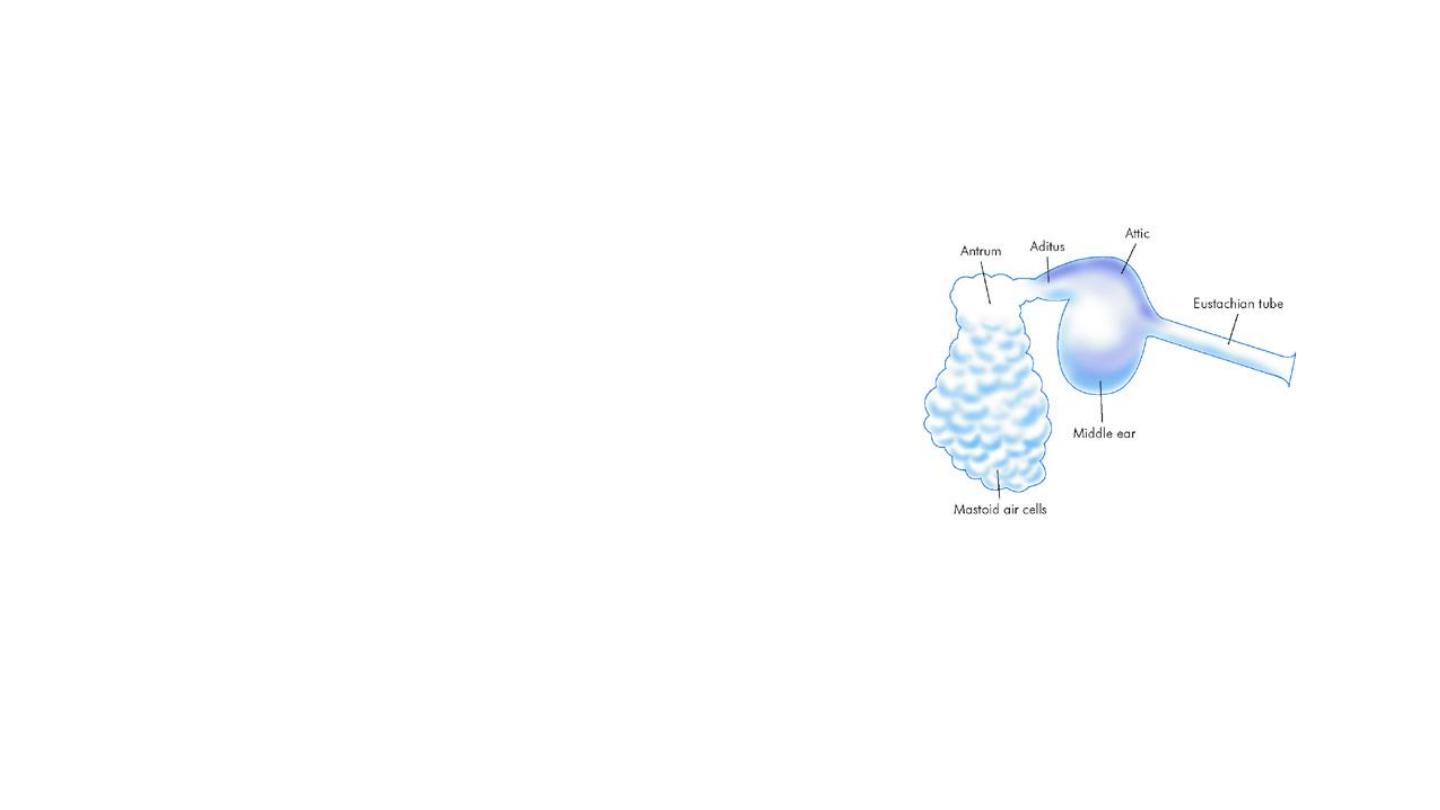
MIDDLE EAR CLEFT
• This is a complex, air-filled structure running posteriorly, from
the bony eustachian tube through the tympanic cavity and the
aditus, mastoid antrum (itself a single large cavity), and then
connected to a variable number of mastoid air cells.
• Lining mucosa: modified respiratory epithelium. Squamous, but
non-keratinizing lining.
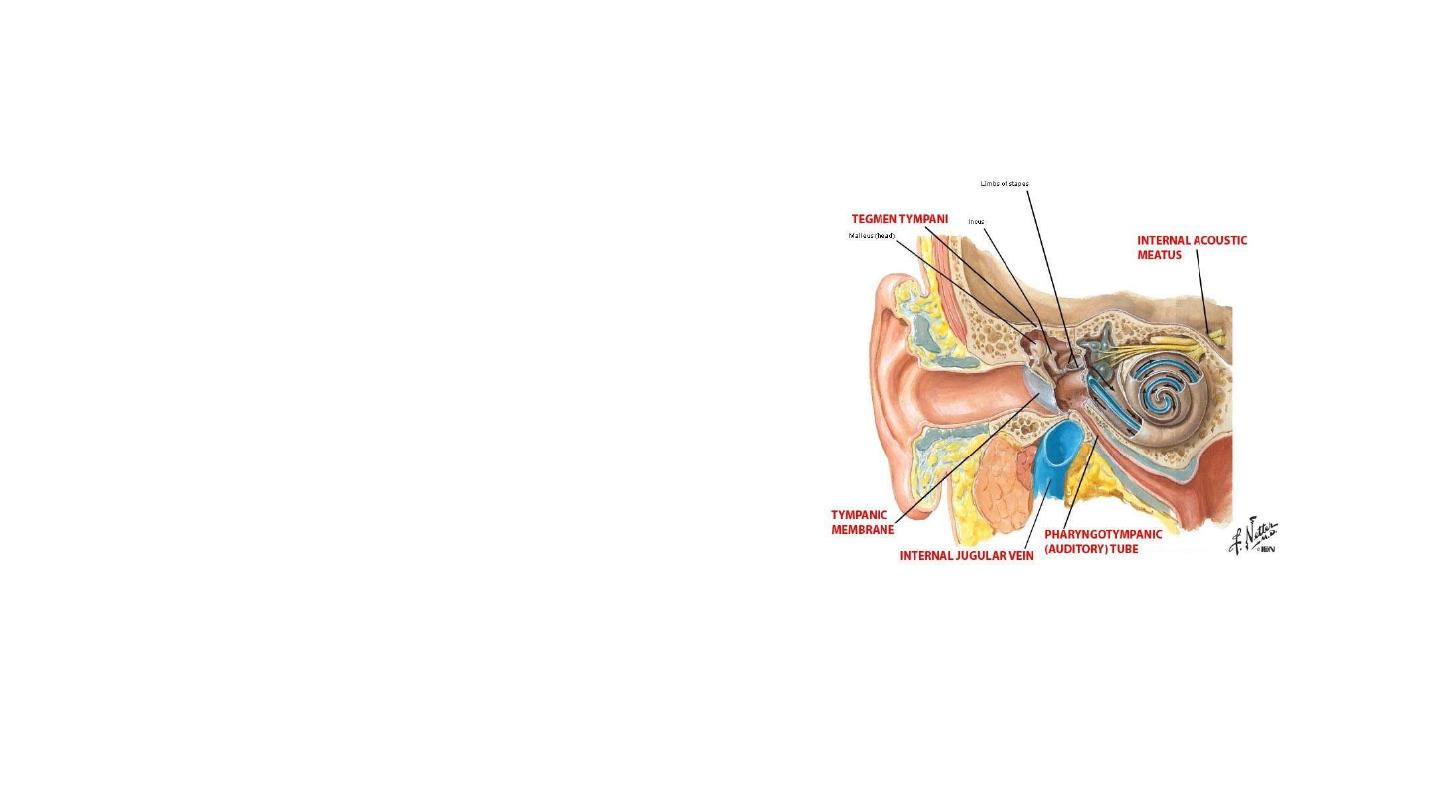
Borders of the middle ear
Roof
– the space superior to the level of the
tympanic membrane is called epitympanic recess or
attic and the roof of this space is formed by a thin
bone from the petrous part of the temporal bone (
tegmen tympani). It separates the middle ear from
the temporal lobe in the middle cranial fossa.
Floor
(jugular wall): thin layer of bone separates the
middle ear from the internal jugular vein
Lateral wall
– made up of the tympanic membrane
and the lateral wall of the epitympanic recess
(scutum)
Medial wall
– formed by the lateral wall of
the inner ear. It contains:
•
Promontory: a prominent bulge houses basal turn of
the cochlea
•
facial nerve
•
oval and round windows. The first accommodates the
stapes footplate.
Anterior wall
– a thin bony plate with two
openings; for the auditory tube and the
tensor tympani muscle. It separates the
middle ear from the internal carotid artery.
• Posterior wall
(mastoid wall)
– it consists of a
bony partition between the tympanic cavity and
the mastoid air cells
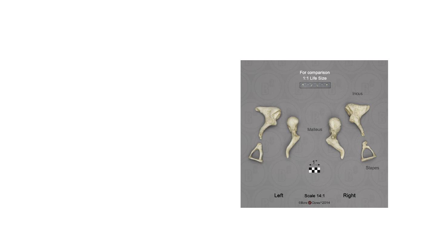
Ossicles
• The malleus
•
Largest
•
Head, neck, handle, lateral and anterior process
• The incus
•
Body, short and long process.
•
The body articulates with the malleus, the short limb attaches to
the posterior wall , and the long limb joins the stapes.
•
The incus is less stable. Skull trauma causing conductive loss
usually indicates incus dislocation
• The stapes
•
smallest bone in the human body
•
It is stirrup-shaped, with a head, two limbs, and a base.
•
The head articulates with the incus, and the base joins the oval
window.
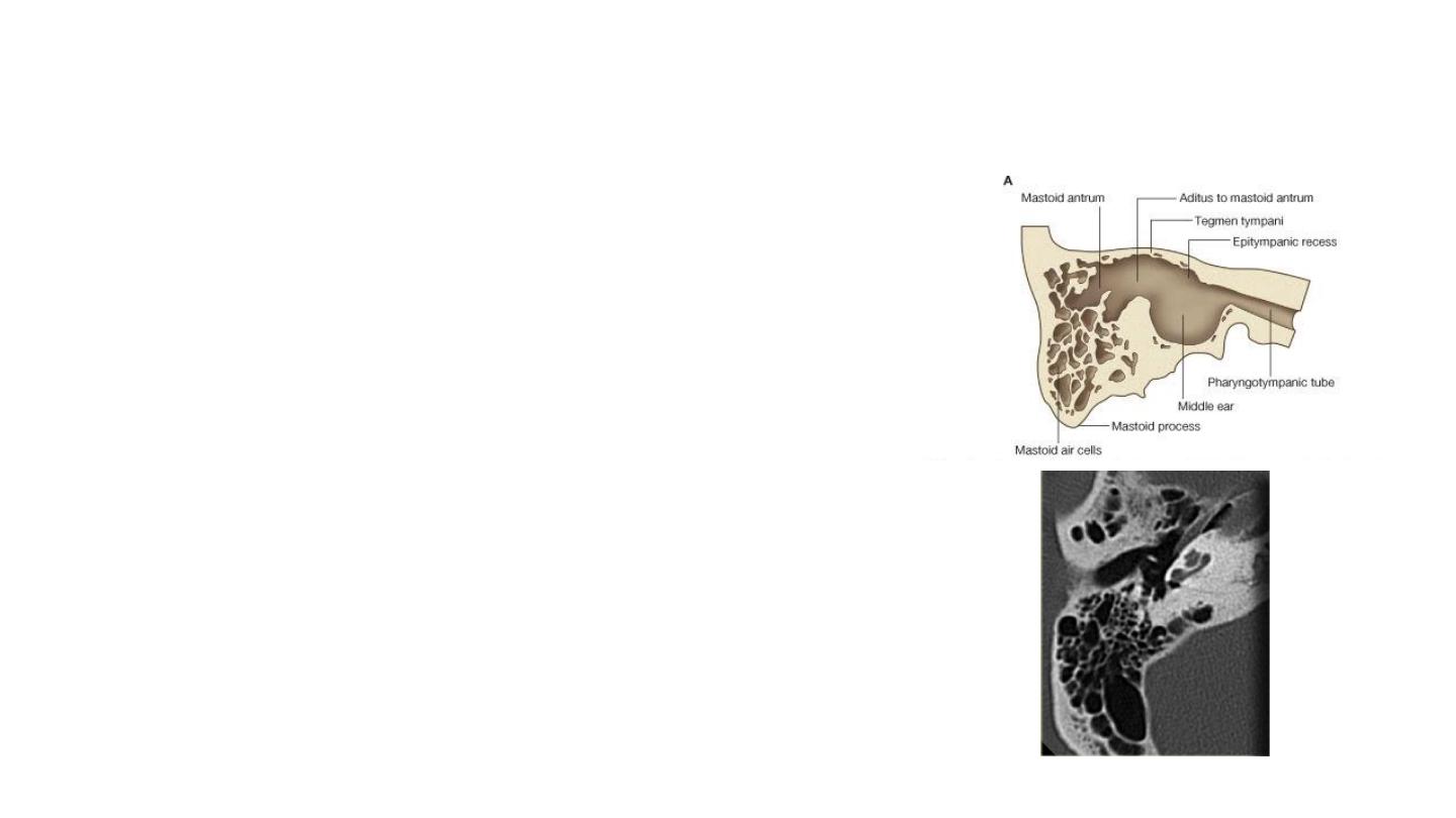
The mastoid air cells
• Located posterior to epitympanic recess.
• They are a collection of air-filled spaces in the mastoid process
of the bone.
• The air cells are contained within a cavity called the mastoid
antrum.
• The mastoid antrum communicates with the middle ear via
the aditus.
• The mastoid air cells act as a ‘buffer system‘ of air – releasing
air into the tympanic cavity when the pressure is too low.
Muscles
• Serve a protective function in the middle ear
• They contract in response to loud noise, inhibiting the
vibrations of the malleus, incus and stapes, and reducing
the transmission of sound to the inner. This action is known
as the acoustic reflex.
• Tensor tympani muscle originates from the auditory tube
and attaches to the handle of malleus. It is innervated by
the tensor tympani nerve, a branch of
the mandibular nerve.
• The stapedius muscle attaches to the stapes, and is
innervated by the facial nerve.

Eustachian tube
• The auditory tube (eustachian tube) connects the middle ear
to the nasopharynx.
• It acts to equalise the pressure of the middle ear to that of
the external auditory meatus.
• Two parts: cartilaginous (proximal two-thirds) and bony
• The tube is shorter and straighter in children, therefore
middle ear infections tend to be more common in children
than adults
The inner ear
• Houses the vestibulocochlear organs.
• It has two main functions:
•
To convert mechanical signals from the middle ear
into electrical signals, which can transfer information to the
auditory pathway in the brain.
•
To maintain balance by detecting position and motion.
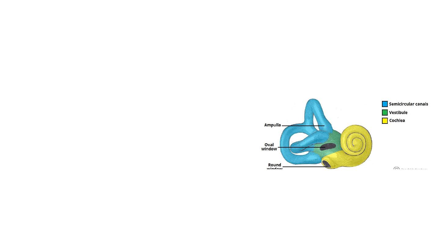
The inner ear has two main components
–
Bony labyrinth: cochlea, vestibule and three semi-circular canals
Membranous labyrinth: it is a continuous system of ducts filled
with endolymph. It lies within the bony labyrinth, surrounded by
perilymph. It is composed of the cochlear duct, three semi-circular
ducts, saccule and the utricle.

Cochlea
• Spiral structure resemble a snail shell.
• The ascending spiral takes its two-and-one-half turns
around the central bony modiolus.
• Spiral lamina
• Contain three chambers:
• Scala vestibuli (upper chamber)
• Scala media (cochlear duct)
• Scala tympani (lower chamber)
• SV & ST meet at the apex of the spiral, the helicotrema
• Both SV& ST are filled with the same fluid, known
as perilymph (essentially the same in composition as the
extracellular fluid bathing most of the nervous system),
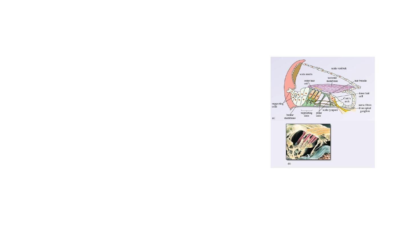
Cochlea
• The scala media is filled with endolymph (with very high
potassium and low sodium concentrations).
• Organ of Corti
• Receptor organ for hearing
• Located in the scala media
• It resides on the basilar membrane, a stiff membrane
separating the scala tympani and scala media
• Contains hair cells covered by tectorial membrane
• OHC:12,000-20,000 cells, IHC: 3,500 cells
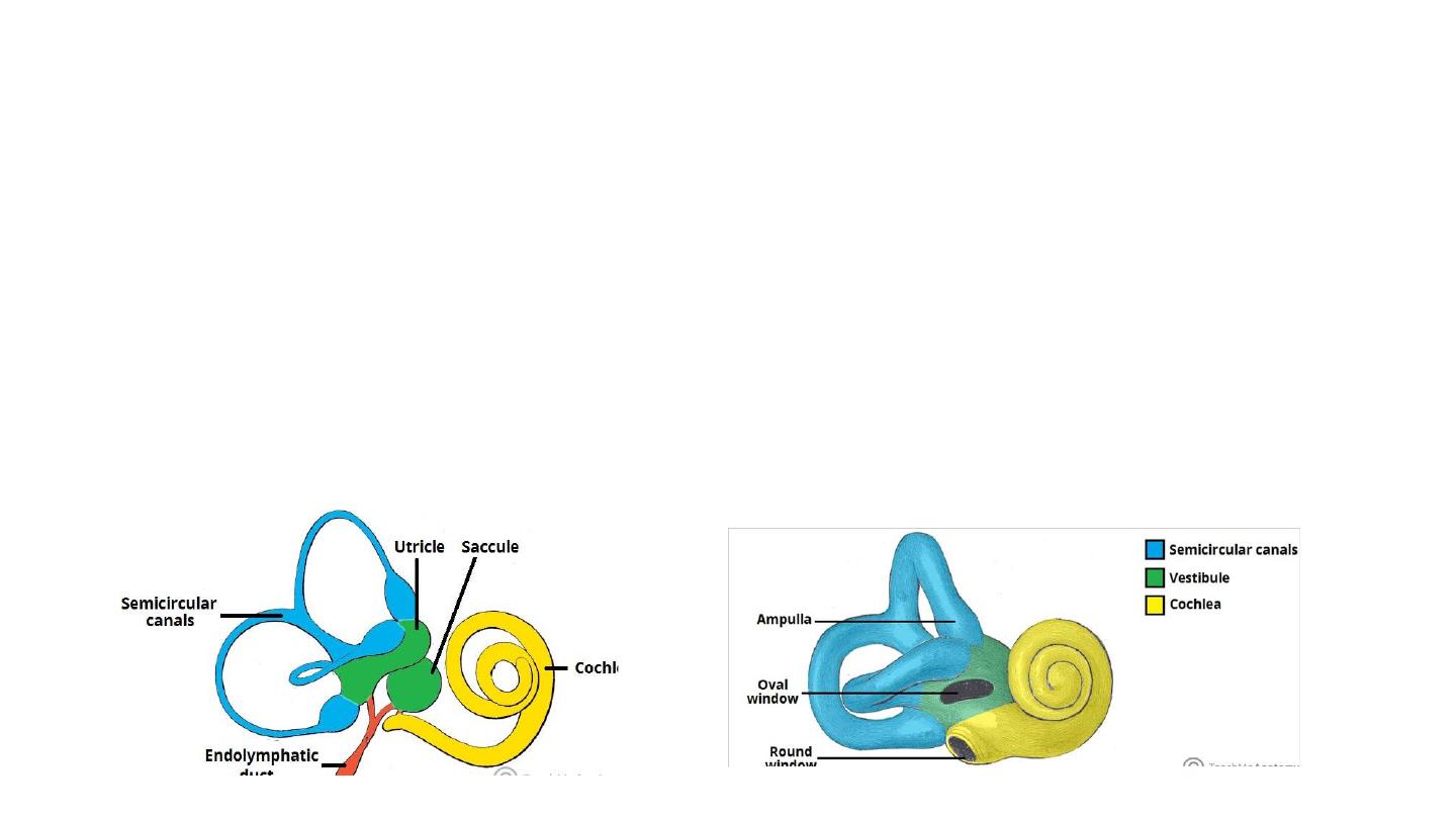
Vestibule
• The vestibule is the central part of the bony labyrinth. It is separated from the middle ear by the oval
window, and communicates anteriorly with the cochlea and posteriorly with the semi-circular
canals.
• Two parts of the membranous labyrinth; the saccule and utricle, are located within the vestibule.
• They are organs of balance which detect movement or acceleration of the head in the vertical and
horizontal planes, respectively.
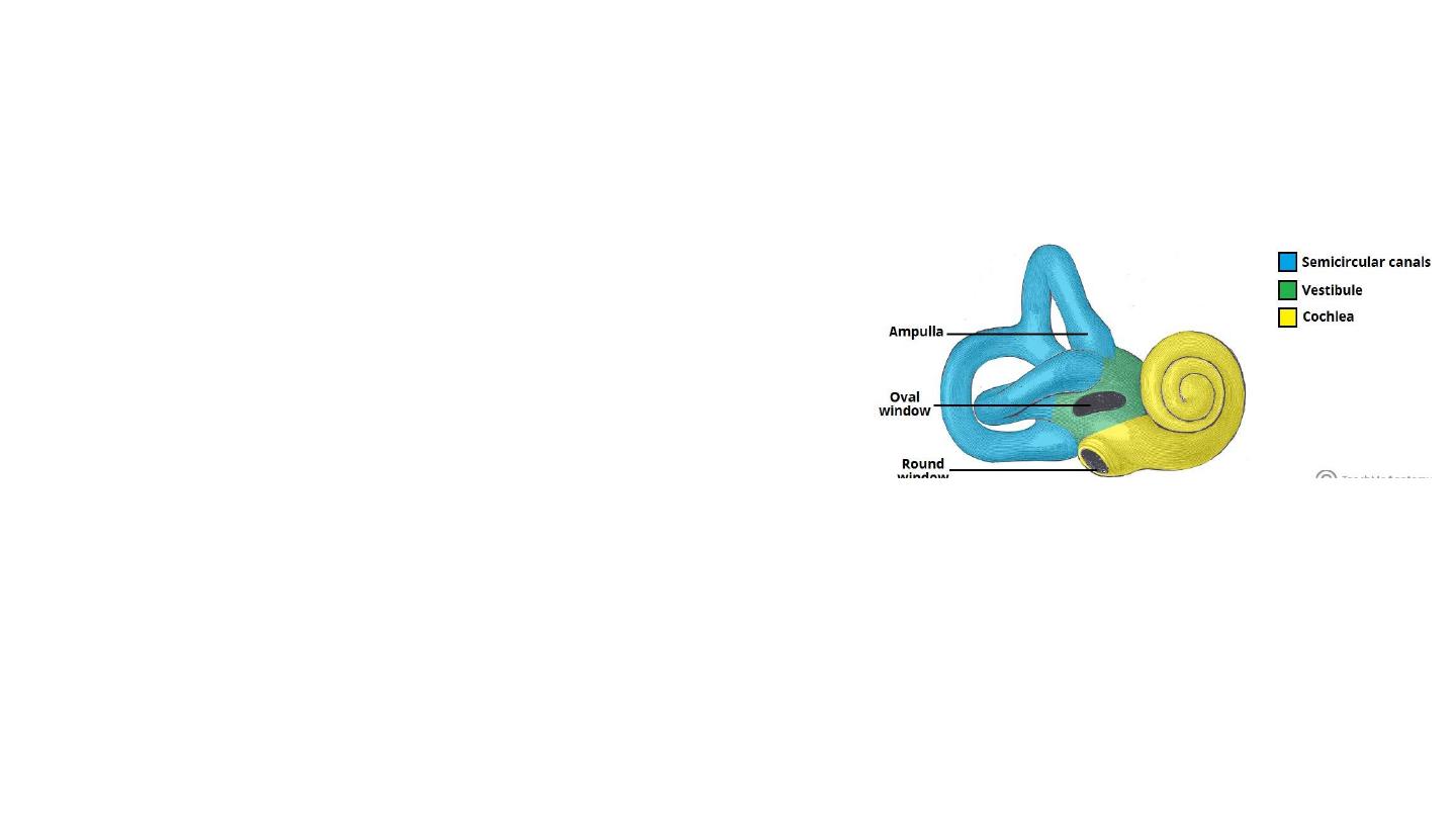
Semi-circular Canals
• There are three semi-circular canals: anterior, lateral and
posterior. They contain the semi-circular ducts, which
are responsible for balance (along with the utricle and
saccule).
• The canals are situated superoposterior to the vestibule,
at right angles to each other. They have a swelling at one
end, known as the ampulla.

Innervation
• The inner ear is innervated by the vestibulocochlear nerve (CN VIII).
• It enters the inner ear via the internal acoustic meatus, where it divides into the vestibular
nerve (responsible for balance) and the cochlear nerve (responsible for hearing):
• The facial nerve CN VII, also passes through the inner ear, but does not innervate any of the
structures present.