Dr.Mushtaq Talib Hussein
F.I.B.M.S(ortho.) C.A.B.O(ortho.)
SUPRACONDYLAR FRACTURES
These are among the commonest fractures in children. The distal fragment may be
displaced either posteriorly (95%) or rare anteriorly.
Mechanism of injury
Usually due to a fall on the outstretched hand, the distal fragment is pushed backwards
and (because the forearm is usually in pronation) twisted inwards.
Anterior displacement is rare; it is thought to be due to direct violence (e.g. a fall on the
point of the elbow) with the joint in flexion.
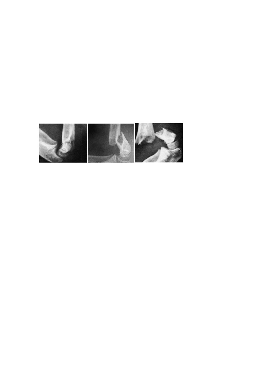
Classification
Type I is an undisplaced fracture.
Type II is an angulated fracture with the posterior cortex still in continuity.
Type III is a completely displaced fracture (although the posterior periosteum is usually
still preserved, which will assist surgical reduction).
Type I Type II Type III
Clinical features
Following a fall, the child is in pain and the elbow is swollen; with a posteriorly
displaced fracture the S-deformity of the elbow is usually obvious and the bony
landmarks are abnormal. It is essential to feel the pulse and check the capillary return;
passive extension of the flexor muscles should be pain-free. The wrist and the hand
should be examined for evidence of nerve injury.
X-ray
Ap , lat. Views, fat pad sign(a triangular lucency in front of the distal humerus, due to the
fat pad being pushed forwards by a haematoma.) in case of undisplaced type.
On a normal lateral x-ray, a line drawn along the anterior cortex of the humerus should
cross the middle of the capitulum. If the line is anterior to the capitulum then a Type II
fracture is suspected.
Treatment
If there is even a suspicion of a fracture, the elbow is gently splinted in 30 degrees of
flexion to prevent movement and possible neurovascular injury during the x-ray
examination.
TYPE I: UNDISPLACED FRACTURE
The elbow is immobilized at 90 degrees and neutral rotation in a light-weight splint or
cast and the arm is supported by a sling. It is essential to obtain an x-ray5–7 days later to
check that there has been no displacement. The splint is retained for 3 weeks and
supervised movement is then allowed.
TYPE II: POSTERIORLY ANGULATED FRACTURE
The same regime above, if MUA failed to reduce the fracture accurately, the surgery is
the best treatment in which we use closed reduction fluoroscopy with percutaneous k-
wire fixation.
TYPES III: ANGULATED AND MALROTATED
These are usually associated with severe swelling, are difficult to reduce and are often
unstable; moreover, there is a considerable risk of neurovascular injury or circulatory
compromise due to swelling. The fracture should be reduced under general anaesthesia
as soon as possible, by the method described above, and then held with percutaneous
crossed K-wires; this obviates the necessity to hold the elbow acutely flexed.
OPEN REDUCTION
This is sometimes necessary for (1) a fracture which simply cannot be reduced closed;
(2) an open fracture; or (3) a fracture associated with vascular damage.
Complications
EARLY
Vascular injury The great danger of supracondylar fracture is injury to the brachial
artery, More commonly the injury is complicated by forearm oedema and a mounting
compartment syndrome which leads to necrosis of the muscle and nerves without
causing peripheral gangrene.
Nerve injury The radial nerve, median nerve(particularly the anterior interosseous
branch) or the ulnar nerve may be injured.
LATE
Malunion Malunion is common. However, backward or sideways shifts are gradually
smoothed out by modelling during growth and they seldom give rise to visible deformity
of the elbow.
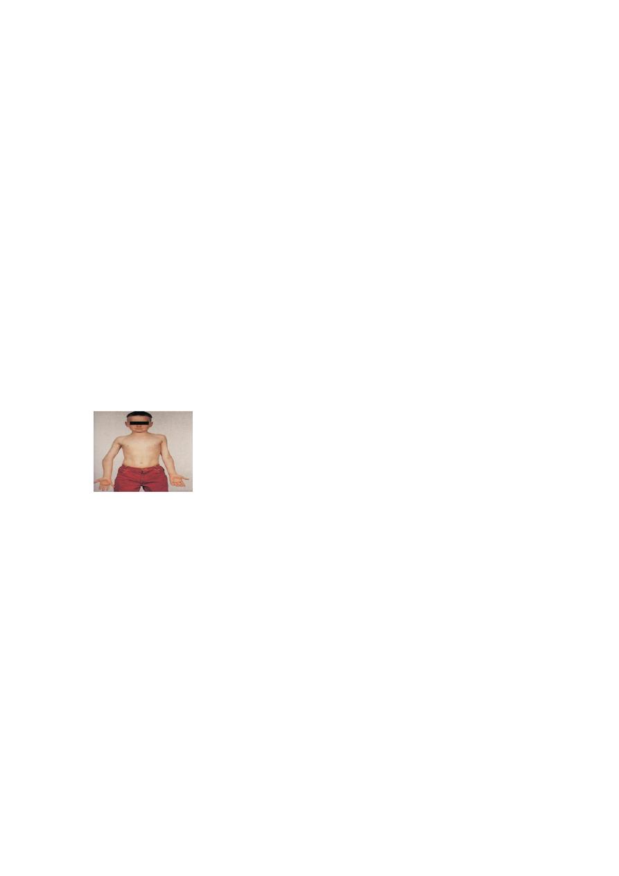
Uncorrected sideways tilt (angulation) and rotation are much more important and may
lead to varus(gunstock deformity) (or rarely valgus) deformity of the elbow; this is
permanent and will not improve with growth
Elbow stiffness and myositis ossifficans Stiffness is an ever-present risk with elbow
injuries. Extension in particular may take months to return.
SUBLUXATION OF THE RADIAL HEAD(‘PULLED ELBOW’)
It is sometimes called subluxation of the radial head; more accurately, it is a subluxation
of the orbicular ligament which slips up over the head of the radius into the
radiocapitellar joint.
A child aged 2 or 3 years is brought with a painful, dangling arm: there is usually a
history of the child being jerked by the arm and crying out in pain. The forearm is held in
pronation and extension, and any attempt to supinate it is resisted. There are no x-ray
changes. A dramatic cure is achieved by forcefully supinating and then flexing the elbow;
the ligament slips back with a snap.
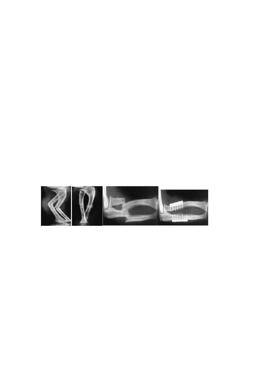
FRACTURES OF THE RADIUS AND ULNA
Mechanism of injury and pathology
Fractures of the shafts of both forearm bones occur quite commonly. A twisting force
(usually a fall on the hand) produces a spiral fracture with the bones broken at different
levels. An angulating force causes a transverse fracture of both bones at the same level.
A direct blow causes a transverse fracture of just one bone, usually the ulna.
Clinical features
The fracture is usually quite obvious, but the pulse must be felt and the hand examined
for circulatory or neural deficit. Repeated examination is necessary in order to detect an
impending compartment syndrome.
X-RAY
Both bones are broken, either transversely and at the same level or obliquely with the
radial fracture usually at a higher level. In children, the fracture is often incomplete
(greenstick) and only angulated. In adults, displacement may occur in any direction –
shift, overlap, tilt or twist. In low-energy injuries, the fracture tends to be transverse or
oblique; in high-energy injuries it is comminuted or segmental.
Treatment
CHILDREN
In children, closed treatment is usually successful because the tough periosteum tends
to guide and then control the reduction. The fragments are held in a well-moulded full-
length cast, from axilla to metacarpal shafts (to control rotation). with the elbow at 90
degrees.
The position is checked by x-ray after a week and, if it is satisfactory, splintage is
retained until both fractures are united (usually 6–8 weeks).
if the fracture cannot be
reduced or if the fragments are unstable. Fixation with intramedullary rods is preferred
,alternatively, a plate or K-wire fixation can be used.
Childhood fractures usually remodel well, but not if there is any rotational deformity or
an angular deformity of more than 15 degrees.
ADULTS
Unless the fragments are in close apposition, reduction is difficult and re-displacement
in the cast almost invariable. So predictable is this outcome that most surgeons opt for
open reduction and internal fixation from the outset.
Complications
EARLY
Nerve injury Nerve injuries are rarely caused by the fracture, but they may be caused by
the surgeon! Exposure of the radius in its proximal third risks damage to the posterior
interosseous nerve.
Vascular injury Injury to the radial or ulnar artery seldom presents any problem, as the
collateral circulation is excellent.
Compartment syndrome Fractures (and operations) of the forearm bones are always
associated with swelling of the soft tissues, with the attendant risk of a compartment
syndrome.
LATE
Delayed union and non-union Most fractures of the radius and ulna heal within 8–12
weeks; high energy fractures and open fractures are less likely to unite.
Malunion With closed reduction there is always a risk of malunion, resulting in
angulation or rotational deformity of the forearm, cross-union of the fragments, or
shortening of one of the bones and disruption of the distal radio-ulnar joint.
Complications of plate removal Removal of plates and screws is often regarded as a
fairly innocuous procedure.
MONTEGGIA FRACTUREDISLOCATION OF THE ULNA
The injury described by Monteggia in the early nineteenthth century (without benefit of
x-rays!) was a fracture of the shaft of the ulna associated with dislocation of the
proximal radio-ulnar joint; the radiocapitellar joint is inevitably dislocated or
subluxated as well. More recently the definition has been extended to embrace almost
any fracture of the ulna associated with dislocation of the radio-capitellar joint,
including trans-olecranon fractures in which the proximal radioulnar joint remains
intact.
Mechanism of injury
Usually the cause is a fall on the hand; if at the moment of impact the body is twisting, its
momentum may forcibly pronate the forearm. The radial head usually dislocates
forwards and the upper third of the ulna fractures and bows forwards.
Clinical features
The ulnar deformity is usually obvious but the dislocated head of radius is masked by
swelling. A useful clue is pain and tenderness on the lateral side of the elbow. The wrist
and hand should be examined for signs of injury to the radial nerve.
X-ray With isolated fractures of the ulna, it is essential to obtain a true anteroposterior
and true lateral view of the elbow. In the usual case, the head of the radius(which
normally points directly to the capitulum) is dislocated forwards, and there is a fracture
of the upper third of the ulna with forward bowing.
Treatment
The key to successful treatment is to restore the length of the fractured ulna;
only then can the dislocated joint be fully reduced and remain stable. In adults, this
means an surgery(open reduction and internal fixation of ulnar fracture)
In a child, closed reduction and plaster is usually satisfactory.
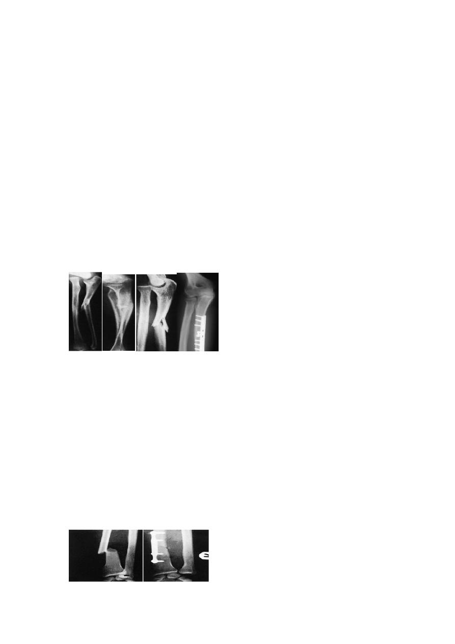
GALEAZZI FRACTURE-DISLOCATION OF THE RADIUS
Mechanism of injury
This injury was first described in 1934 by Galeazzi. The usual cause is a fall on the hand;
probably with a superimposed rotation force. The radius fractures in its lower third and
the inferior radio-ulnar joint subluxates or dislocates.
Clinical features
The Galeazzi fracture is much more common than the Monteggia. Prominence or
tenderness over the lower end of the ulna is the striking feature. It may be possible to
demonstrate the instability of the radio-ulnar joint by ‘ballotting’ the distal end of the
ulna (the ‘piano-key sign’) or by rotating the wrist. It is important also to test for an
ulnar nerve lesion, which may occur.
X-ray A transverse or short oblique fracture is seen in the lower third of the radius, with
angulation or overlap. The distal radio-ulnar joint is subluxated or dislocated.
Treatment
As with the Monteggia fracture, the important step is to restore the length of the
fractured bone. In children, closed reduction is often successful; in adults, reduction is
best achieved by open operation and compression plating of the radius. An x-ray is
taken to ensure that the distal radio-ulnar joint is reduced.