
IMMEDIATE
DENTURE
Part II
Dr. Inas Aziz M. Jawad
UNIVERSITY OF MOSUL
COLLEGE OF DENTISTRY
2020-2021
Department of
Prosthodontics
Department of:
HERE

1.Conventional immediate denture
(CID):
1- Used as a long term prosthesis after
the healing period by relining it.
2- Teeth extracted in two surgical phases
- posterior teeth extracted initially,
followed by the anteriors.
3- indicated if patient can function without
posterior teeth for 3-4 weeks as the
posterior ridge heals.
2. Interim (transitional) immediate
denture (IID):
1- Replaced by a new definitive
prosthesis after the healing period
2- All teeth - anterior and posterior
extracted in single visit.
3- Indicated if a patient is unwilling to
function without any teeth.
U N I V E R S I T Y O F M O S U L
C O L L E G E O F D E N T I S T R Y
TYPES OF ID
(according to time of teeth extraction and
denture purpose)
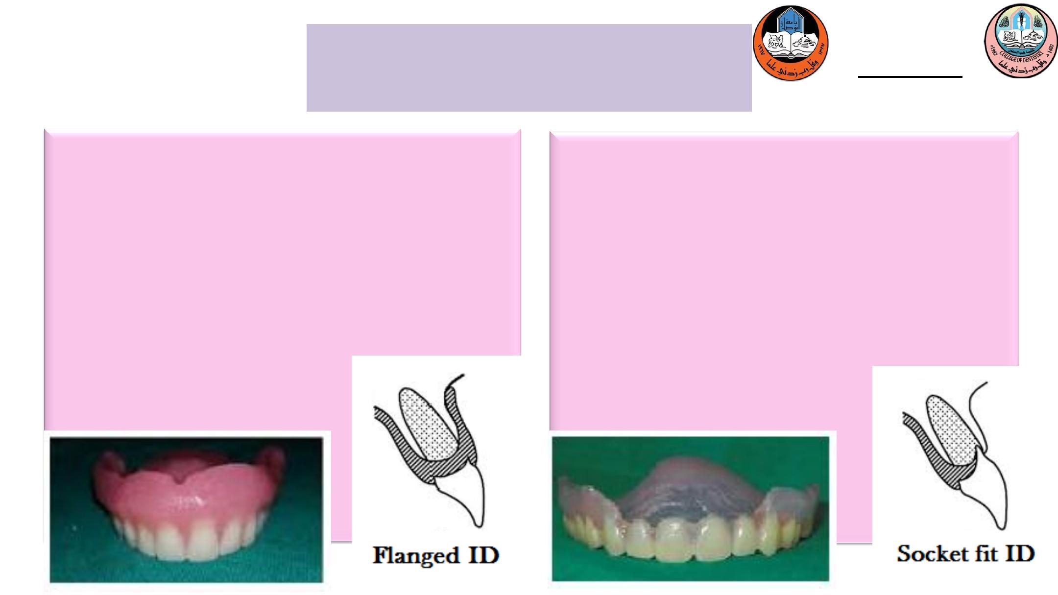
1. Flanged Denture:
a. Retentive
b. Easier to reline and rebase
c. May be difficult to place where there is
an undercut.
d. Wherever possible, a flange denture
should always be designed.
2. Socket fit Denture (Open face):
a. The teeth sit into sockets of the extracted teeth,
gives more natural appearance.
b. Esthetically good initially
c. Contraindicated in mandible because of poor
stability of lower denture during function.
d. Prone to loss of esthetic as resorption continues
e. Difficult to reline/rebase or changed to flanged
type.
f. Have poor retention
U N I V E R S I T Y O F M O S U L
C O L L E G E O F D E N T I S T R Y
TYPES OF ID
(according to flange shape)

U N I V E R S I T Y O F M O S U L
C O L L E G E O F D E N T I S T R Y
Department of:
HERE
The examination should be done to determine:
1.The suitability of the patient for immediate dentures.
2.Any influencing factors
3.Mental attitude.
4.To find out local or systemic factors that may contraindicate surgery or make extraction difficult.
The health of the oral and facial tissues must be assessed :
i. Soft tissues: basic periodontal evaluation, probing depth give an indication of initial
collapse/retraction of soft tissues, pre-extraction scaling and polishing to reduce the
inflammation and improves healing and accuracy.
ii. Hard tissue: edentulous area, charting of teeth, use orthopantomograph and periapical
radiographs of the teeth to be removed are examined for hypercementosis, multiple or curved
roots, or other pathologies that can cause problems during extraction and subsequent
treatment.
DIAGNOSIS AND
TREATMENT PLANNING

U N I V E R S I T Y O F M O S U L
C O L L E G E O F D E N T I S T R Y
Department of:
HERE
The patient should be educated about:
1. The difficulties with immediate denture provision .
2. Cleared explained of technique.
3. Visits to be planned.
4. To know which teeth are to be removed.
5. Motivation.
Facebow mounted articulated casts are also used to aid treatment planning.
DIAGNOSIS AND
TREATMENT PLANNING

U N I V E R S I T Y O F M O S U L
C O L L E G E O F D E N T I S T R Y
Department of:
HERE
CHOICES OF TREATMENT PLANNING
For a few teeth immediate denture when no denture is present:
1. Primary and/or master impressions, usually in alginate.
2. Select shape and shade of tooth.
3. Extraction of tooth/teeth and delivery of dentures.
For a few teeth addition to an existing denture:
1. Impression of mouth with denture in situ.
2. Addition of denture tooth/teeth as soon as possible.
3. Extraction of tooth/teeth and delivery of denture.
For multiple teeth immediate denture, one of following option is possible:
1. Extract all teeth at one time and insert immediate dentures (IID).
2. Extract posterior teeth prior to making immediate dentures to replace anterior teeth (CID).
DIAGNOSIS AND
TREATMENT PLANNING

U N I V E R S I T Y O F M O S U L
C O L L E G E O F D E N T I S T R Y
1. Primary impressions in alginate with or without impression compound
(sectional impression technique).
2. Master impression in alginate.
3. Occlusal record rims for existing edentulous area record vertical dimension
and jaw relation:
a) If all teeth are present enough to be articulated, no need to construct bite
rim.
b) If remaining teeth are scattered , bite rim is constructed.
4. Trial stage.
a) If remaining anterior and posterior teeth, try -in cannot be made.
b) If only anterior teeth remaining jaw relation and vertical dimension can
be made.
c) If only anterior teeth to be extracted, try-in cannot be made.
CLINICAL PROCEDURE

U N I V E R S I T Y O F M O S U L
C O L L E G E O F D E N T I S T R Y
5. Surgery and insertion of the denture:
The first step
: preparing the patient for the surgery in case patients that
suffering from systemic diseases such as diabetes and hypertension and
other systemic diseases precautions should be taken before this surgical
procedure so the appropriate dental managements for each patient with
systemic diseases should be preformed.
The second step
: Extraction of the teeth that were decided to be extracted in
the first appointment and in case of multiple extraction of the posterior
teeth start extracting of the posterior teeth toward the anterior teeth to
prevent damaging the wound and to allow healing of posterior area and
improve the adaptation of the denture over the alveolus and tuberosity.
CLINICAL PROCEDURE

U N I V E R S I T Y O F M O S U L
C O L L E G E O F D E N T I S T R Y
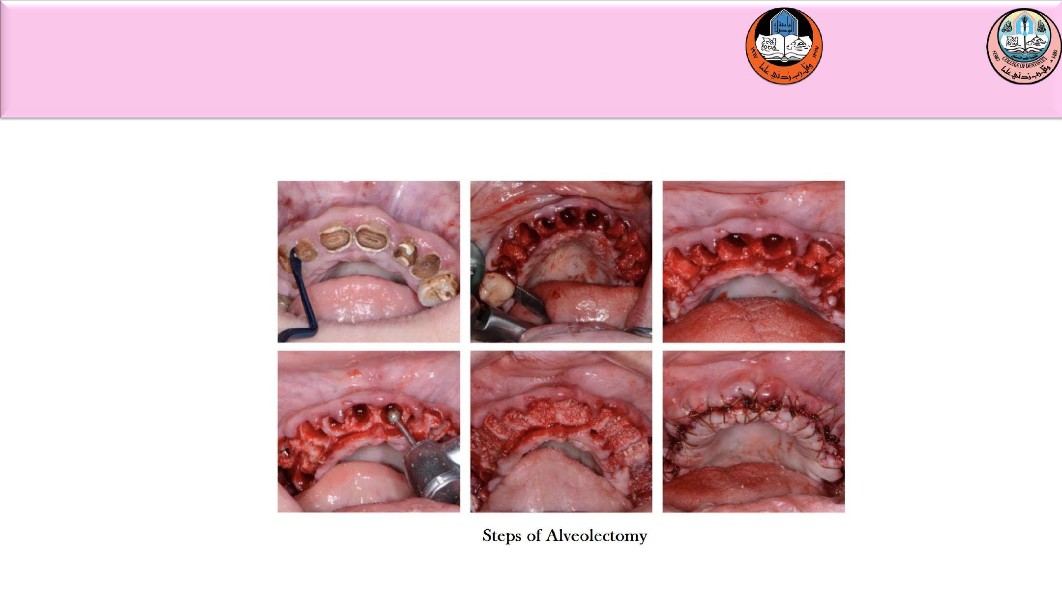
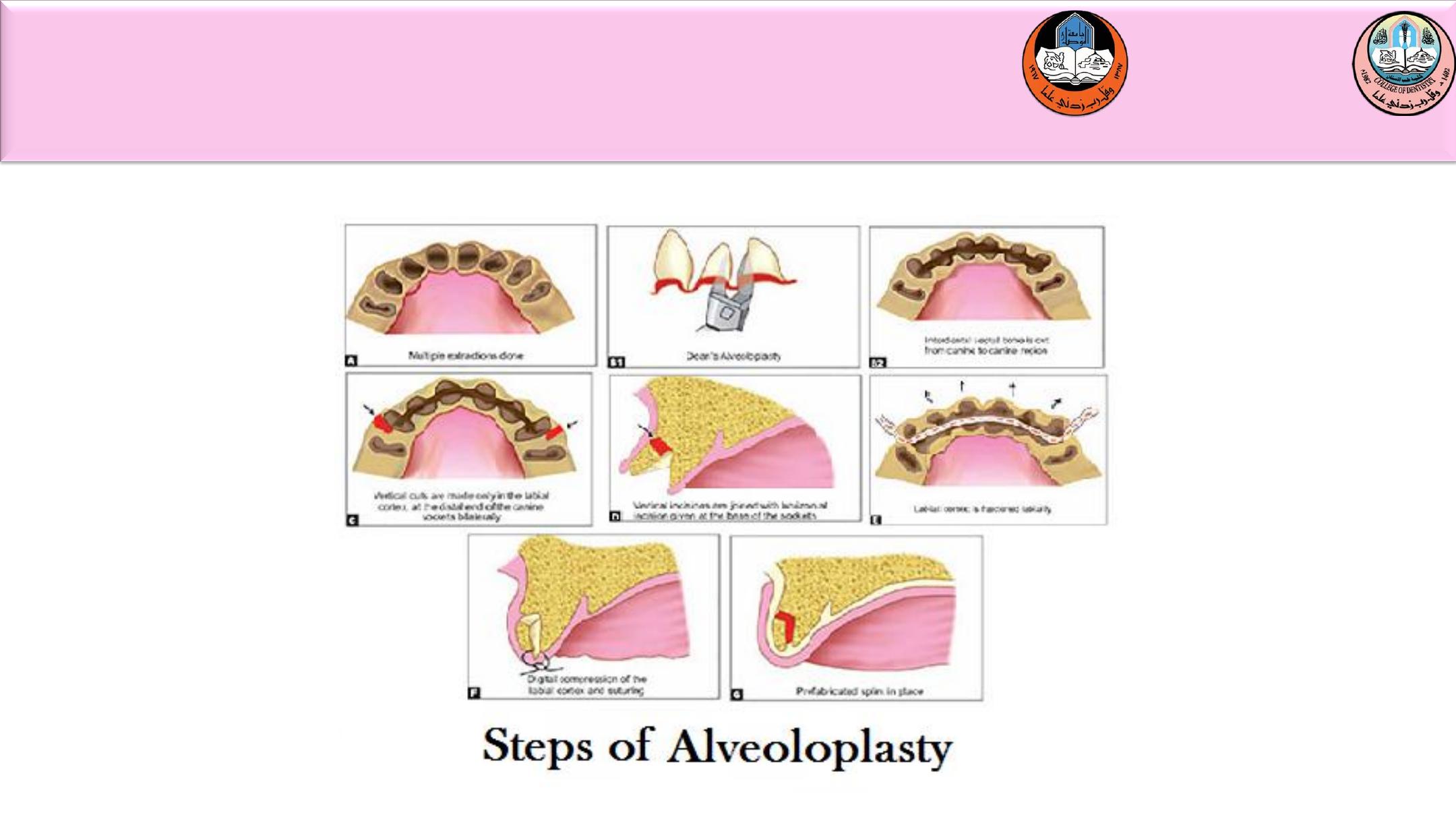
The third stage:
After extraction, alveolectomy is done in the area by simple
recontouring or an interseptal aleveoloplasty preserving as much as possible
of the vertical height and cortical bone that bony recotouring and
elimination of gross irregularity is completed. The tissue is approximated
with digital pressure and surgical guide is inserted and any area of tissue
blanching or irregularity are then reduce until surgical guide is adapted to
the alveolar ridge in all areas.
CLINICAL PROCEDURE
U N I V E R S I T Y O F M O S U L
C O L L E G E O F D E N T I S T R Y
CLINICAL PROCEDURE
U N I V E R S I T Y O F M O S U L
C O L L E G E O F D E N T I S T R Y
CLINICAL PROCEDURE

U N I V E R S I T Y O F M O S U L
C O L L E G E O F D E N T I S T R Y
The forth stage:
Incision are closed with continuous or interrupted suture
and the use of suture will depend on the number of extracted teeth.
The fifth stage:
Use of tissue conditioner in the denture for better retention
and faster healing.
The final step:
Insertion of the immediate denture.
CLINICAL PROCEDURE

U N I V E R S I T Y O F M O S U L
C O L L E G E O F D E N T I S T R Y
6.Review appointment.
It is important to review a patient with an immediate denture at regular
intervals especially in the first few weeks and months. The initial days
are primarily concerned with the postoperative care of the healing tooth
sockets, while the later reviews are directed at the management of
resorption.
A simple time table for reviewing a patient is as following:
at 24 hours, a general check is made of the over all comfort and borders
of the dentures and to ensure no major ulceration has occurred and that
the socket are healing well, try to avoid occlusal adjustment.
CLINICAL PROCEDURE

U N I V E R S I T Y O F M O S U L
C O L L E G E O F D E N T I S T R Y
at 48 hours, patient is seen for sore spot.
at 1 week, a more detailed check and occlusal adjustment of dentures
can be made, removal of suture and changing of tissue conditioning
material.
at 1 month, the socket has healed and chair side temporary reline may
required.
at 3 to 6 month, the management of loss of fit of the denture owing to
bone resorption is undertaken, this may involve relines and /or rebases,
which are taken either chair side or with aid of the laboratory.
at 1 year, a new denture is made.
CLINICAL PROCEDURE
U N I V E R S I T Y O F M O S U L
C O L L E G E O F D E N T I S T R Y
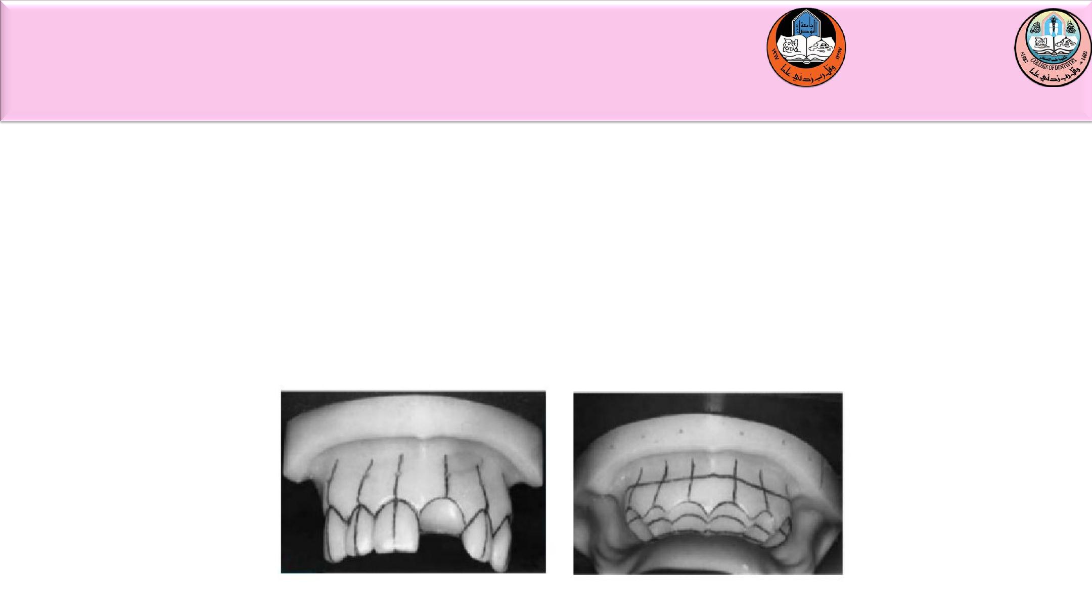
Trimming of casts between try-in and before processing of dentures. The
cast must be prepared by the dental surgeon as he/she alone has seen the
patient and undertaken the clinical examination.
In socket fit denture the cast is marked with a pencil to show the gingival
margin, the long axis of the teeth and the length of the teeth.
The long axis of the teeth are drawn to help in placement of the artificial
teeth.
LABORATORY PROCEDURE
U N I V E R S I T Y O F M O S U L
C O L L E G E O F D E N T I S T R Y
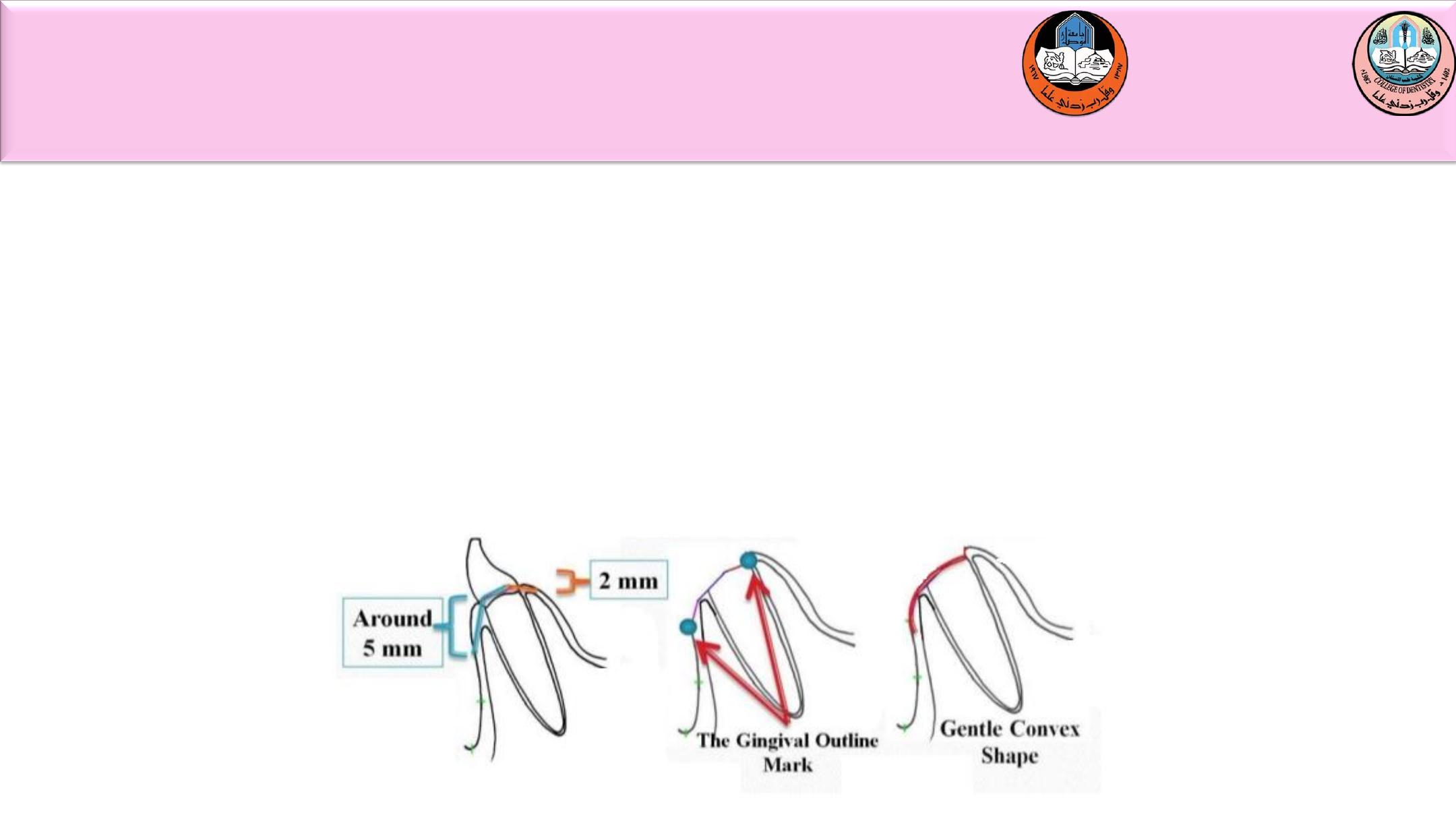
In a socket fit denture, prepared root socket (5mm depth on the stone
model in the direction of the root labially and about 2mm depth lingually)
and then the neck of the artificial teeth is placed in the preparation and at
the time of insertion the neck of the artificial teeth will just enter socket
of the tooth after extraction (see the figure bellow).
In a flanged denture, the stone is trimmed to simulate the ridge following
tooth extraction.
LABORATORY PROCEDURE

U N I V E R S I T Y O F M O S U L
C O L L E G E O F D E N T I S T R Y
You must leave your dentures in your mouth for the first 24 hours.
Removing the dentures will not decrease pain due to the extractions.
Swelling may occur, and if you remove your dentures, you may not be able
to reinsert them.
Holding ice packs against your face in the area of the extractions (no more
than 20 minutes/hour for the first 24 hours may reduce swelling).
After 24 hours, use a wet heat compress).
Take prescribed medications as directed.
The denture will act as a bandage and help to limit bleeding and prevent
breakdown of the blood clots that form in the sockets.
Although bleeding is normally minimal, you must remember that a few
drops of blood will color your saliva pink.
Your diet for the first 24 hours should be restricted to liquids or soft
foods.
POST OPERATIVE INSTRUCTION

U N I V E R S I T Y O F M O S U L
C O L L E G E O F D E N T I S T R Y
After the 24 hour appointment, your dentures should be removed for
cleaning after meals.
Always hold the dentures over a sink partially filled with water while
cleaning them (should you accidentally drop them, the water will break
their fall and damage will be less likely).
Scrub the tissue surface (inside) of the dentures with a denture brush,
liquid soap, and water. Brush the external surfaces and the teeth of the
dentures and, for maximal cleanliness, brush your tongue and the roof of
your mouth.
Always keep the dentures wet while they are out of your mouth.
After 24 hours, you should begin removing your dentures at night.
Removing the dentures allows small blood vessels to enlarge and provide
nourishment to the tissues supporting the dentures.
POST OPERATIVE INSTRUCTION

U N I V E R S I T Y O F M O S U L
C O L L E G E O F D E N T I S T R Y
2020-2021