
The Muscles of Facial Expression
and Mastication
Firas Al-Hameed
Thi-Qar Medical School

Muscles of Facial Expression
• The muscles of facial expression are located in the subcutaneous
tissue
• Originate from bone or fascia, and inserting onto the skin.
• They are the only group of muscles that insert into skin.
• By contracting, the muscles pull on the skin and exert their effects.
• These muscles have a common embryonic origin – the 2nd
pharyngeal arch.
• All the muscles of facial expression are innervated by the facial nerve.
• The facial muscles can broadly be split into three groups: orbital,
nasal and oral.
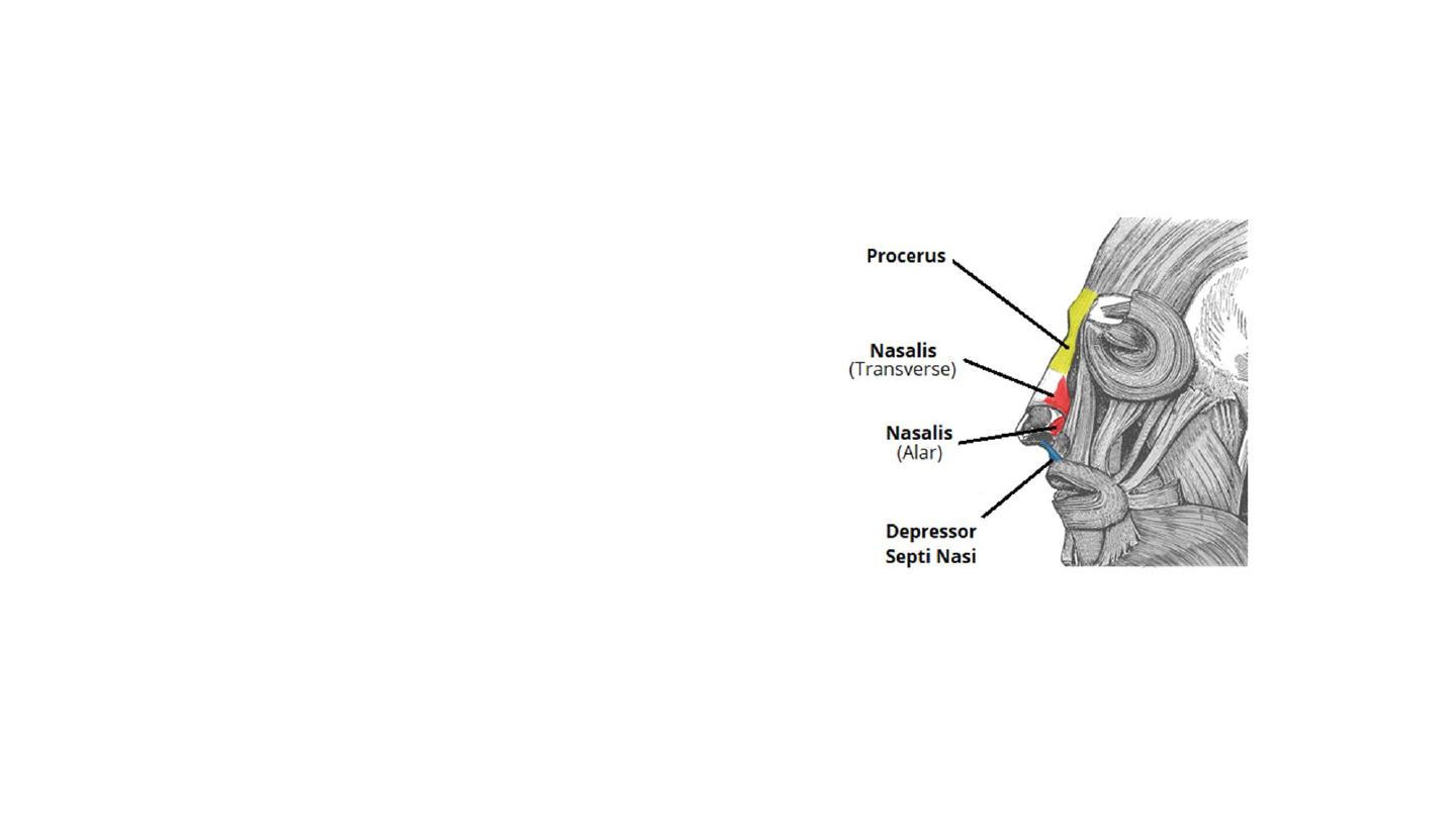
Nasal Group
• The nasal group of facial muscles are
associated with movements of the
nose, and the skin around it.
• There are three muscles in this group
• all innervated by the facial nerve.
• Function:
• Movements of the nose and the
surrounding skin.
• Contributes to the expression of frowning.
• Widens the nasal aperture
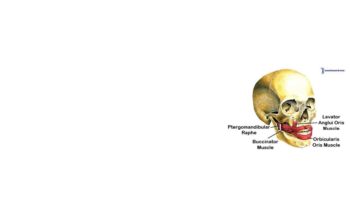
Oral Group
• Responsible for movements of the mouth and lips.
• The oral group of muscles consists of the orbicularis oris,
buccinator, and various smaller muscles.
Orbicularis Oris
• Enclose the opening to the oral cavity.
• Attachments: Arises from the maxilla and from the other
muscles of the cheek. It inserts into the skin and mucous
membranes of the lips.
• Action: Purses the lips.
• Innervation: Facial nerve.
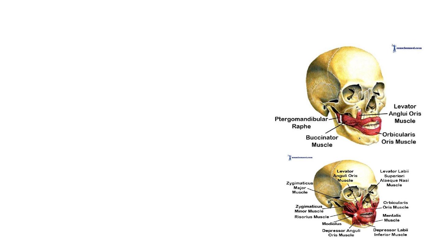
• Buccinator
• This muscle is located between the mandible and
maxilla, deep to the other muscles of the face.
• Attachments: It originates from the maxilla and
mandible. The fibres run in an inferomedial direction,
blending with the orbicularis oris and the skin of the
lips.
• Actions: The buccinator pulls the cheek inwards against
the teeth, preventing accumulation of food in that
area.
• Innervation: Facial nerve.
• Other Oral Muscles
• There are other muscles that act on the lips and
mouth.
• The lower group contains the depressor anguli oris,
depressor labii inferioris and the mentalis.
• The upper group contains the risorius, zygomaticus
major, zygomaticus minor, levator labii superioris,
levator labii superioris alaeque nasi and levator anguli
oris.

The Muscles of Mastication
• The muscles of mastication are associated with movements of the jaw
(temporomandibular joint).
• Masseter
• Temporalis
• Medial pterygoid
• Lateral pterygoid
• The muscles of mastication develop from the first pharyngeal arch.
Thus, they are innervated by a branch of the trigeminal nerve (CN V),
the mandibular nerve.
• All the muscles are bilateral structures.
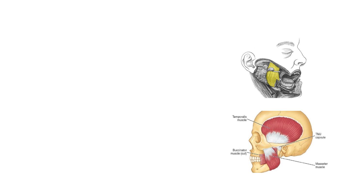
Masseter
• The most powerful muscle of mastication.
• It is quadrangular in shape and has two parts:
deep and superficial.
• Attachments:
• The superficial part originates from maxillary process
of the zygomatic bone.
• The deep part originates from the zygomatic arch of
the temporal bone.
• Both parts attach to the ramus of the mandible.
• Actions: Elevates the mandible, closing the
mouth.
• Innervation: Mandibular nerve (V3).
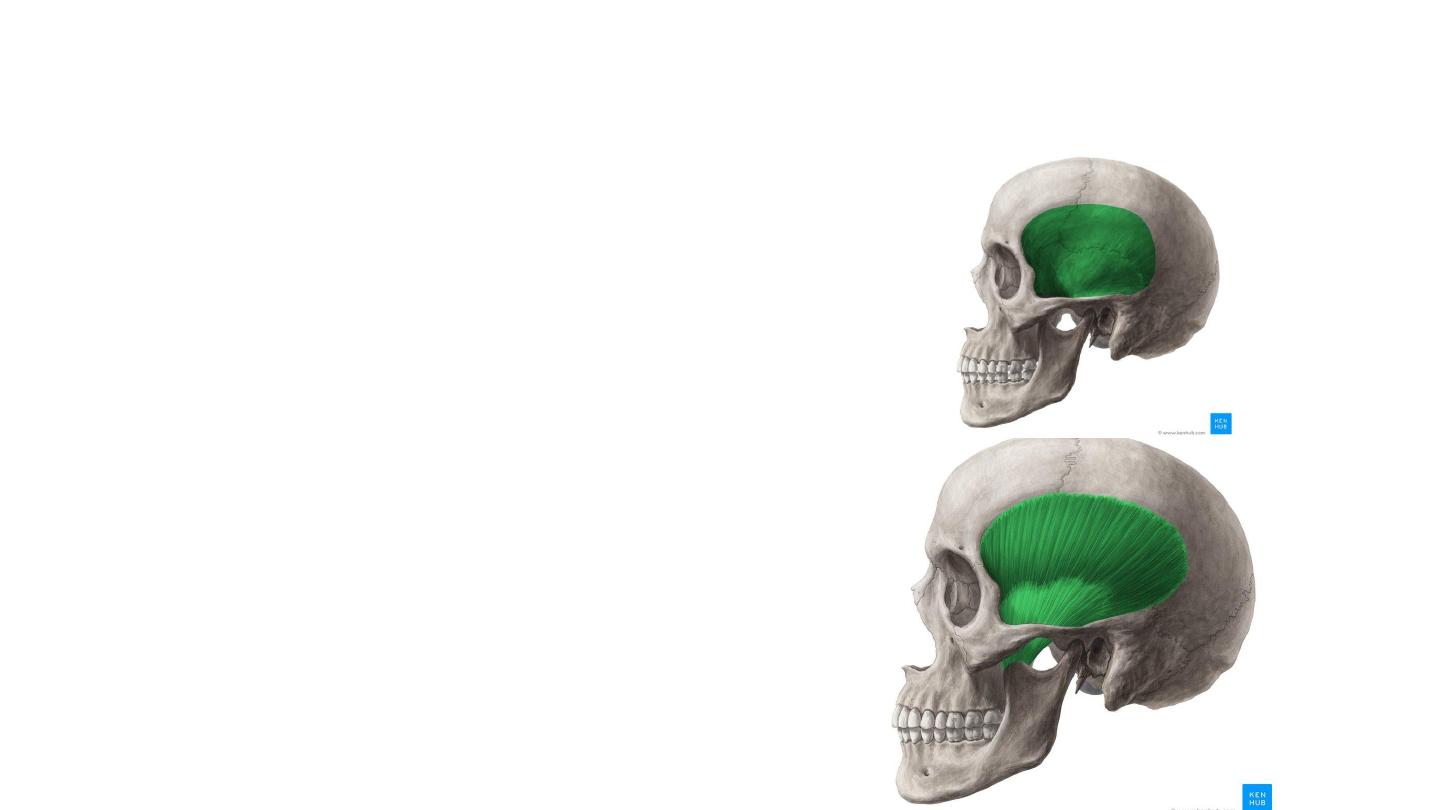
Temporalis
• Attachments: Originates from the
temporal fossa. It condenses into a
tendon, which inserts onto the coronoid
process of the mandible.
• The muscle is covered by tough fascia
• Actions: Elevates the mandible, closing
the mouth. Also retracts the mandible,
pulling the jaw posteriorly.
• Innervation: Mandibular nerve (V3).
Pterygoid muscles
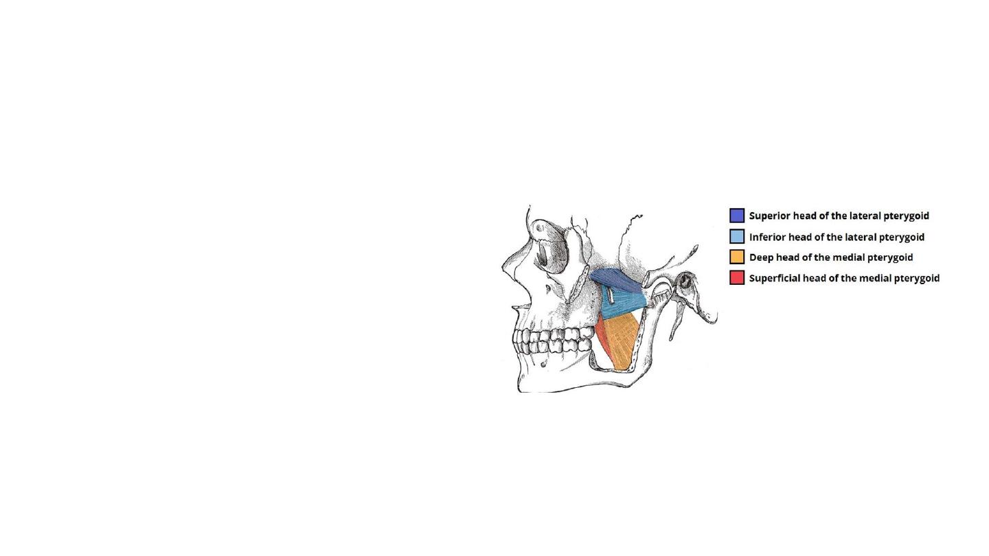
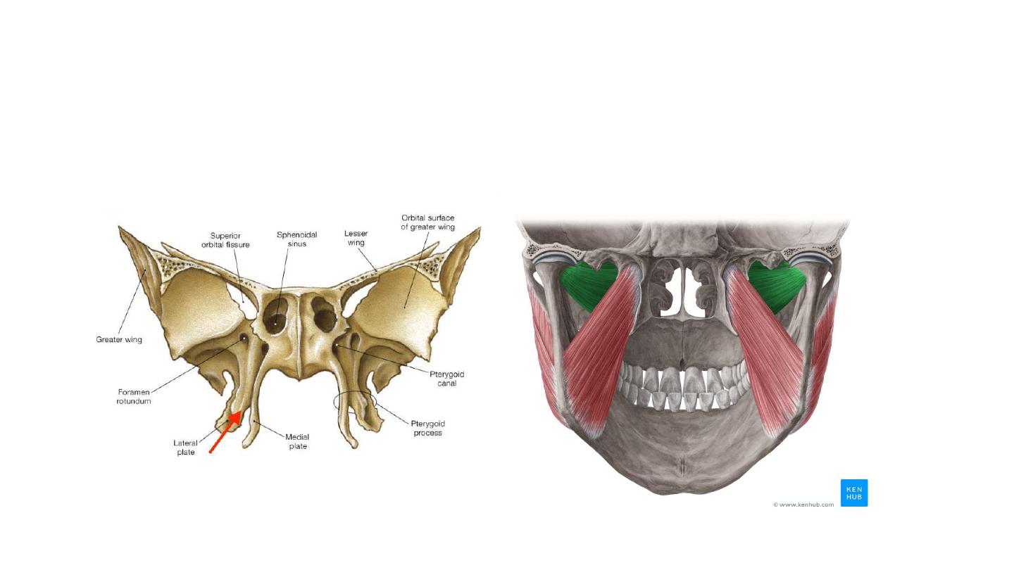
Lateral Pterygoid
• Triangular shape with two heads: superior and
inferior.
• Attachments:
• The superior head originates from the greater
wing of the sphenoid.
• The inferior head originates from the lateral
pterygoid plate of the sphenoid.
• The two heads converge into a tendon which
attaches to the neck of the mandible.
• Actions: Acting bilaterally, the lateral
pterygoids protract the mandible, pushing the
jaw forwards. Unilateral action produces the
‘side to side’ movement of the jaw.
• Innervation: Mandibular nerve (V3).
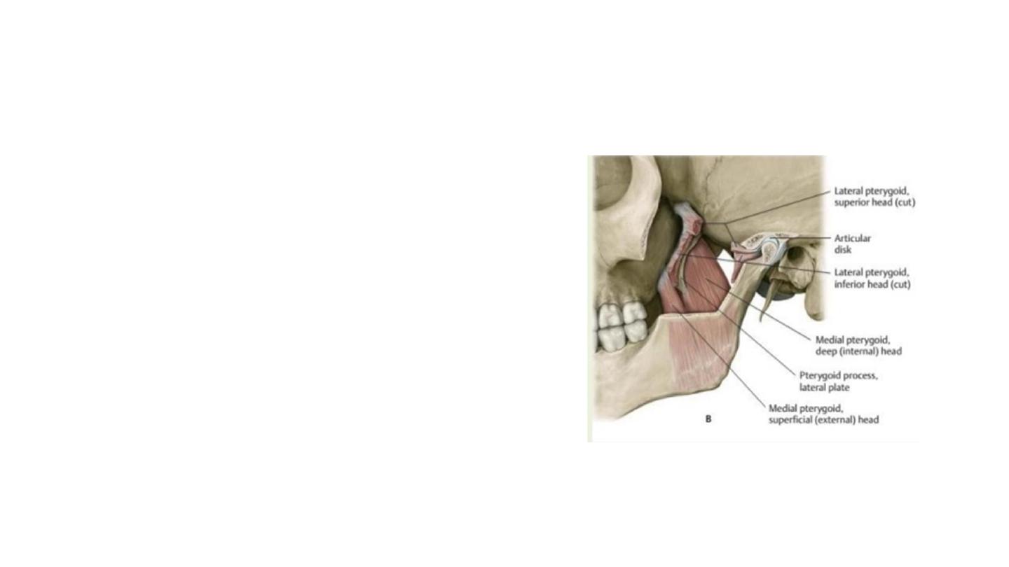
Medial Pterygoid
• The medial pterygoid muscle has a quadrangular
shape with two heads: deep and superficial.
• It is located inferiorly to the lateral pterygoid.
• Attachments:
• The superficial head originates from the maxilla and
the palatine bone.
• The deep head originates from the medial aspect of
the lateral pterygoid plate of the sphenoid bone.
• Both heads attach to the ramus of the mandible
near the angle of mandible.
• Actions: Elevates the mandible, closing the
mouth.
• Innervation: Mandibular nerve (V3).