
Neck (2)
Firas Al-Hameed
Thi-Qar Medical School
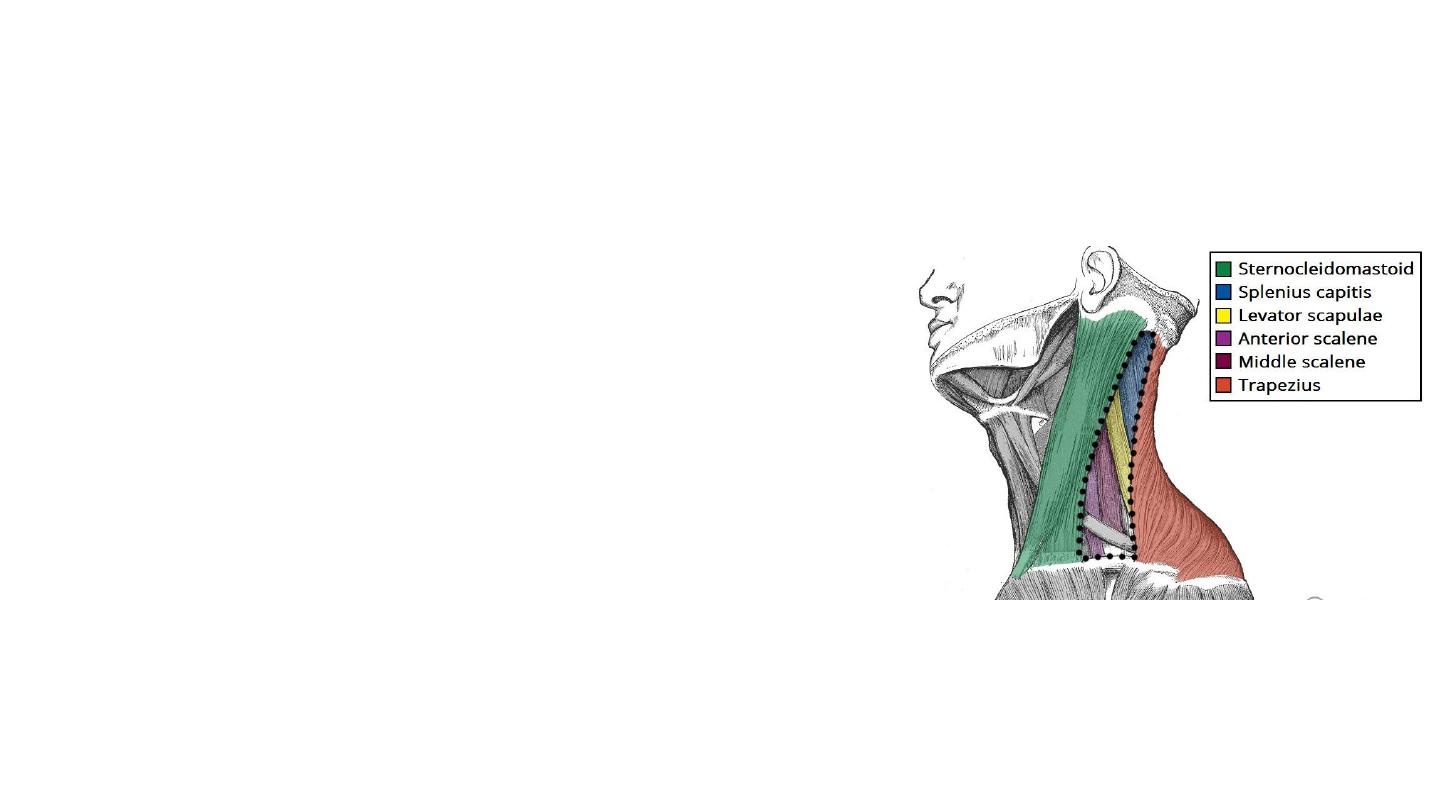
The posterior triangle of the neck
The posterior triangle of the neck is an anatomical
area located in the lateral aspect of the neck.
Borders
• Anterior – posterior border of the sternocleidomastoid.
• Posterior – anterior border of the trapezius muscle.
• Inferior – middle 1/3 of the clavicle.
• The posterior triangle of the neck is covered by the
investing layer of fascia, and the floor is formed by
the prevertebral fascia.
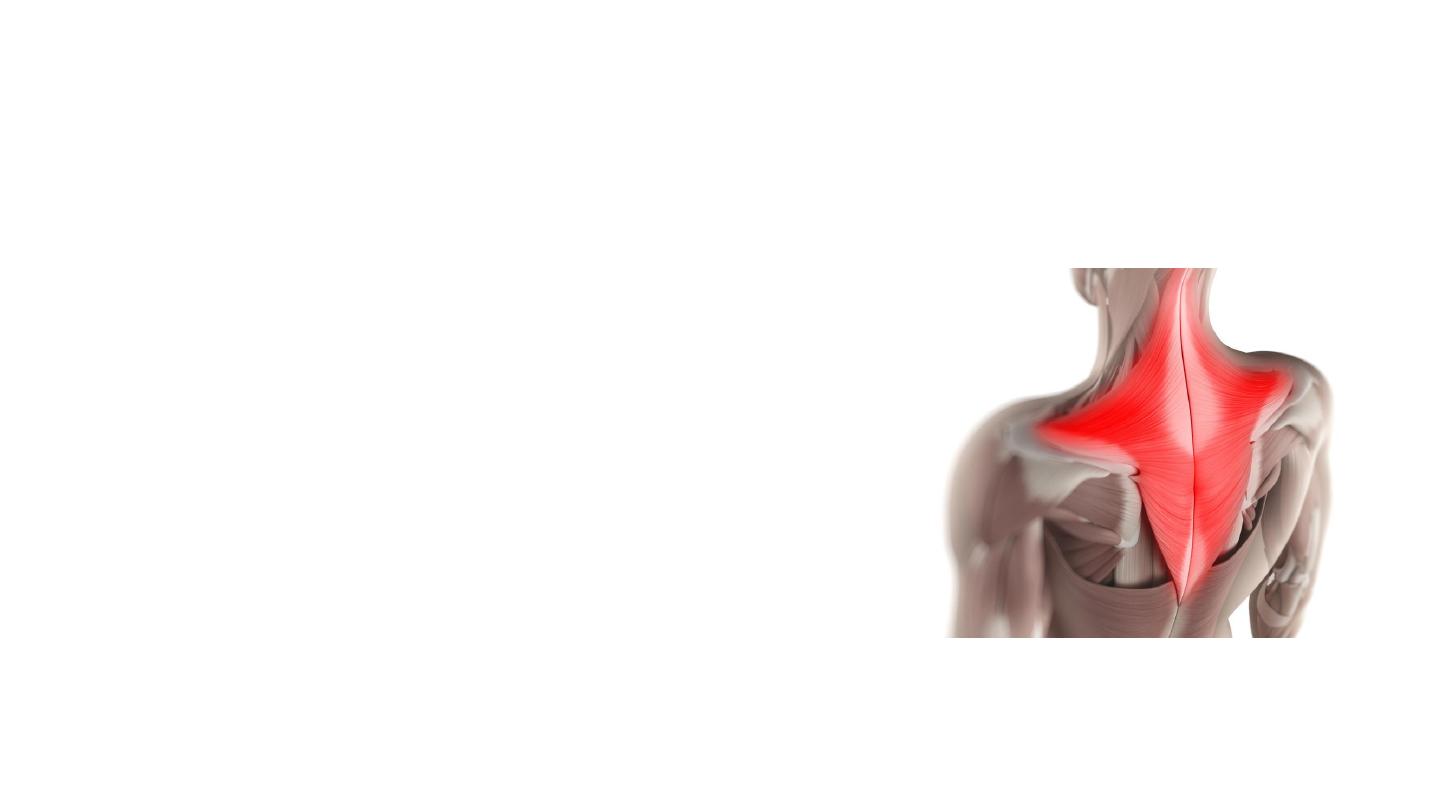
Trapezius Muscle
• Flat diamond-shaped muscle at back of
neck and chest , formed from 3 parts
upper , middle & lower fibers
• Origin: occipital bone and spines of C7 –
T12
• Insertion: Lateral 1/3 of clavicle, acromion,
spine of scapula
• Functions:
• Its main function is to stabilize and move the
scapula.
• Nerve supply the accessory nerve CN 11
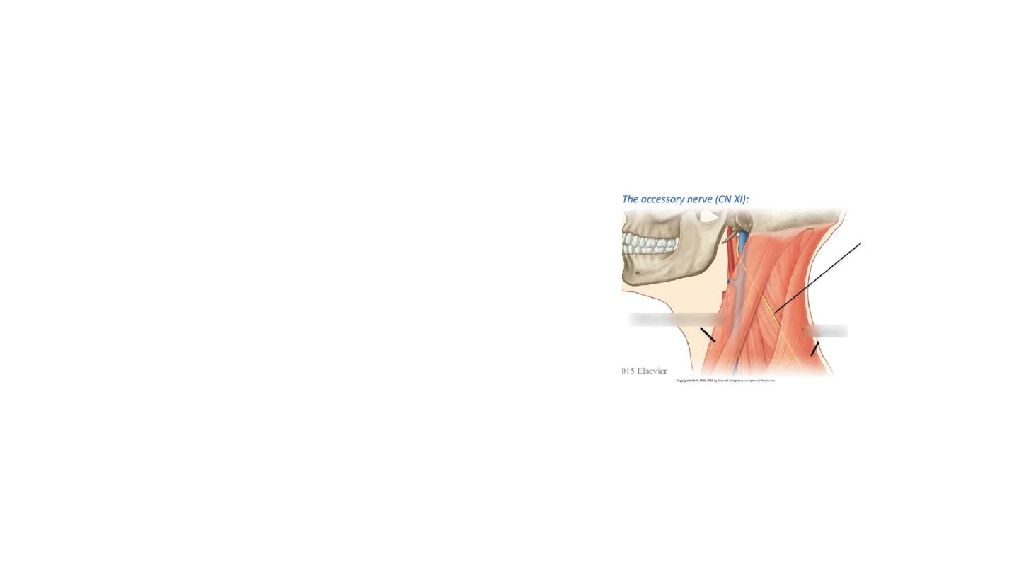
The accessory nerve (CN XI)
• Innervates sternocleidomastoid and enters
the posterior triangle.
• It crosses the posterior triangle in an oblique,
inferoposterior direction, within the
investing layer of fascia. It lies relatively
superficial in the posterior triangle, leaving it
vulnerable to injury.
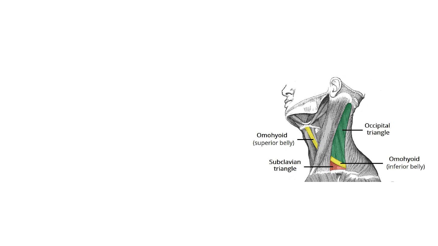
Subdivisions
• The omohyoid muscle splits the
posterior triangle of the neck into two:
• The larger, superior part is termed the
occipital triangle.
• The inferior triangle is known as the
subclavian triangle and contains the
distal portion of the subclavian artery.

Contents
Muscles
• The inferior belly of the omohyoid muscle
• A number of vertebral muscles (covered by
prevertebral fascia) form the floor of the
posterior triangle:
• Splenius capitis
• Levator scapulae
• Anterior, middle and posterior scalenes
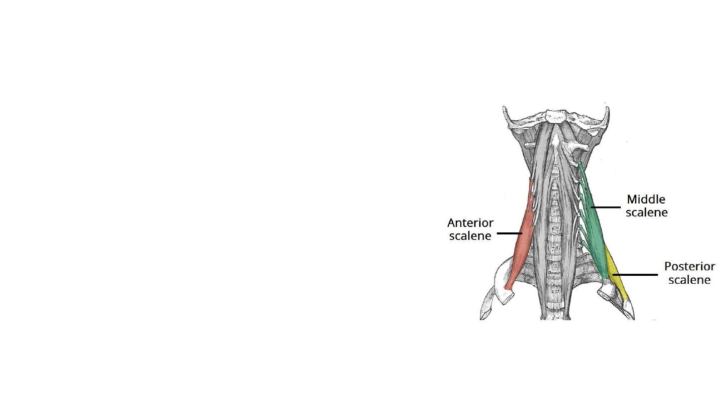
The scalene muscles
• The scalenes act as accessory muscles of respiration, and perform
flexion at the neck.
Anterior Scalene
• It lies deep to the prominent sternocleidomastoid muscle.
• Attachments: Originates from the transverse processes of C3-C6,
and attaches onto the inner border of the first rib.
• Function: Elevation of the first rib. Ipsilateral contraction causes
ipsilateral lateral flexion of the neck, and bilateral contraction
causes anterior flexion of the neck.
• Innervation: anterior rami of C5-C6
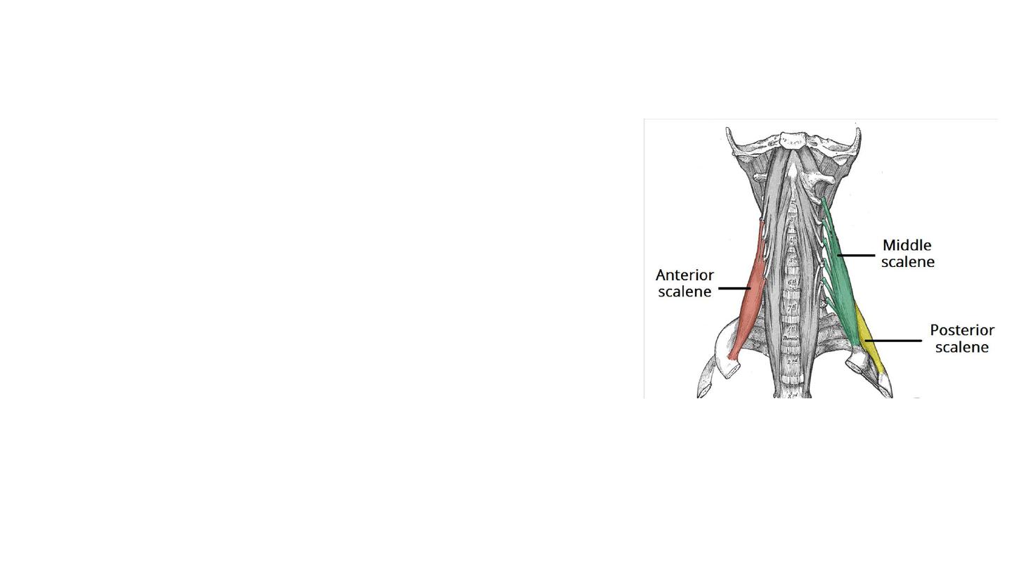
Middle Scalene
• It is the largest and longest of the three scalene muscles.
• Attachments: Originates from the transverse processes of C2-C7. It
has several long, thin muscles bellies which converge into one large
belly that inserts into the first rib.
• Function: Elevation of the first rib. Ipsilateral contraction causes
ipsilateral lateral flexion of the neck.
• Innervation: Anterior rami of C3-C8.
Posterior Scalene
• It is the smallest and deepest of the scalene muscles.
• Attachments: Originates from the transverse processes of C5-C7,
and attaches into the
second rib
.
• Function: Elevation of the second rib, and ipsilateral lateral flexion
of the neck.
• Innervation: Anterior rami of C6-C8.
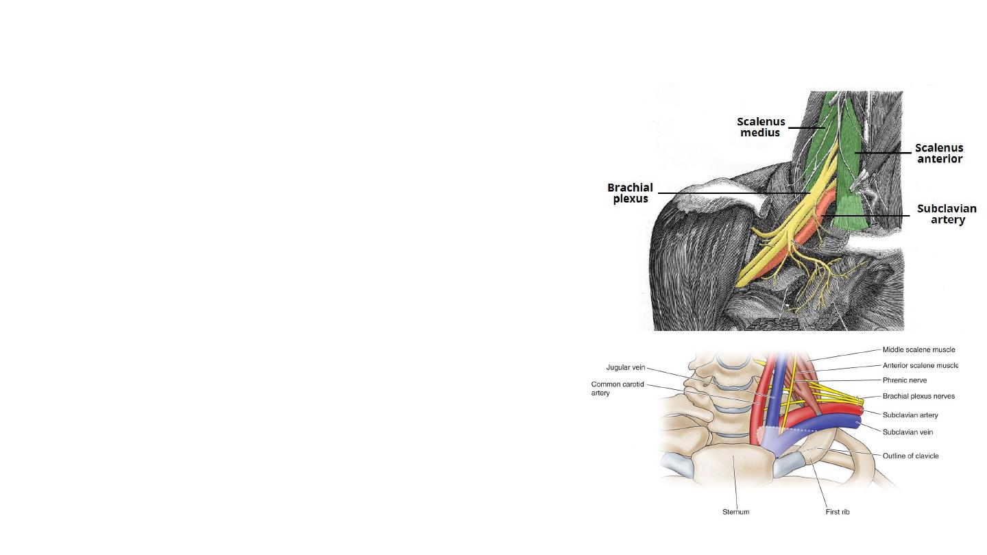
Anatomical Relationships of scalene muscles
• The brachial plexus and subclavian artery pass
between the anterior and middle scalene muscles.
• This provides an important anatomical landmark in
anaesthetics for performing an interscalene block.
• The subclavian artery is located posterior to the anterior
scalene.
• The subclavian vein and phrenic nerve pass anteriorly
to the anterior scalene – the subclavian vein courses
horizontally across it, while the phrenic nerve runs
vertically down the muscle.
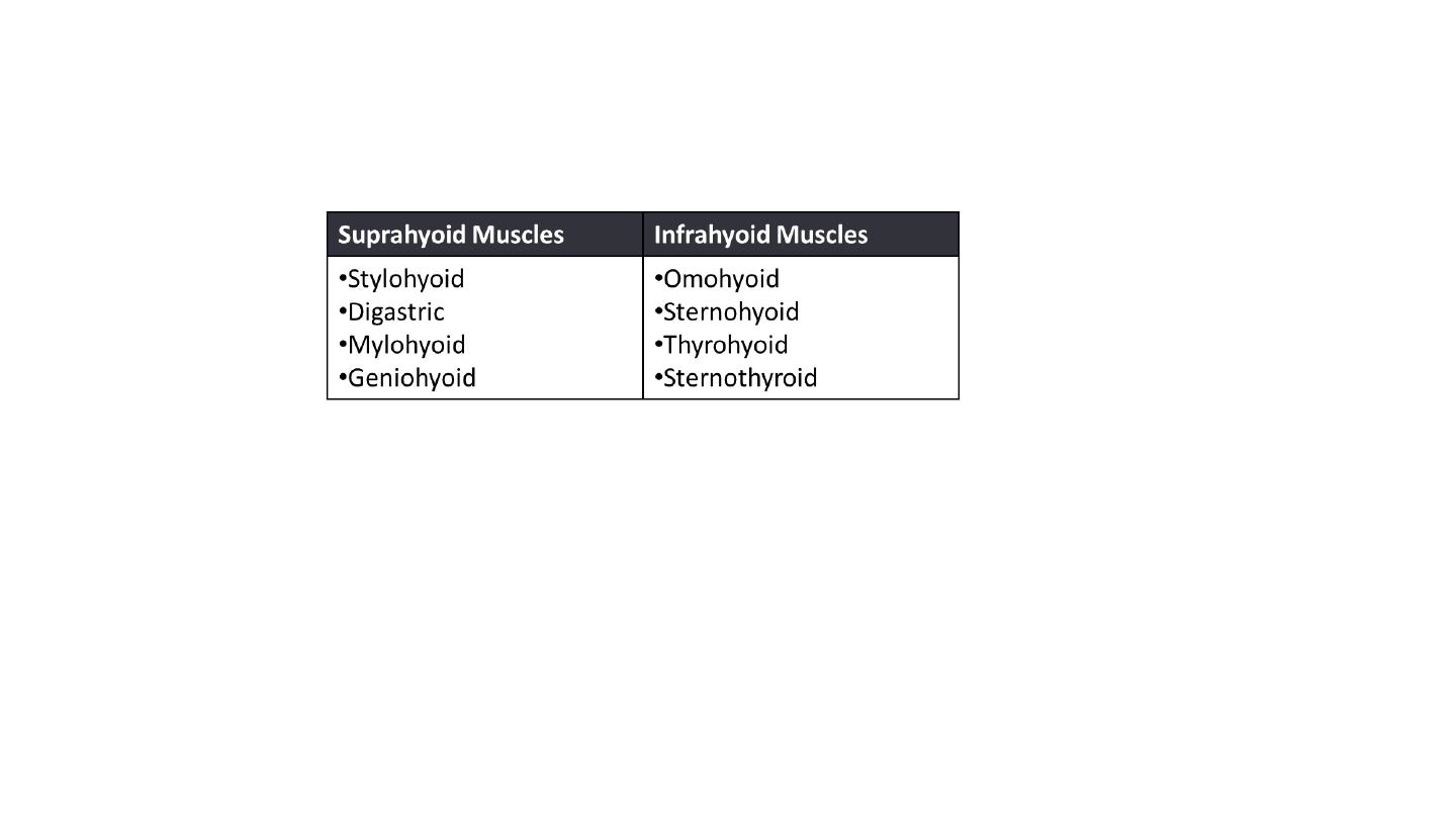
Suprahyoid and infrahyoid muscles
The suprahyoid muscles
They all act to elevate the hyoid bone – an action involved in swallowing.
The arterial supply to these muscles is via branches of the facial artery, occipital
artery, and lingual artery.
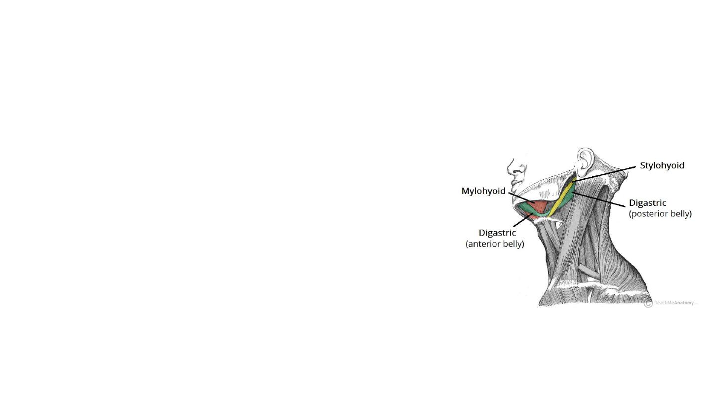
Digastric
• The digastric is comprised of two muscular bellies, which are
connected by a tendon
• Attachments:
• The anterior belly arises from the digastric fossa of the mandible.
• The posterior belly arises from the mastoid process of the temporal
bone.
• The two bellies are connected by an intermediate tendon, which is
attached to the hyoid bone via a fibrous sling.
• Actions: Depresses the mandible and elevates the hyoid
bone.
Innervation:
• The anterior belly is innervated by a branch of the
mandibular nerve (which is derived from the trigeminal
nerve, CN V).
• The posterior belly is innervated by a branch of the facial
nerve, CN 7)
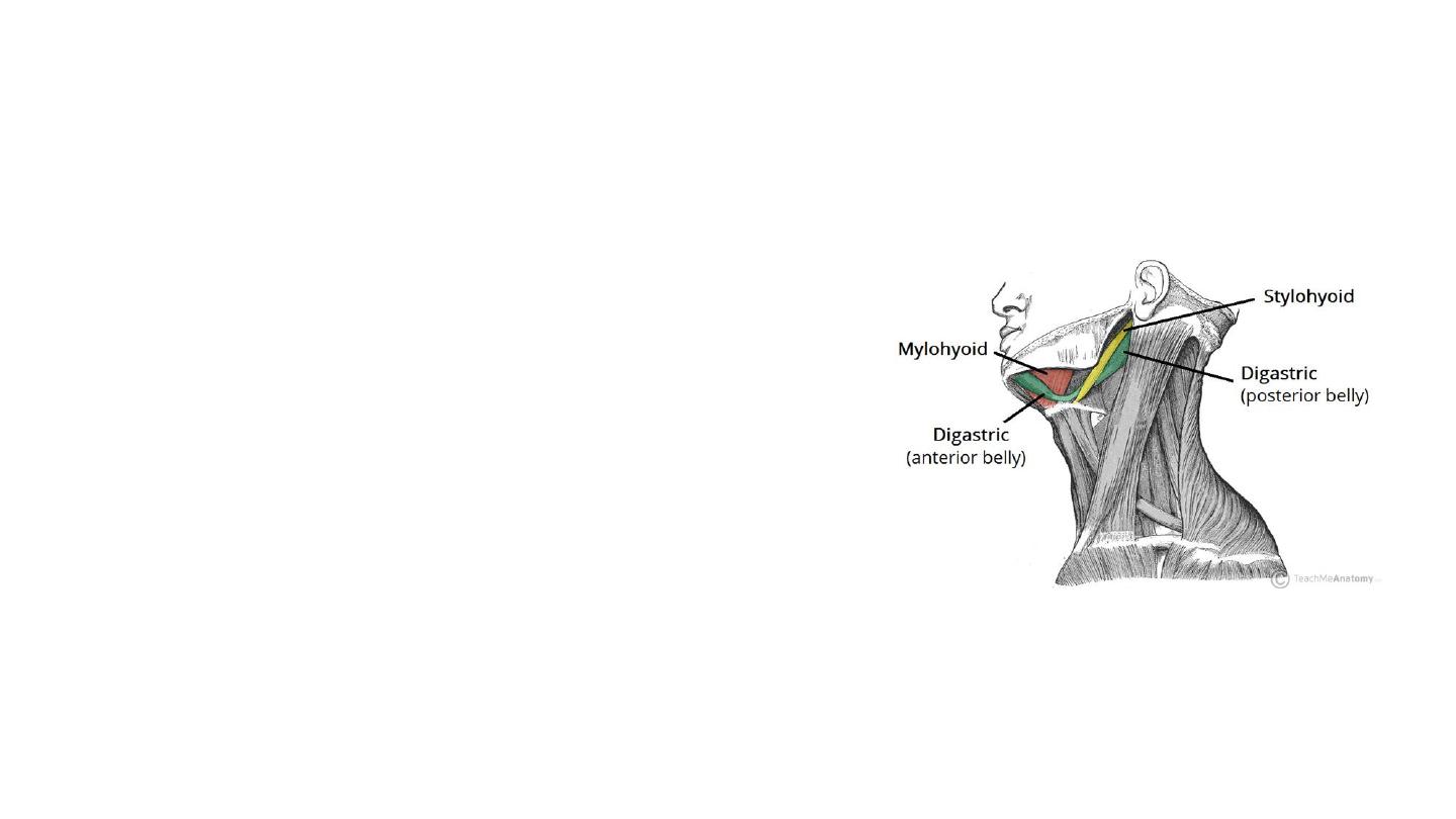
Stylohyoid
• Located superiorly to the posterior belly of the
digastric muscle.
• Attachments: Arises from the styloid process of
the temporal bone and attaches to the lateral
aspect of the hyoid bone.
• Actions: pull the hyoid bone in a posterior and
superior direction.
• Innervation: Stylohyoid branch of the facial
nerve (CN VII).
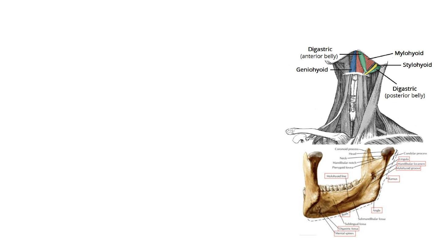
Mylohyoid
• The mylohyoid is a broad, triangular shaped
muscle. It forms the floor of the oral cavity and
supports the floor of the mouth.
• Attachments: Originates from the mylohyoid line
of the mandible, and attaches onto the hyoid
bone.
• Actions: Depresses the mandible, elevates the
hyoid bone and the floor of the mouth.
• Innervation: a branch of the mandibular nerve
(which is derived from the trigeminal nerve, CN
V).
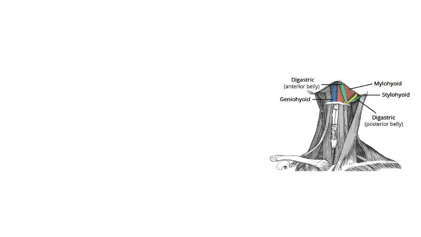
Geniohyoid
• The geniohyoid is located close to the midline
of the neck, deep to the mylohyoid muscle.
• Attachments: Arises from the mandible. It
inserts onto the hyoid bone.
• Actions: Depresses the mandible and elevates
the hyoid bone.
• Innervation: C1 nerve roots that run within
the hypoglossal nerve.