Types of proteinuria
Ass. Prof.Dr. Amin Turki
Nephrology and urology
Transient proteinuria:
• It characterized by positive test for proteinuria by urinary dipsticks but it will resolve with repeated dipsticks tests with normal urinary protein excretion.• It occurs in approximately 10% of children
• It is more common in adolescent than in younger children.
The cause is unclear, but it might associated with:
• temperature >38.3°C (101°F)• exercise
• dehydration
• cold exposure
• heart failure
• seizures
• stress
proteinuria usually does not exceed 1-2+ on the dipstick.
It is benign condition, no evaluation or therapy is needed.
Orthostatic (Postural) proteinuria:
• Is the most common cause of persistent proteinuria in school-age children and adolescents.• It is asymptomatic, and discovered by routine urinalysis.
• Occurring in up to 60% of children with persistent proteinuria.
• While in upright position, urinary protein excretion increased 10-fold, reaching 1,000 mg/24 hr.
• In supine position there is normal or minimally increase in protein excretion
• There are no hematuria, hypertension, hypoalbuminemia, edema, and renal dysfunction.
• The cause is unknown, but increased body mass index is recognized as a strong correlation factor.
• It is benign condition. No further evaluation is necessary
• Patients should be monitored for the development of non-orthostatic proteinuria, particularly in the presence of hematuria, hypertension, or edema which may indicate underlying kidney disease.
Nephrotic syndrome (NS)
Glomerular diseases characterizes by:• Heavy (nephrotic-range) proteinuria
• Hypoalbuminemia (≤2.5 g/dL)
• Edema
• Hyperlipidemia
(cholesterol >200 mg/dL)
proteinuria >3.5 g/24 hr
or
> 40 mg/m2/ hr
or
Urine protein : creatinine ratio >2.
or
Urinary dipstick albumin > 3+
Without treatment, nephrotic syndrome associated with a high risk of death, most commonly from infections.
Nephrotic range proteinuria :
3. Membranous nephropathy.
Causes of nephrotic syndrome:I. Idiopathic (primary) NS:
1. Minimal change disease.
2. Focal segmental glomerulosclerosis.
5. Metabolic Disorders With or Without NS: Alagille syndrome α1-Antitrypsin deficiency, Glycogen storage disease, Sickle cell disease
4. Mitochondrial disorders
3. Congenital NS with lung and skin involvement
2. Denys-Drash syndrome
1. Finnish-type congenital NS
II. Congenital NS:
III. Secondary NS:
4. Associated With Malignant Disease:
1. Infections:
Endocarditis,Hepatitis B and C, HIV-1, IMN, Malaria, Syphilis (congenital and secondary), Toxoplasmosis
2. Drugs:
Captopril, Penicillamine, Gold, NSAID drugs, Interferon, Mercury, Heroin, Lithium
3. Immunologic or Allergic Disorders
Vasculitis syndromes, Food allergens, Serum sickness.
Lymphoma, Leukemia, Solid tumors.
Pathogenesis:
increased glomerular capillary wall permeabilityhypoalbuminemia
massive proteinuria
Edema
• Immune mechanism.
Increase glomerular capillary permeability might due to:• Abnormal function of the podocytes which are epithelial cells located on the outside of the glomerular capillary loops
Mechanisms of clinical squeal of NS:
• The exact mechanism of edema formation is uncertain.1. Edema:
• Is the most common presenting symptom of children with NS.
• There are 2 opposing theories:
A. The underfill hypothesis:
leakage of plasma water
into the interstitium
nephrotic-range
proteinuria
Hypoproteinemia
IV oncotic
pressure
Edema
tubular Na and
water retention
IV volume
vasopressin and atrial
natriuretic factor secretions
PLUS
aldosterone effect
This hypothesis does not fit the clinical picture of some patients with edema caused by nephrotic syndrome who have clinical signs of intravascular volume overload and not volume depletion.
Clinical weakness of this hypothesis:
B. The overfill hypothesis
Distal tubular Na channel defectprimary sodium retention
IV volume expansion
leakage of excess fluid into the interstitium
Edema
• Numerous nephrotic patients are presented with an obvious clinical picture of IV volume depletion such as low blood pressure, tachycardia, and elevated hemoconcentration.
• Amiloride, (sodium channel blocker), is not sufficient to induce adequate diuresis if it use alone.
Clinical weakness of this hypothesis:
2. Hyperlipidemia:
• serum lipid levels (cholesterol, triglycerides, low-density lipoprotein, and very-low-density lipoproteins) are elevated for 2 reasons:• Hypoalbuminemia stimulates generalized hepatic protein synthesis, including synthesis of lipoproteins.
• Urinary losses of lipoprotein lipase will decrease lipid catabolism.
3. Increased susceptibility to infections:
• Haemophilus influenza• There is increase risk of infections (sepsis, peritonitis, pyelonephritis)
• The causative MO are:
• Streptococcus pneumoniae (most common MO)
• Gram-negative bacteria e.g. E. coli.
Causes:
• Edema or ascites acting as a potential culture medium.• loss of complement factor C3 and C5 and C3b, opsonins such as properdin factor B, and immunoglobulins in the urine.
• Use of immunosuppressive medications in treatment of nephrotic syndrome.
• Malnutrition
4. Hypercoagulability:
• Urinary losses of antithrombotic factors such as antithrombin III and protein S.
• Vascular stasis from hemoconcentration and IV volume depletion
• Increased platelet number and aggregability
• Increase hepatic production of fibrinogen and other clotting factors
• Is NS manifesting at birth or within the 1st 3 mo of life.
Classification:I. Primary Congenital NS(Inherited autosomal recessive disorders):
• Finnish-type (absence of nephrin). Affected infants present at birth with: Edema and enlarged placenta (>25% of the infant’s weight). Prenatal diagnosis by elevated maternal and amniotic α-fetoprotein levels.
Congenital NS:
2. Denys-Drash syndrome (abnormal podocyte function). Patients present with early-onset nephrotic syndrome, progressive renal insufficiency, ambiguous genitalia, and Wilms tumors.
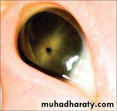
3. Pierson syndrome: include congenital NS and bilateral microcoria (fixed narrowing of the pupil).
II. Secondary Congenital NS:
• Congenital infections (CMV, syphilis, hepatitis B and C, HIV)• Infantile systemic lupus erythematous (SLE)
• Exposure to mercury.
Clinical Features:
• Severe generalized edema
• Poor growth and nutrition
• Hypoalbuminemia
• Increased susceptibility to infections
• Hypothyroidism (from urinary loss of thyroxin-binding globulin
• Increased risk of thrombosis
Prognosis: is very poor and most infants have progressive renal insufficiency
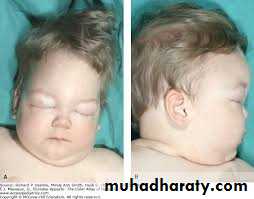
Congenital NS
Eye of Pierson syndromeIdiopathic NS:
• Its occur in approximately 90% of children with nephrotic syndrome.• Is associated with primary glomerular disease without an identifiable causative disease or drug.
Histological types:
1. Minimal change NS (MCNS):
• >95% of children with MCNS respond to corticosteroid therapy.
• About 85% of idiopathic NS
• Glomeruli appear normal or show a minimal increase in mesangial cells and matrix.
• Approximately 50% of patients respond to C.S. therapy.
2. Mesangial proliferation• Characterized by a diffuse increase in mesangial cells and matrix on light microscopy.
• 10-20% of primary NS
• The disease is often progressive, involving all glomeruli and ultimately leads to end-stage renal disease in most patients.
3. focal segmental glomerulosclerosis (FSGS):
• There is mesangial cell proliferation and segmental scarring on light microscopy.
• Only 20% respond to C.S therapy.
Thank you for your attention
• 5-15% of primary NS