TESTICULAR TUMOUR
TESTICULAR TUMOUR• 1% of all Malignant Tumour
• Affects young adults - 20 to 40 yrs - when Testosterone Fluctuations are maximum
• 90% to 95% of all Testicular tumours from germ cells
• 99% of all Testicular Tumours are malignant.
• Causes Psychological & Fertility Problems in young
Survival in Testicular Tumours
Improved overall survival in last 15 to 20 years due to -Better understanding of Natural History and Pathogenesis of disease
Reliable Tumour Markers
Cis-platinum based chemotherapy
CROSS SECTION OF TESTIS
TestisStroma Seminiferous Tubules
(200 to 350 tubules)
Interstitial Cells Supporting Spermatogonia
Leydig or(Androgen) Sertoli Cell
EPIDEMIOLOGY
Incidence : 1% of all Malignant TumorAge : 3 Peaks
- 20-40 yrs. Maximum
- 0 - 10 yrs.
- After - 60 yrs.
Bilaterality : 2 to 3% Testicular Tumor
CLASSIFICATION
I. Primary Neoplasma of Testis.A. Germ Cell Tumour
B. Non-Germ Cell Tumour
II. Secondary Neoplasms.
III. Paratesticular Tumours.
I. PRIMARY NEOPLASMS OF TESTIS
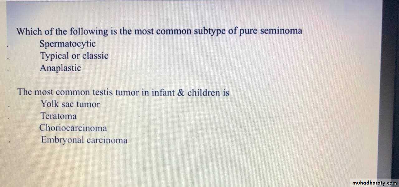
• Germinal Neoplasms : (90 - 95 %)1. Seminomas - 40%
(a) Classic Typical Seminoma(b) Anaplastic Seminoma
(c) Spermatocytic Seminoma
2. Non seminomatous GCT 1. Embryonal carcinoma- 20-25% 2. Teratoma- 25-35% (a) mature (b) Immature 3. choriocarcinoma -1% 4. Yolk sac tumor
I. PRIMARY NEOPLASMS OF TESTIS
B. Nongerminal Neoplasms : ( 5 to 10% )1. Specialized gonadal stromal tumor
(a) Leydig cell tumor
(b) Other gonadal stromal tumor
2. Gonadoblastoma
3. Miscellaneous Neoplasms
(a) Adenocarcinoma of the rete testis
(b) Mesenchymal neoplasms
(c) Carcinoid
(d) Adrenal rest “tumor”
A. Adenomatoid
B. Cystadenoma of Epididymis
C. Mesenchymal Neoplasms
D. Mesothelioma
E. Metastases
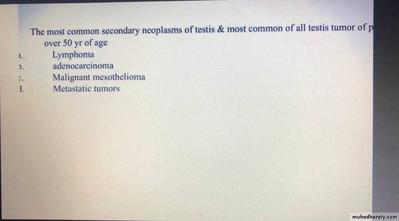
II. SECONDARY NEOPLASMS OF TESTIS
A. Reticuloendothelial Neoplasms
B. Metastases
III. PARATESTICULAR NEOPLASMS
AETIOLOGY OF TESTICULAR TUMOUR
• 1. Cryptorchidism• 2. Carcinoma in situ
• 3. Trauma
• 4. Atrophy
CRYPTORCHIDISM & TESTICULAR TUMOUR
Risk of Carcinoma developing in undescended testis is14 to 48 times the normal expected incidence
CRYPTORCHIDISM & TESTICULAR TUMOUR
The cause for malignancy are as follows:
Abnormal Germ Cell Morphology
Elevated temperature in abdomen & Inguinal region as opposed to scrotum
Endocrinal disturbances
Gonadal dysgenesis
A-Seminoma
-35% of GCT.-3 types classic, spermaocytic & anaplastic
-The classic seminoma is most common(85%) of
all seminoma & is most common in the 4th decade
of life.
-Microscopically monotonous sheets of large cells with clear cytoplasm & densely staining nuclei are seen.
-Syncytiotrophoblastic elements are seen in approximately 10-15% of cases, an incidence of
hCG production in seminoma.
B-Embryonal cell ca
-20%*2 variants of embryonal cell ca are common
-adult type &
-infantile type (yolk sac tumor) which is the most common testicular tumor of infant & children.
C-Teratoma
-May be seen in both adult & children.
-They contain more than one germ cell layer in various stages of maturation & differentiation.
-Grossly the tumor appears lobulated & contains variable size cysts filled with gelatinous or mucinous material.
Mature teratoma
may have elements resembling structures derived from (ectoderm, mesoderm, &endoderm)while immature teratoma consist of undifferentiated primitive tissue.
-Mature teratoma doesn’t attain the same degree of differentiation as teratoma of the ovary.
D- Choriocarcinoma <1%
-lesions tend to be small within the testis-usually demonstrate central hemorrhage on gross inspection.
*Microscopically syncytio & cytotrophoblast must be visualized.
*Clinically choriocarcinoma behave in an aggressive fashion characterized by early hematogenous spread.
-Paradoxically, small intratesticular lesion can be associated with wide spread metastatic disease.
E- Mixed cell type 40%
Within the mixed cell types most are teratocarcinomas about 25% of all testicular
tumor.
Which are combination of teratoma & embryonal
cell ca.
Treatment of mixture testicular tumor SGCT & NSGCT similar to that of NSGCT.
F- Carcinoma in situ (CIS)
CIS is demonstrated in 5.2% of the contralateral testis.
If diagnosed CIS is usually treated by external beam radiation therapy.
Pattern of metastatic spread.
With the exception of choriocarcinoma which demonstrates early hematogenous spread to the lung, germ cell tumor typically spread in stepwise lymphatic fashion.In paraaortic LN it extend from T1 to L4 but are concentrated at the level of renal hilum because of their common embryologic origin of the kidney.
Certain factors may alter the primary drainage of the testis neoplasm.
-Invasion of epididymis or spermatic cord may allow spread to external iliac & obturator LN,-scrotal violation or invasion of tunica albuginea may result in inguinal metastases.
-Visceral metastases may be seen in advanced disease. Lung, liver, brain, bone, or adrenal may be involved.
TNM staging of testicular carcinoma
TX-Primary tumor cannot be assessedT0-No evidence of primary tumor
Tis-Carcinoma in situ
T1-Tumor has not spread beyond the testicle and epididymis
T2-Tumor has spread to blood vessels, lymphatics, or tunica vaginalis
T3-Tumor invades spermatic cord
T4-Tumor invades scrotum
NX-Regional nodes cannot be assessed
N0-No nodal metastasis
N1-Metastasis to single lymph node < 2 cm in greatest diameter
N2-Metastasis to single lymph node > 2 cm, but < 5 cm, or multiple nodes < 5 cm
N3-At least one node > 5 cm
MX-Distant metastasis cannot be assessed
M0-No distant metastasisM1-Distant metastasis is present
S1
LDH < 1.5× normal,
b-hCG < 5000 mIU/ml,
a-FP < 1000 ng/ml
S2
LDH 1.5–10× normal,
b-hCG 5000–50 000 mIU/ml,
a-FP 1000–10 000 ng/ml
S3
LDH >10× normal,
b-hCG >50 000 mIU/ml,
a-FP 10 000 ng/ml
Clinical findings.
-Painless testicular swelling is the most common presentation, the enlargement is usually gradual its usually firm & nontender
-testicular heaviness is not unusual.
-The typical delay of treatment from initial recognition of the lesion by the pt to definitive therapy (orchiectomy) range from 3-6 months.
-The length of delay correlate with incidence of metastases.
10% of pt presented with acute testicular pain may be the result of intratesticlar infarction or hemorrhage
10% presented with symptom related to metastatic disease,
-back pain (retroperitoneal metastases involving nerve roots) is the most common symptom.
-Other symptoms include cough or dyspnea (pulmonary metastases),
-anorexia nausea or vomiting (retro duodenal metastases),
-bone pain(skeletal metastases),
-lower extremity swelling (venacaval obstruction).
10% of pt are asymptomatic at presentation tumor detected incidentally like after trauma.
5-10% of testicular tumor may presented with hydrocele & help to camouflage it.*Gynecomastia present in
-5% of all GCT &
-30-50% of sertoli& leydig cell tumor due to complex hormonal interaction.
DICTUM FOR ANY SOLID SCROTAL SWELLINGS
All patients with a solid, Firm Intratesticular Mass that cannot be Transilluminated should be regarded as Malignant unless otherwise provedTumour Markers
TWO MAIN CLASSES
Onco-fetal Substances : AFP & HCG
Cellular Enzymes : LDH & PLAP
( AFP - Trophoblastic Cells
HCG - Syncytiotrophoblastic Cells )
AFP –( Alfafetoprotein )
• NORMAL VALUE: Below 16 ngm / ml• HALF LIFE OF AFP – 5 and 7 days
• Raised AFP :
• Pure embryonal carcinoma
• Teratocarcinoma
• Yolk sac Tumour
• Combined Tumour
REMEMBER: AFP Not raised is Pure Choriocarcinoma or Pure Seminoma
HCG – ( Human Chorionic Gonadotropin )
Has and polypeptide chainNORMAL VALUE: < 1 ng / ml
HALF LIFE of HCG: 24 to 36 hoursRAISED HCG -
100 % - Choriocarcinoma
60% - Embryonal carcinoma
55% - Teratocarcinoma
25% - Yolk Cell Tumour
7% - Seminomas
ROLE OF TUMOUR MARKERS
Helps in Diagnosis - 80 to 85% of Testicular Tumours have Positive MarkersMost of Non-Seminomas have raised markers
Only 10 to 15% Non-Seminomas have normal marker level
After Orchidectomy if Markers Elevated means Residual Disease or Stage II or III Disease
Elevation of Markers after Lymphadenectomy means a STAGE III Disease
ROLE OF TUMOUR MARKERS cont...
Degree of Marker Elevation Appears to be Directly Proportional to Tumour BurdenMarkers indicate Histology of Tumour:
If AFP elevated in Seminoma - Means Tumour has Non-Seminomatous elements
Negative Tumour Markers becoming positive on follow up usually indicates -
Recurrence of Tumour
Markers become Positive earlier than X-Ray studies
Imaging
Primary testicular tumor can be rapidly & accurately assessed by scrotal ultrasound.
This technique can determine
-whether the mass is truly intratesticular,
-can be used to distinguish the tumor from epididymal pathology, &
-may also facilitate testicular examination in the presence of hydrocele.
Once the diagnosis is established by inguinal orchiectomy,
-chest x-ray & abdominal CT used to assessthe 2 most common sites of metastatic spread
( the lung & retro peritoneum).
Differential diagnosis
1-Epididymoorchitisis the most common misdiagnosis,
history of acute onset of symptoms, fever, urethral discharge, & irritative voiding symptoms make the diagnosis of epididymitis more likely.
2-Hydrocele
trnsillumination of scrotum readily distinguish between fluid filled hydrocele & solid testicular tumor.Since 5-10% of testicular tumor may be associated with hydrocele ultrasound study of the scrotum is mandatory.
3-Others like spermatocel, hematocele, granulomatous orchitis, most commonly result from tuberculosis.
Treatment
Inguinal exploration with cross clamping of the spermatic cord vasculature & delivery of the testis into the field is the main stay of exploration for possible testicular tumor.Scrotal approach & open testicular biopsy should be avoided.
Further therapy depend on the histologic characteristic of the tumor as well as the tumor stage. SGCT are radiosensitive & chemosensitive while NSGCT are chemosensitive.