
Spacing in Orthodontics:
Ass.Prof.Dr.Zaid Al.Dewachi
Spacing is a common condition in deciduous dentition and constitutes a
very important feature of the dentition, as it is an indicator for the
favorable development of permanent teeth. Lack of spacing suggests
Severe risk for crowding in the permanent dentition (Foster and Grundy,
1986),
The incidence of spacing in primary was reported around 90% Spacing is
more common in the maxilla rather than the mandible And in boys
rather than girls, Arch size seems to play a more important role than
teeth size; Spacing is more common in wider arches beside that found
that mandibular incisors and canines were smaller in children with
spacing, whereas in children who had only primate spaces, only
mandibular incisors were statistically smaller. However, a general finding
was that children with any type of spacing in the primary dentition
Had wider jaws.
SPACING IN THE PERMANENT DENTITION:
A dentition with spaces is considered a normal type of occlusion, which
is found in almost one third of the population, (21.4%) of the general
Population presented spacing in both arches,(50%) of the population
had spacing in one arch and In individuals with spacing in only one of
the arches, the condition was twice as frequent in the maxillary arch.
Maxillary spacing is more common in the anterior part of the maxilla
In studies of young populations, spacing in both arches was more
common in boys than in girls.
However, more thorough observation of gender differences reveals that,
in ages older than 16-18 years, spacing incidence is the same in both
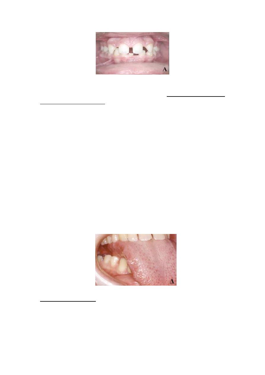
boys and girls. Concerning the median maxillary diastema, found that its
incidence is 36.8%. Median diastema was the only spacing present in a
mere 1.6% of individuals with spacing, Most large spaces are mesial and
distal to the canines, followed by the diastema located between central
and lateral incisors . It is true that spacing tends to decrease in the
permanent dentition as age increases.
It is reported that during dental maturation spaces distal to the canines
tend to close, while new spaces, mesial to this tooth, usually appear.
This may be due to third molar eruption or to molar tendency for mesial
migration and premolar and canine tendency for distal migration. The
stability of the space mesial to first premolars is greater than that of the
distal one, while the maxillary median diastema seems to be the most
stable.

ETIOLOGY OF SPACING
The causes of spacing may be hereditary, acquired or functional.
Hereditary causes include tooth size - arch size discrepancies,
congenitally missing teeth, Macroglossia, supernumerary teeth, small
teeth and hypertrophic upper lip frenum. Functional causes include bad
oral habits, whereas acquired causes include pathologic conditions
Increasing tongue size, missing teeth, delayed eruption of permanent
teeth and periodontal disease.
Tooth size - jaw size discrepancy
In spacing cases caused by tooth size - jaw size discrepancy), the
problem lies with jaw size. It has been found that individuals with bigger
faces and jaws usually have spacing and not crowding, in male patients
with spacing:
a.)intercanine and interpremolar distances were greater only in the
maxilla.
b) Mean dental width in men did not differ between those with and
those without spacing.
c) In contrast, in female patients with spacing, central incisors, canines
and all posterior teeth were found to be significantly narrower.
d) In women, dental arch size was not related to spacing. Thus, it may
be concluded that most frequently spacing is mainly due to greater jaw
size and not to smaller teeth.
Congenitally missing teeth
Regarding the congenitally missing teeth, several genetic and
environmental factors have been implicated as the cause of Spacing.
Existing teeth are often smaller, with an atypical conical shape and
create esthetic and functional problems worsening the spacing problem;
congenitally missing teeth may be either an isolated clinical sign or a
syndromic feature, especially in cases of more than 6 missing teeth, thus
resulting in extensive spacing.
Second mandibular premolars are the most common congenitally
missing teeth, followed by maxillary lateral incisors and second Maxillary
premolars, other teeth, such as upper central incisors, upper and lower
canines or first molars are rarely missing congenitally and, if so, this is
usually a syndromic feature.
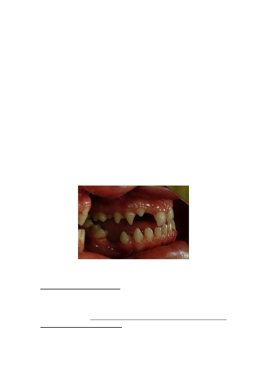
Macroglossia
True macroglossia is a condition where the tongue is bigger than normal.
Macroglossia constitutes an etiological factor for spacing, open bite and
protrusion of both jaws. A large tongue may also compromise the
stability of orthodontic treatment outcome and cause masticatory,
swallowing, respiratory and speech problems, the causes of true
macroglossia may be hereditary or acquired, Macroglossia diagnosis may
be performed when the tongue occupies the entire oral cavity, when
impressions of the lingual surfaces of mandibular teeth are present at
the lateral tongue margins or when the patient is capable of touching
the chin or the nose tip with her/his tongue. Tongue size can be
estimated with direct measurement, indirect measurement through an
impression and, finally, with magnetic tomography. Certain
cephalometric measurements may also aid in diagnosing macroglossia,
however, due to lack of practical methods for measuring tongue size, it
is sometimes difficult to assess to what extent macroglossia is
responsible for malocclusion.
Pseudomacroglossia is also an etiologic factor for spacing. Tongue size is
normal, but it appears larger than other anatomical features because
certain causes force the tongue to an anterior position. This condition
results in spacing, which is more pronounced in the anterior dental arch
(Macroglossia )
Supernumerary teeth
Supernumerary teeth constitute one of the causes for local interdental
spaces, as they interfere with the eruption of neighboring teeth or
displace them out of the arch. Incidence in the permanent dentition
ranges between 0.5% and 3.8%, whereas in the deciduous dentition
The condition is rarer with an incidence of 0.35- 0.6%, approximately
75% of supernumerary teeth are located in the maxilla, the most
common supernumerary teeth are maxillary mesiodentes (46.9%),
followed by premolars (24.1%) and molars (18%).It was also found that
patients with supernumerary teeth have larger teeth in general. This
leads to lack of space for the eruption of the remaining teeth even
After the supernumerary one is removed.
Small teeth and teeth with crown anomalies
Small teeth usually result in generalized spacing, Small teeth and teeth
with smaller and anomalous crowns may also be the cause of localized
spacing, approximately 5% of the population presents some degree of
discrepancy concerning tooth sizes .It has also been found that
oligodontia and microdontia occur more often in women, whereas
megalodontia and supernumerary teeth are more common in men
Developmental anomalies that result in changes of tooth shape and size
are found in all permanent teeth ranking in the following order of
frequency: third molars, maxillary lateral incisors and mandibular second
premolars . The cause of dental shape or size anomaly may be congenital
or acquired.
Hypertrophic upper lip frenum
Hypertrophic upper lip frenum has long been held responsible for
median diastema, however, diastemata, which sometimes create severe
esthetic problems due to their location, may also be due to other causes.
The latter include incomplete fusion of the two osseous parts of the
premaxilla at the suture, congenitally missing lateral incisors,
supernumerary teeth at the midline, small teeth or even the
combination of suture deficiency at the midincisor area and congenitally
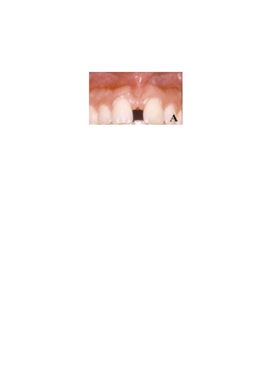
missing lateral incisors. It must be stressed, however, that the median
diastema is often a normal feature of the stomatognathic system
development, especially during the initial phase of permanent upper
Central incisor eruption (the “ugly duckling” stage)
Deleterious oral habits
Harmful oral habits constitute another cause of generalized spacing or
localized interdental spaces usually appearing among anterior teeth
Pathological causes of tongue augmentation
The main pathological conditions leading to tongue augmentation
Are acromegaly, myxedema, lymphangioma, amyloidosis, tertiary
syphilis, cysts or tumors affecting the tongue and nerve injury.
Lost teeth – Permanent teeth extractions
It is well known that the percentage of individuals with spacing is clearly
higher among people with a dental history of permanent teeth
extractions.
The residual spaces after first molar extraction are distributed mainly at
the posterior and partially at the anterior dental arches in both jaws. On
The other hand, the residual spaces after first molar extractions are
distributed over the whole of the dental arch in the mandible, whereas
in the maxillary arch these spaces are limited between canines and
second permanent molars. In the anterior maxillary area, a correlation
has been found between spacing and extractions of permanent teeth
mesial to first molars.
Delayed eruption of permanent teeth
Chronic and Aggressive periodontitis

Treatment of spacing:
Factors to be considered in a comprehensive treatment plan for spacing
include
1. Initial cause of the problem
2. Patient age
3. Location and extent of spacing,
4. Number and status of existing teeth,
5. Periodontal tissue condition,
6. Free inter-maxillary space,
7. Possible malocclusion,
8. Patient expectations
9. Certain socioeconomic factors
Alternative therapeutic approaches for spacing include:
(1) No treatment or esthetic restoration with composite resins
Individuals with few; small spaces who feel their dental
appearance is satisfactory may be left without any treatment.
This is usually the case when spaces are distal to the canine or
when they are not visible during speech and smiling, these cases
are acceptable when the risk for malocclusion development due
to tooth migration is excluded. In other cases it is possible to
close small spaces with tooth reshaping using composite resins
.It should be noted that, within treatment context, a small
residual space, especially distal to the lateral incisors, may be
considered acceptable in certain cases.
(2). Orthodontic space closure :Orthodontic space closure has always
been considered the most appropriate treatment alternative, as
prosthetic restorations used for spacing treatment may
sometimes create periodontal problems .). Furthermore, fixed
prostheses always involve the loss of healthy dental tissue.
Finally, finances should also be considered, since there may be a
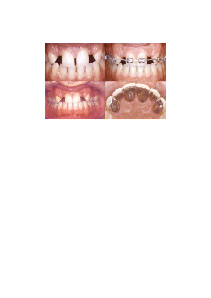
need to replace the prosthetic restoration two or three times
during a patient’s life.