[1]
Disorders of the adrenal glands
ADRENAL CORTICAL ADENOMA AND CARCINOMA
Adrenal cortical adenomas and carcinomas may nonfunctioning or
functioning. The term functioning refers to metabolically active
tumors that produce excessive amounts of adrenal cortical
hormones. The most common clinical syndromes associated with
a functioning adrenal cortical adenoma and functional carcinoma
are primary hyperaldosteronism or Cushing's syndrome.
however, many patients also have evidence of virilization or
Feminization
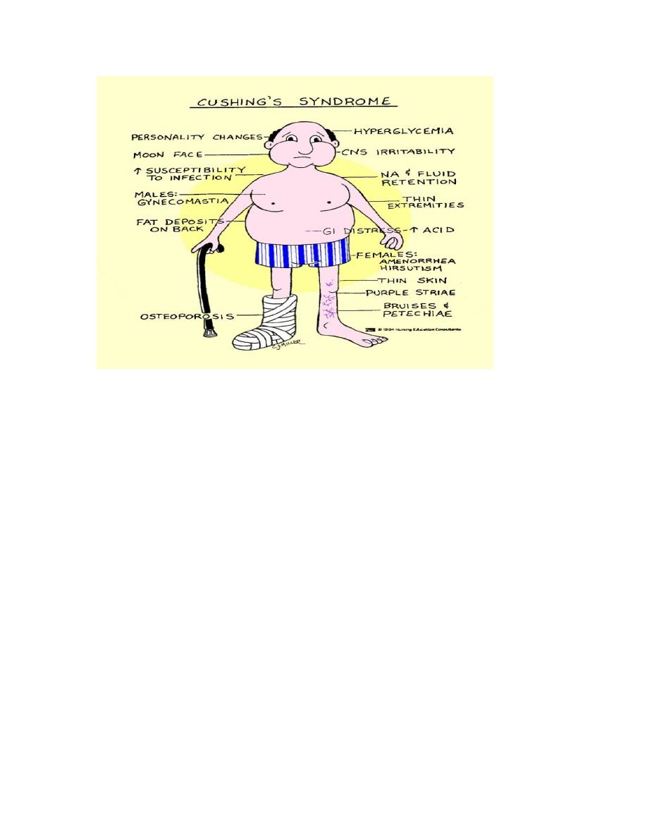
Cushing's syndrome
Cushing's syndrome is caused by excessive adrenal secretion of
corticosteroid with a resulting characteristic clinical presentation
of truncal obesity, impotence or gynecomastia in the male,
increased bruising andstriae,hypertension,osteoporosis,peripheral
extremity muscle wasting

[2]
Cushing's syndrome may be due to:
• a pituitary adenoma (Cushing's disease, 70%),
• an ectopic ACTH-producing tumor 10%
• a primary adrenal cortical tumor (20%).
Radiographic imaging tests
CT, MRI, or ultrasonography.
In patients with Cushing's syndrome due to a primary adrenal
tumor, the underlying pathology may be a benign adenoma or an
adrenal cortical carcinoma. An adrenal adenoma is more likely
when the size of the lesion is < 6 cm and when pure Cushing's
syndrome is present

[3]
treatment for Cushing's disease
• Cushing's disease of pituitary origin treated by
transsphenoidal hypophysectomy
•IF treatment is ineffective then bilateral surgical adrenalectomy
• Cushing's syndrome due to a primary adrenal tumor treated
by Surgical adrenalectomy
Clinical presentation of adrenal cortical carcinoma :
• 50% functioning with symptoms related to excessive adrenal
cortical steroid production.
• 50% are nonfunctioning tumors and these patients present
with nonspecific symptoms such as abdominal pain, mass,
fatigue, and weight loss.
Treatment : Complete surgical excision
PRIMARY ALDOSTERONISM
It is a secondary cause of hypertension characterized by excessive
and unregulated secretion of aldosterone.
Etiology
• An adrenal cortical adenoma
• bilateral adrenal hyperplasia
This distinction is important because adenomas respond to
surgery, whereas hyperplasia is treated medically.

[4]
Clinical features
• Hypertension is a central feature of the disease.
• Other symptoms are nonspecific and may include polyuria,
nocturia, proximal muscle weakness, and headaches
The biochemical features of primary aldosteronism.
1. Hypokalemia
2. High plasma aldosterone
3. Low plasma renin activity
4. Metabolic alkalosis
Screened for the disease
A Hypertensive patients with:
• Spontaneous hypokalemia
• Moderately severe hypokalemia after conventional diuretic
therapy
• Refractory hypertension
How is the diagnosis confirmed?
The best way to confirm the diagnosis of primary aldosteronism is
to demonstrate nonsuppressible aldosterone secretion during
prolonged salt repletion.

[5]
The localization procedures
• Computed tomographic (CT) scan of the adrenals,
Bilaterally
enlarged adrenals suggest hyperplasia
• Scintigraphy
• Adrenal vein sampling for aldosterone
Indications for surgery
• primary aldosteronism
• unilateral adenoma.
Spironolactone is used to treat primary aldosteronism medically
PHEOCHROMOCYTOMA
Pheochromocytoma is a tumor derived from chromaffin cells that
is associated with pathologic secretion of catecholamines
(norepinephrine and epinephrine).
Location
• About 90% are located in the adrenal gland;
• 10% may be extra-adrenal.
Most extra-adrenal pheochromocytomas are associated with
sympathetic ganglia in the retroperitoneum
Rule of 10%
10% of tumors are:
Extra-adrenal

[6]
Malignant
Associated with MEN syndromes
Bilateral
Pediatric
Symptoms:
The symptoms are those of excessive catecholamine secretion
and include the classic triad of headaches, sweating, and
palpitations. Pheochromocytoma, however, can present with var-
ious nonspecific symptoms, including tremors, nausea, dyspnea,
fatigue, dizziness, and chest or abdominal pain.
Physical findings
• Hypertension most common which may be sustained or
paroxysmal.
• . signs of catecholamine excess include tachycardia, tremor,
, and Raynaud's phenomenon.
Who should be evaluated?
Priority for evaluation should be given to patients with:
• Headaches, sweating, and palpitations
• Incidental adrenal mass
• Hypertensive crisis with surgery, anesthesia, or parturition
• Family history of pheochromocytoma

[7]
Localization of pheochromocytomas
1. CT scan of the abdomen or pelvis
2. Magnetic resonance imaging (MRI)
3. Metalodobenzylguanidine (MIBG)
Preoperative regimen:
The goal of preoperative management to prevent cardiovascular
morbidity due to severe hypertension. The standard medical
preparation has been to treat patients with the nonslictive a-
adrenergic blocker, phenoxybenzamine, for 4 weeks before
surgery.
beta-blocking drugs are used to control cardiac arrhythmias. In
these cases, a-blockade should be in place first to avoid a
paradoxical hypertensive crisis.
In addition to medications, many of these patients are volume
depleted and require vigorous intravenous hydration the day
before surgery.
NEUROBLASTOMA
• the most common extracranial solid tumor of childhood,
• 80% of children are diagnosed at < 4 years of age.
• These tumors are of neural crest cell origin and can occur
anywhere in the neuroectodermal chain.
• Approximately 50% arise in the adrenal medulla, and most
of the others occur along the sympathetic chain

[8]
Presention:
• Non specific :like fever, abdominal pain, abdominal mass,
weight loss, anemia, bone pain, and/or proptosis and perior-
bital ecchymoses.
• Neuroblastomas may also present on prenatal
Ultrasonography.
Staging System for Neuroblastoma
Stage 1 Tumor confined to organ of origin with grossly complete
excision
Stage 2A Unilateral tumor with gross residual after resection
Stage 2B Unilateral tumor with positive ipsilateral lymph nodes
Stage 3 Tumor crossing the midline or positive contralateral
lymph nodes
Stage 4 Metastatic disease beyond regional lymph nodes
Stage 4S Unilateral tumor with or without positive ipsilateral
lymph nodes with metastatic disease limited to the liver, skin,
and/or bone marrow
Stage 4S reflects a unique expression of metastatic
neuroblastoma. Patients are generally < 1 year of age and have
localized primary tumors, as well as metastases limited to the
liver, skin, and bone marrow. These tumors have a tendency to
resolve with little or no treatment.
urinary catecholamine metabolites measured

[9]
In addition to radiographic evaluation, all patients undergo a 24-
hour urine collection for measurement of catecholamine
metabolites. Urinary homovanillic acid (HMA) and/or vanillyl-
mandelic acid (VMA) levels are elevated in more than 90% of
patients with neuroblastoma.
MIBG scan
(MIBG) is an amine precursor that is concentrated in neuroblas-
tomas and other neuroendocrine tumors. MIBG scans are very
sensitive for detecting neuroblastomas.
Treatment :
• Patients with low-stage favorable tumors may be treated
with surgical excision alone.
• Patients with higher risk tumors require adjuvant multiagent
chemotherapy and sometimes radiotherapy as well.
