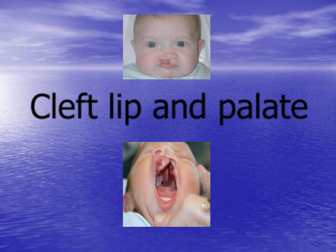
Cleft lip and palate
Cleft lip and cleft palate
are the
second
most
frequently occurring of the major congenital
anomalies they occur in
1:750-1:1000
,club foot
being the most common.
Racial and ethnic variations exist, with clefting
occurring more commonly in Asians and less
frequently in Africans, while whites are
intermediate in occurrence.
1-Medication:
e.g. phyenytoin, steroid, diazepam, retinoic
acid
2-Smoking.
3-Parental age
especially father age, or both mother and
father age over 30 years.
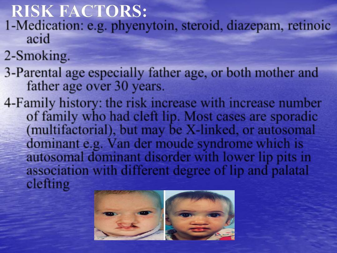
4-Family history:
the risk increase with increase number
of family who had cleft lip. Most cases are sporadic
(multifactorial), but may be X-linked, or autosomal
dominant e.g.
Van der moude syndrome
which is
autosomal dominant disorder with lower lip pits in
association with different degree of lip and palatal
clefting
.
5-Folic acid and B6:
intake during pregnancy may reduce
cleft lip and cleft palate.
6- low socioeconomic status
:
this is possibly related to
inadequate nutrition.
Or associated with syndrome e.g.
Down syndrome
,
there are more than 150 syndromes described in
which the clefting may be a feature.
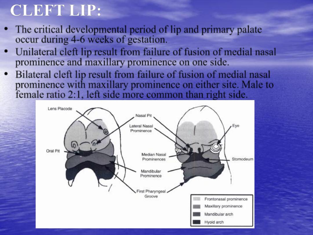
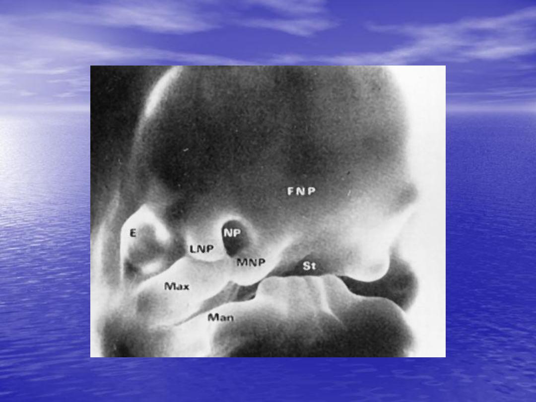
•
The critical developmental period of lip and primary palate
occur during
4-6 weeks
of gestation.
•
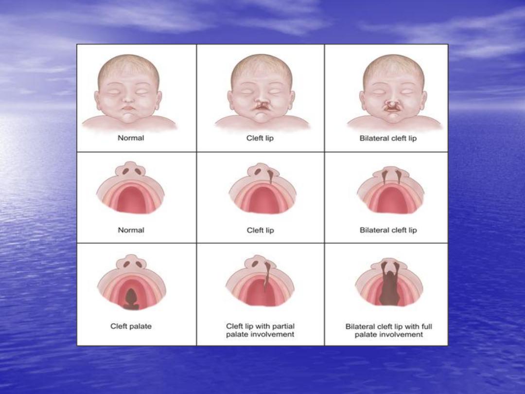
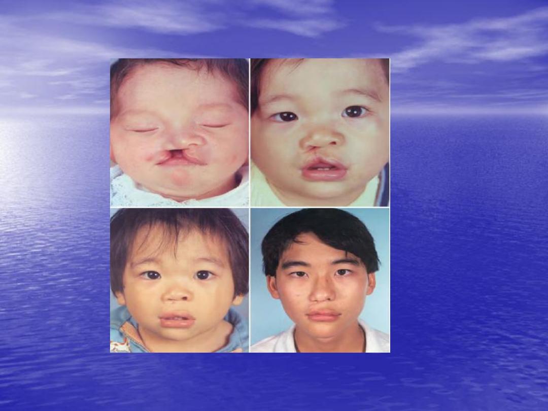
Unilateral cleft lip
result from failure of fusion of medial nasal
prominence and maxillary prominence on one side.
•
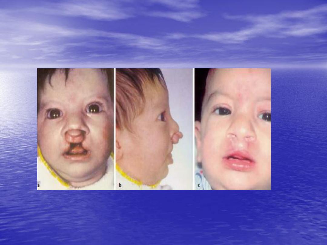
Bilateral cleft lip
result from failure of fusion of medial nasal
prominence with maxillary prominence on either site.
Male to
female
ratio 2:1
, left side more common than right side.
Unilateral cleft lip
Bilateral cleft lip and palate

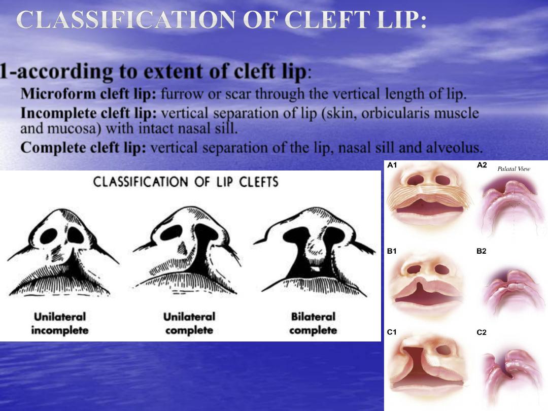
1-according to extent of cleft lip
:
–
Microform cleft lip:
furrow or scar through the vertical length of lip.
–
Incomplete cleft lip:
vertical separation of lip (skin, orbicularis muscle
and mucosa) with intact nasal sill.
–
Complete cleft lip:
vertical separation of the lip, nasal sill and alveolus.
•
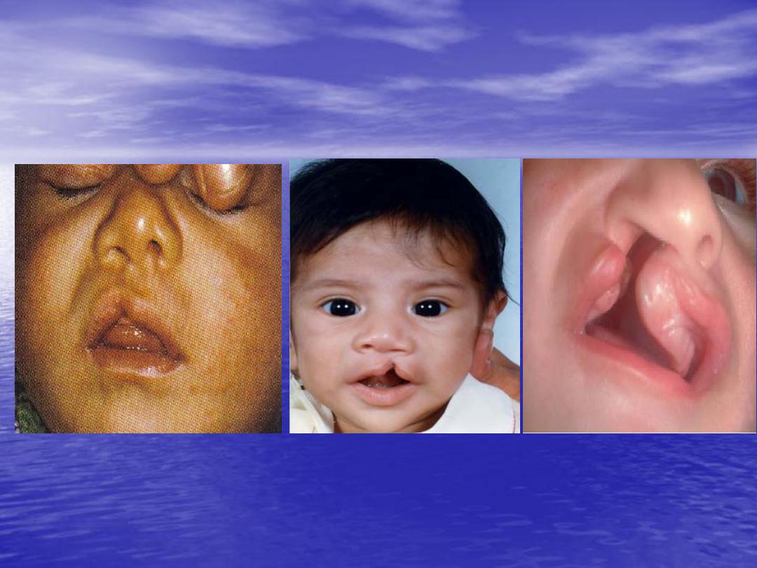
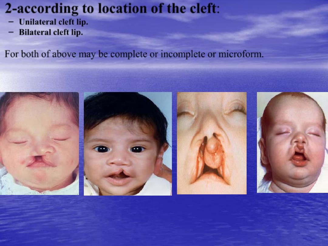
2-according to location of the cleft:
– Unilateral cleft lip.
– Bilateral cleft lip.
•
For both of above may be complete or incomplete or microform.
•
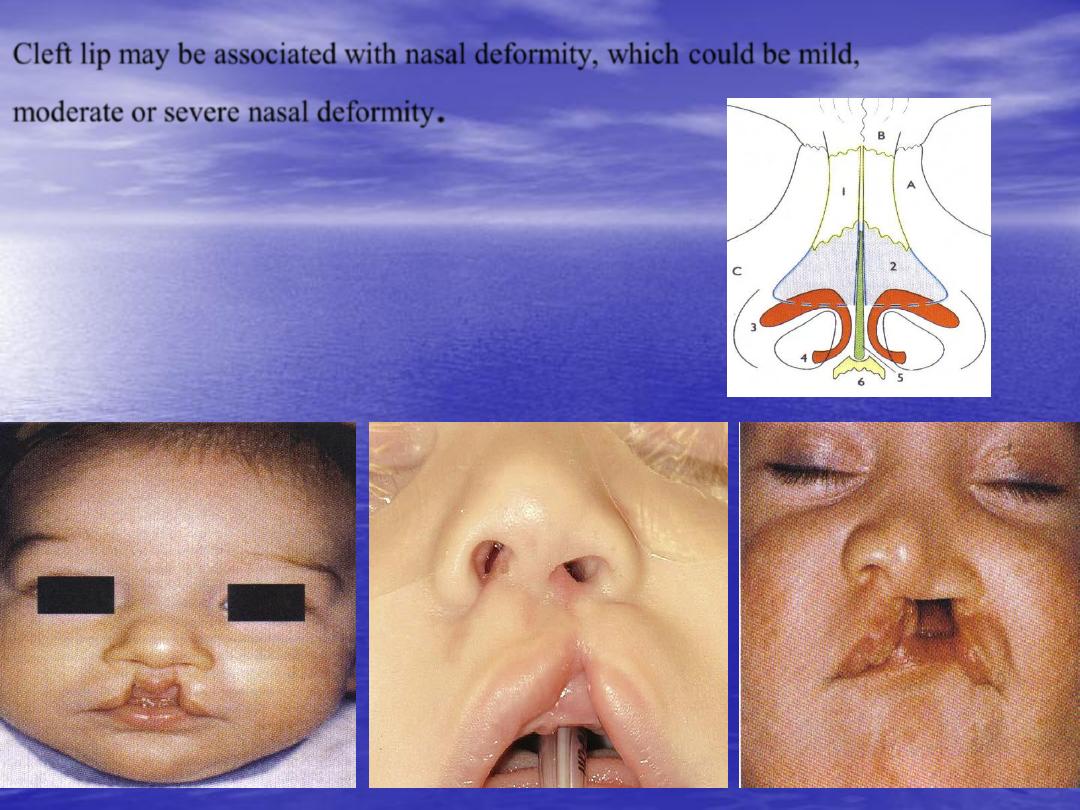
Cleft lip may be associated with nasal deformity, which could be mild,
moderate or severe nasal deformity
.
•
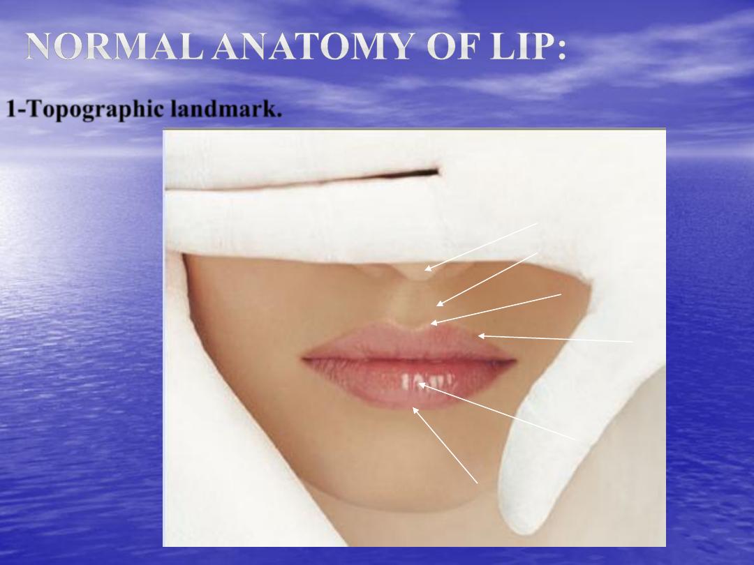
1-Topographic landmark.
Vermillion roll
Wet vermillion
Dry vermillion
Cupid’s bow
Philter columns
Columella

•
2-muscles:
– Orbicularis oris: function as sphincter (deep fibers) and for speech (superficial fibers).
– Levator labii superoris: elevate the upper lip.
– Nasalis or depressor septi nasi muscle: depress the columella down and elevate the
upper lip.
•
3-Arterial blood supply:
by labial artery bilaterally.
•
4-Sensory innervation:
by maxillary branch of trigeminal nerve.
•
5-Motor innervation:
by zygomatic and buccal branches of facial nerve.
•
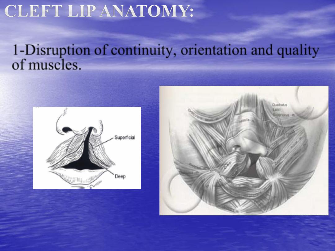
1-
Disruption of continuity, orientation and quality
of muscles.
•
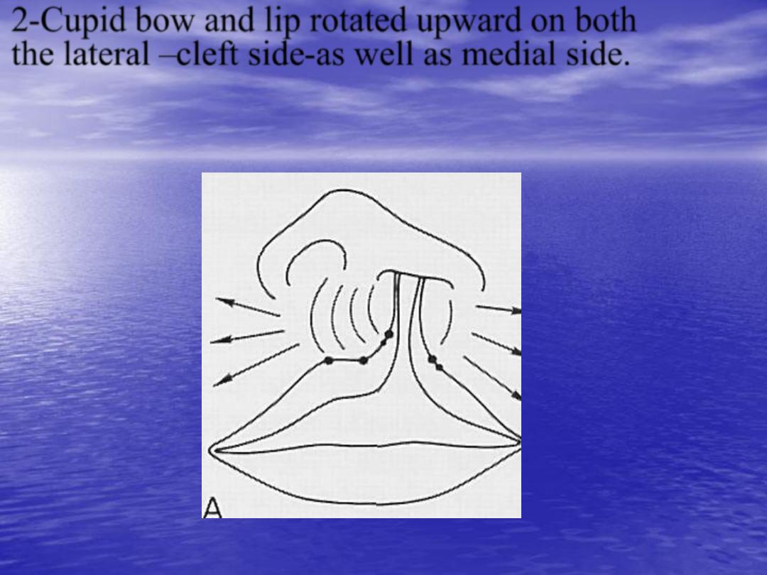
2-
Cupid bow and lip rotated upward on both
the lateral –cleft side-as well as medial side.

•
3-
The alveolus and nostril floor are open in
complete cleft lip.
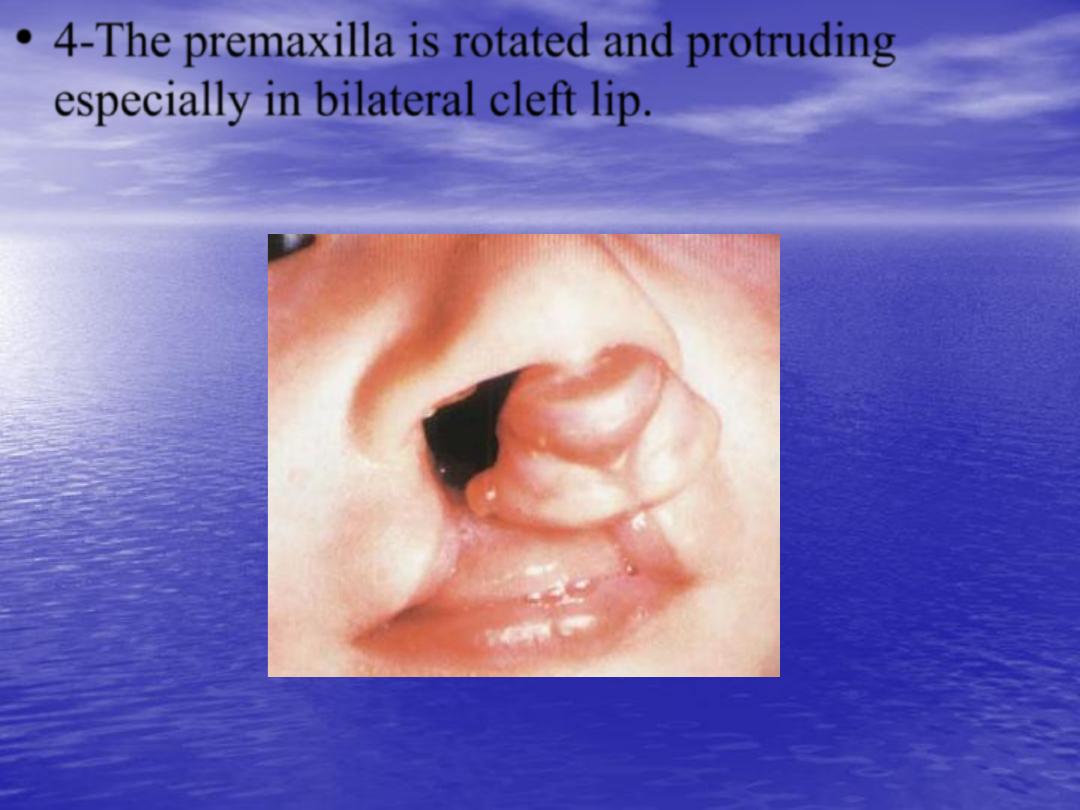
•
4-
The premaxilla is rotated and protruding
especially in bilateral cleft lip.

•
5-
Associated cleft lip nasal deformity e.g.
flatten alar dome on affected side, shortened
columella especially bilateral cases.
Deficient columella
•
The parents should be reassured, and the
newborn should evaluate for associated
anomalies, and the parents should inform about
the stages and operation that expected throughout
the child lifetime.
•
Time of repair:
according to rules of ten:
•
Should be
10 weeks
old.
•
Should be
10 pounds
(4.5kg).
•
Should be
10gm/dl
hemoglobin level.
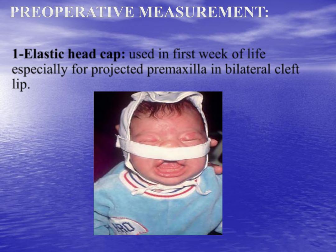
•
1-Elastic head cap: used in first week of life
especially for projected premaxilla in bilateral cleft
lip.
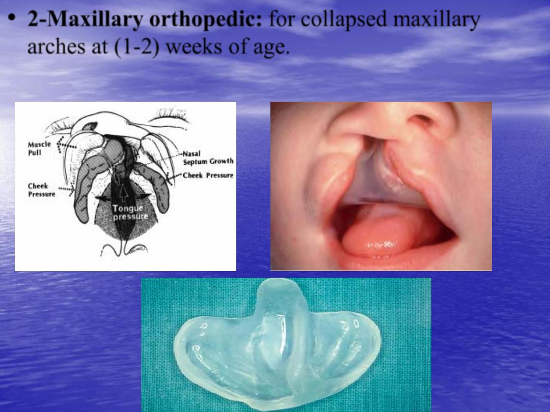
•
2-Maxillary orthopedic: for collapsed maxillary
arches at (1-2) weeks of age.
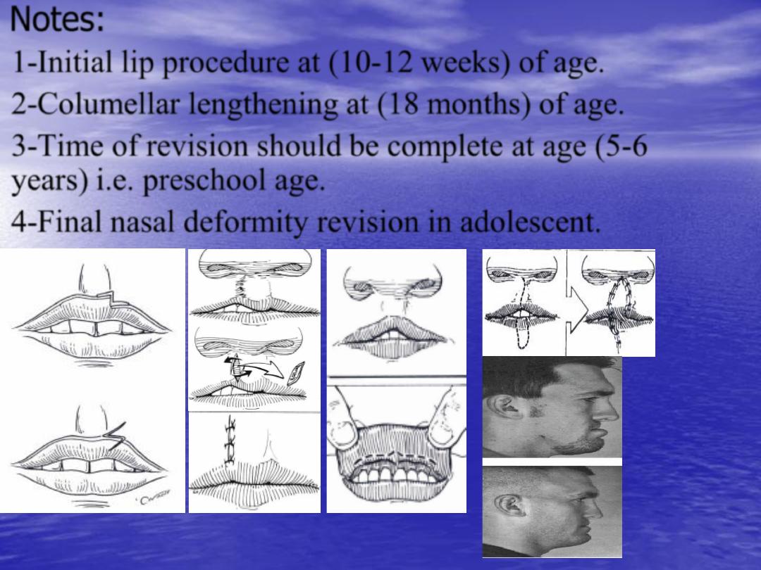
Notes:
•
1-
Initial lip procedure at
(10-12 weeks)
of age.
•
2-
Columellar lengthening at
(18 months)
of age.
•
3-
Time of revision should be complete at age
(5-6
years)
i.e. preschool age.
•
4-
Final nasal deformity revision in adolescent.
Preoperative investigation: chest X-ray to exclude any
chest infection, bleeding profile to exclude any bleeding
tendencies and Hb level.
•
Principal of repair:
•
1-
Produce functional continuity of muscles.
•
2-
Recreate symmetry.
•
3-
Reconstruction Cupid bow.
•
4-
Minimize scarring.
•
5-
Treated the associated nasal deformity.
•
6-
Should repair all layers of lip (skin, muscles, and
mucos).
•
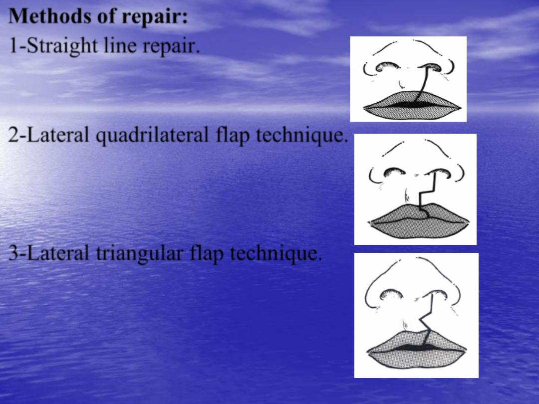
Methods of repair:
•
1-
Straight line repair.
•
2-
Lateral quadrilateral flap technique.
•
3-
Lateral triangular flap technique.
•
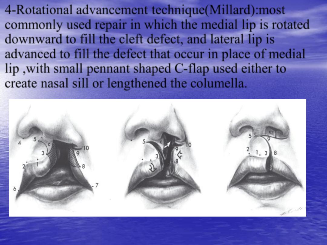
4-
Rotational advancement technique
(Millard):
most
commonly used repair in which the medial lip is rotated
downward to fill the cleft defect, and lateral lip is
advanced to fill the defect that occur in place of medial
lip ,with small pennant shaped C-flap used either to
create nasal sill or lengthened the columella.
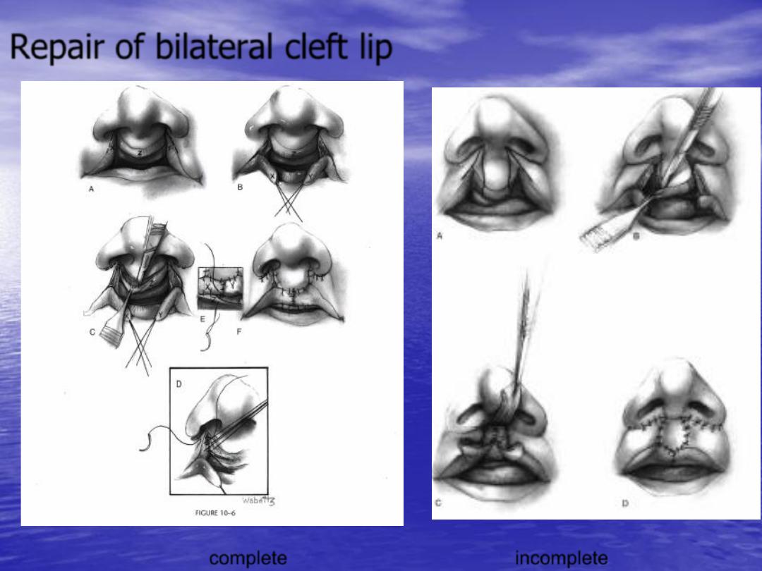
Repair of bilateral cleft lip
incomplete
complete
•
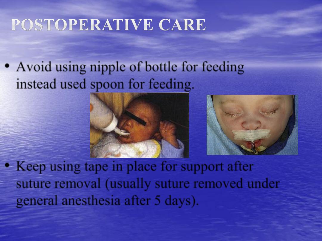
Avoid using nipple of bottle for feeding
instead used spoon for feeding.
•
Keep using tape in place for support after
suture removal (usually suture removed under
general anesthesia after 5 days).
•
It could occur as separated deformity or in combination
with cleft lip deformity. It may be unilateral or bilateral
deformity
.
•
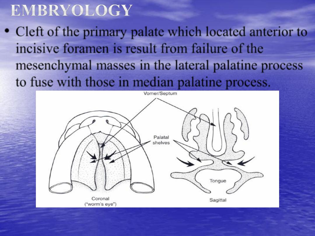
Cleft of the
primary palate
which located anterior to
incisive foramen is result from failure of the
mesenchymal masses in the lateral palatine process
to fuse with those in median palatine process.
•
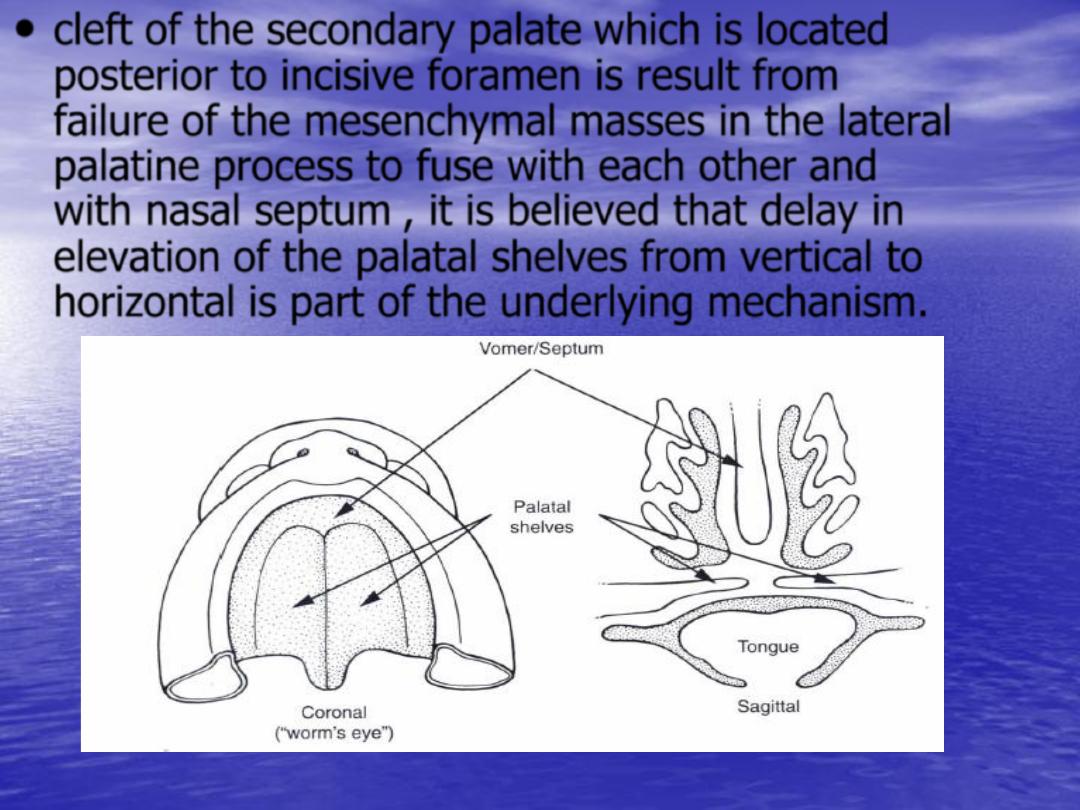
cleft of the
secondary palate
which is located
posterior to incisive foramen is result from
failure of the mesenchymal masses in the lateral
palatine process to fuse with each other and
with nasal septum , it is believed that delay in
elevation of the palatal shelves from vertical to
horizontal is part of the underlying mechanism.
•
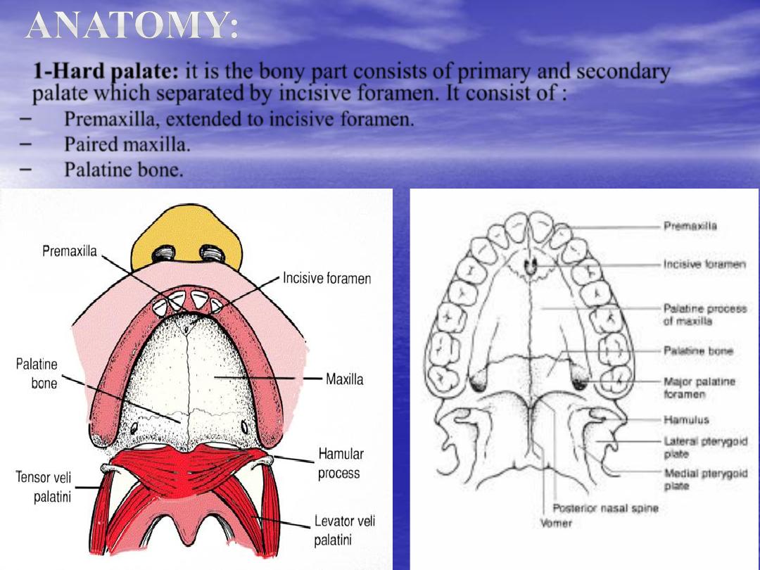
1-Hard palate:
it is the bony part consists of primary and secondary
palate which separated by incisive foramen. It consist of :
–
Premaxilla, extended to incisive foramen.
–
Paired maxilla.
–
Palatine bone.
•
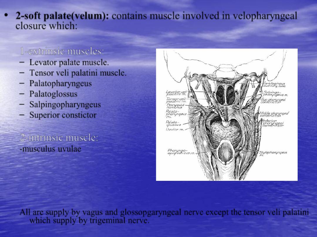
2-soft palate(velum):
contains muscle involved in velopharyngeal
closure which:
1-extrinsic muscles:
– Levator palate muscle.
– Tensor veli palatini muscle.
– Palatopharyngeus
– Palatoglossus
– Salpingopharyngeus
– Superior constictor
2-intrinsic muscle:
-musculus uvulae
All are supply by vagus and glossopgaryngeal nerve except the
tensor veli palatini
which supply by trigeminal nerve.
•
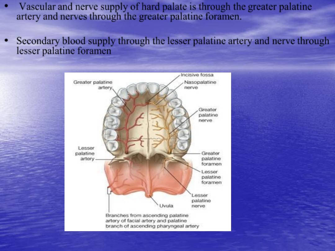
Vascular and nerve supply of hard palate is through the
greater palatine
artery and
nerves
through the greater palatine foramen.
•
Secondary blood supply through the
lesser palatine artery and nerve
through
lesser palatine foramen

•
-
The
prepalatal
structures
(primary palate)
: structure anterior to
incisive foramen (alveolus, lip, nasal floor, and alar cartilage).
•
-
The palatal
structures
(secondary palate)
: those posterior to
incisive foramen.
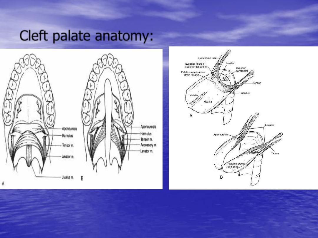
Cleft palate anatomy:
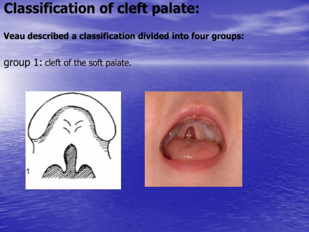
Classification of cleft palate:
Veau
described a classification divided into four groups:
group 1:
cleft of the soft palate.
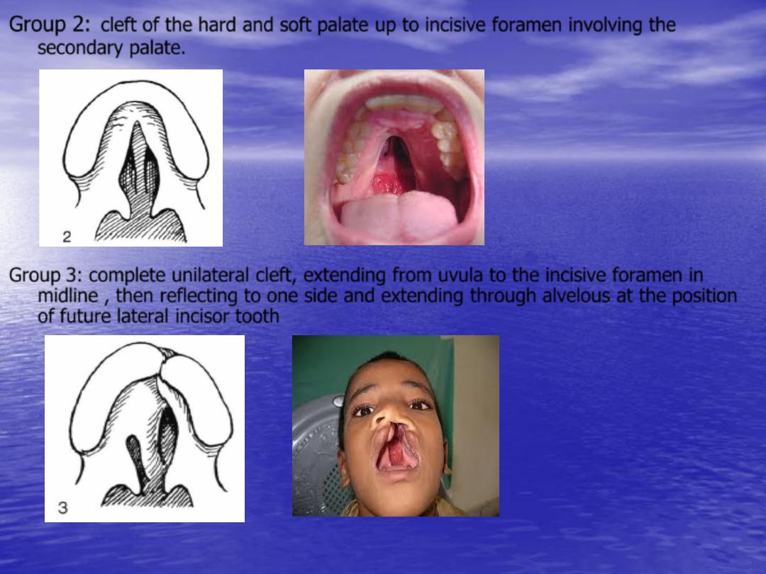
Group 2:
cleft of the hard and soft palate up to incisive foramen involving the
secondary palate.
Group 3:
complete unilateral cleft, extending from uvula to the incisive foramen in
midline , then reflecting to one side and extending through alvelous at the position
of future lateral incisor tooth

Group 4:
complete bilateral cleft, resembling group 3 with two cleft
projecting forward from incisive foramen through alveolus; the small
anterior segment of palate, the premaxilla, remains suspended from
the nasal septum.
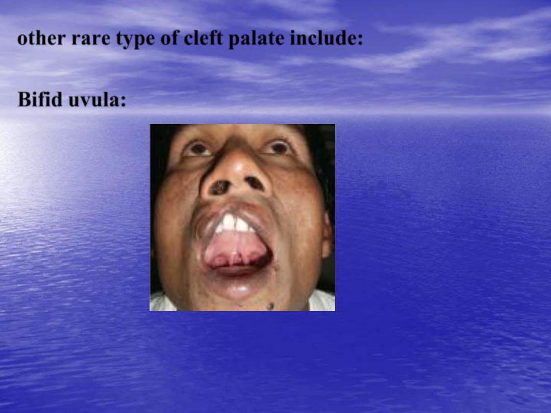
other rare type of cleft palate include:
Bifid uvula:
•
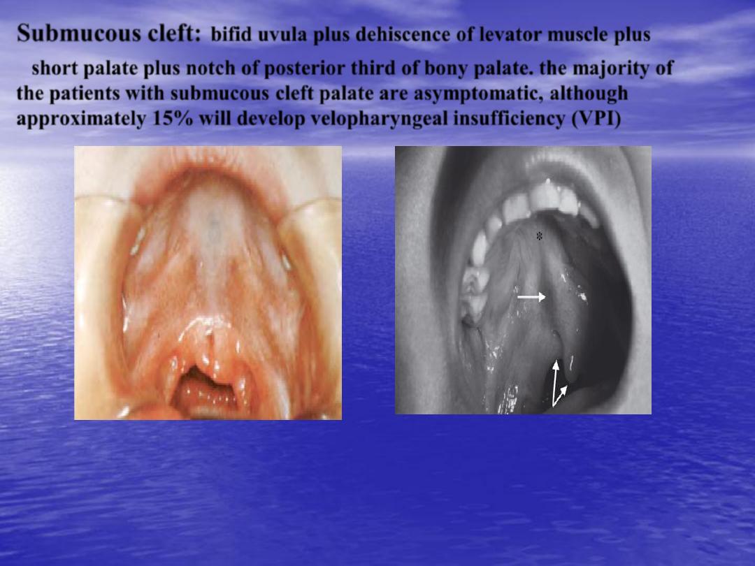
Submucous cleft:
bifid uvula plus dehiscence of levator muscle plus
•
short palate plus notch of posterior third of bony palate. the majority of
the patients with submucous cleft palate are asymptomatic, although
approximately
15%
will develop
velopharyngeal insufficiency (VPI)
1-
Feeding: since the baby with cleft palate is unable to create adequate suction
so that the feeding should be done with nipple with large holes, and baby
should hold in 45° degree to decrease regurgitation into the nose, and
feeding should take longer time.
2-
Maintenance airway by prone position during sleeping.
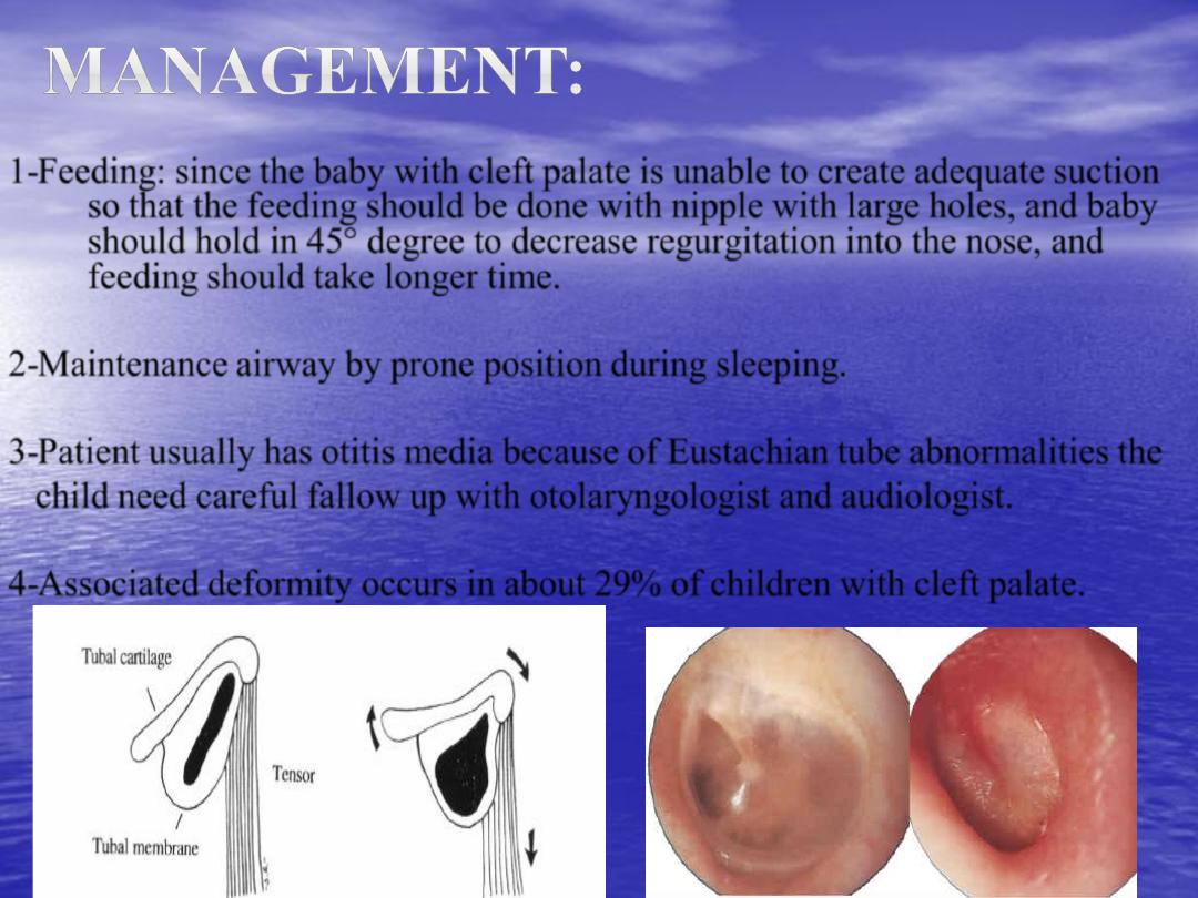
3-
Patient usually has otitis media because of Eustachian tube abnormalities the
child need careful fallow up with otolaryngologist and audiologist.
4-
Associated deformity occurs in about 29% of children with cleft palate.
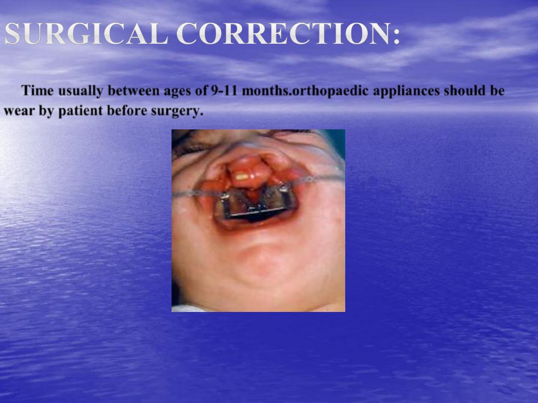
Time usually between ages of 9-11 months.orthopaedic appliances should be
wear by patient before surgery.
Goals of reconstruction:
1- Recreate a continuous hard palate with palatal tissue that allow
no communications between oral and nasal cavities.
2- Reconstruct the muscular anatomy to that of normal to allow
the action of the levator veli palatini muscle to elevate the
mobile soft palate and separate the nose.
These goals should be accomplished without creating significant
scarring of maxilla which increase the incidence of midface
retrusion.
•
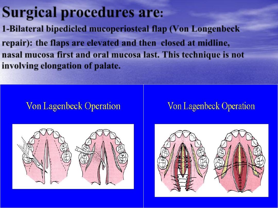
Surgical procedures are
:
•
1-Bilateral bipedicled mucoperiosteal flap (Von Longenbeck
repair):
the flaps are elevated and then
closed at midline,
nasal mucosa first and oral mucosa last. This technique is not
involving elongation of palate.
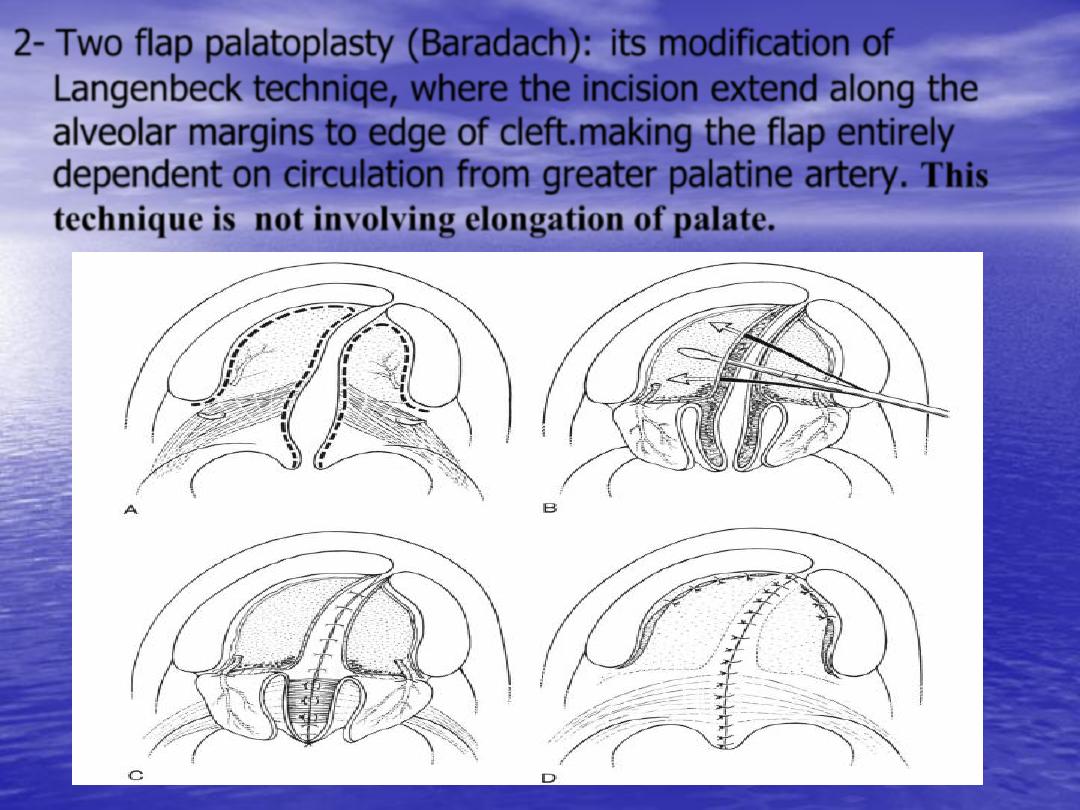
2- Two flap palatoplasty (Baradach):
its modification of
Langenbeck techniqe, where the incision extend along the
alveolar margins to edge of cleft.making the flap entirely
dependent on circulation from greater palatine artery. This
technique is not involving elongation of palate.
•
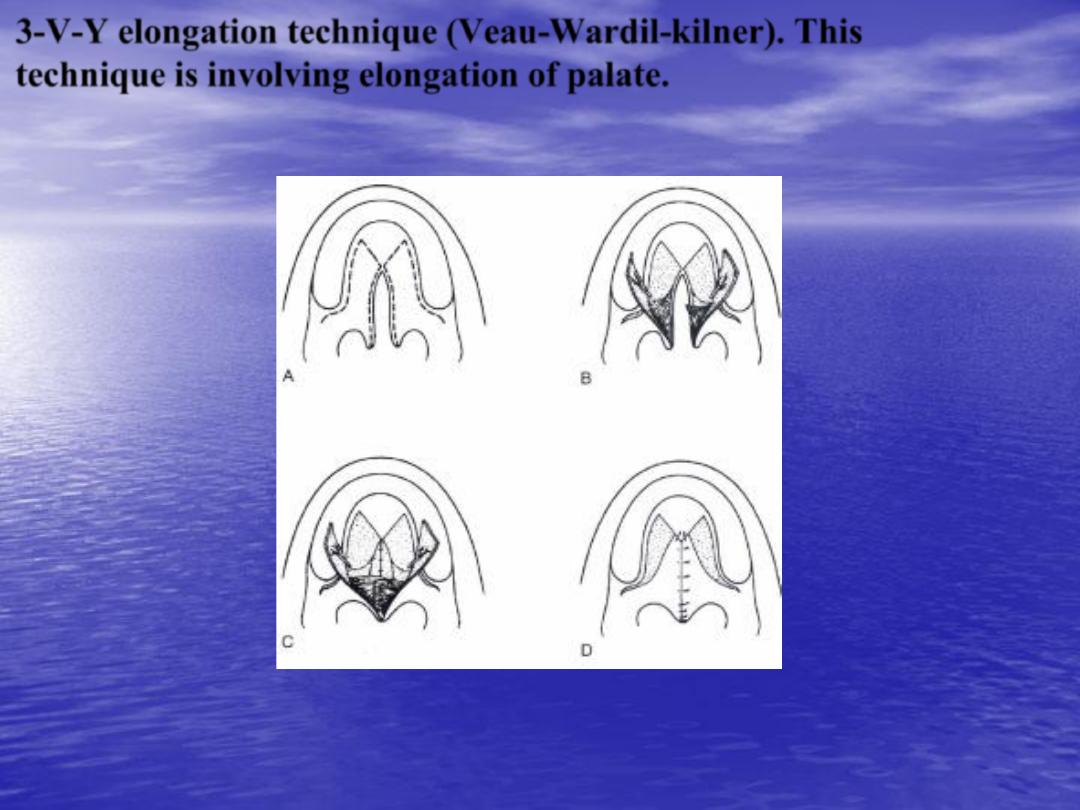
3-V-Y elongation technique (Veau-Wardil-kilner).
This
technique is involving elongation of palate.
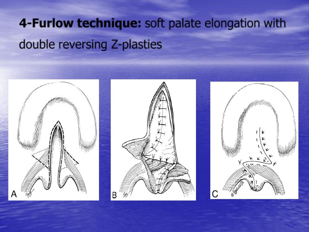
4-Furlow technique:
soft palate elongation with
double reversing Z-plasties
Postoperative care:
-
Elbow restrain are worn continually for 4-6 weeks.
-
The airway is observed and the child placed on
oxygen monitor.
-
Patient are given liquid only diet for 3 week and child
is transitioned to soft diet for an additional 3 weeks.
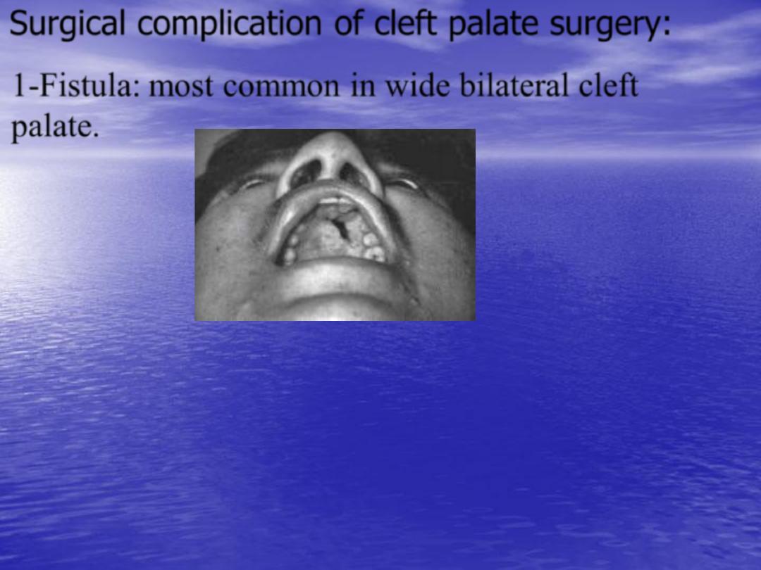
Surgical complication of cleft palate surgery:
•
1-Fistula:
most common in wide bilateral cleft
palate.
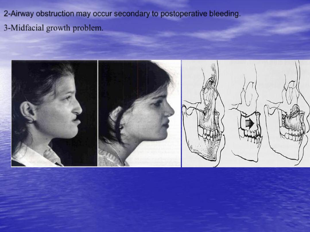
3-
Midfacial growth problem.
2-
Airway obstruction may occur secondary to postoperative bleeding.
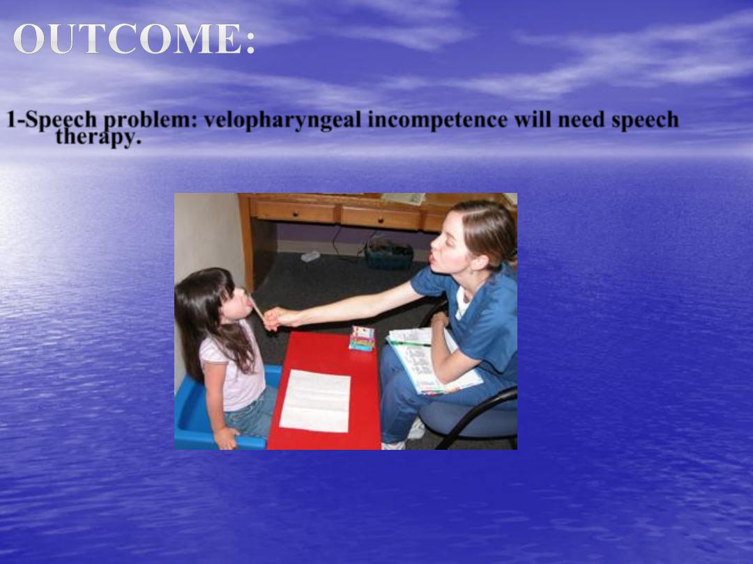
1-
Speech problem: velopharyngeal incompetence will need speech
therapy.
•
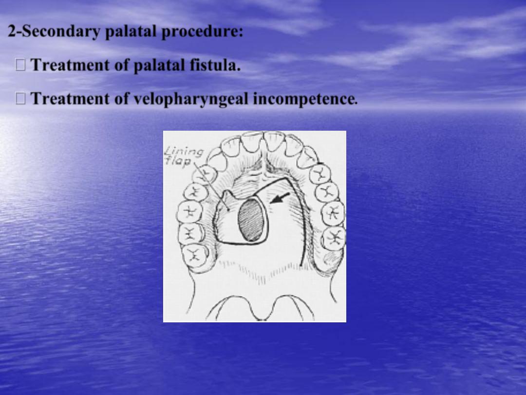
2-
Secondary palatal procedure:
Treatment of palatal fistula.
Treatment of velopharyngeal incompetence
.
•
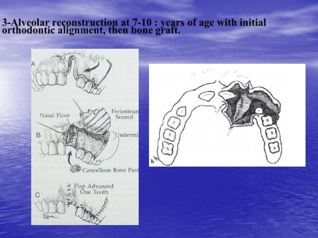
3-
Alveolar reconstruction at 7-10 : years of age with initial
orthodontic alignment, then bone graft.

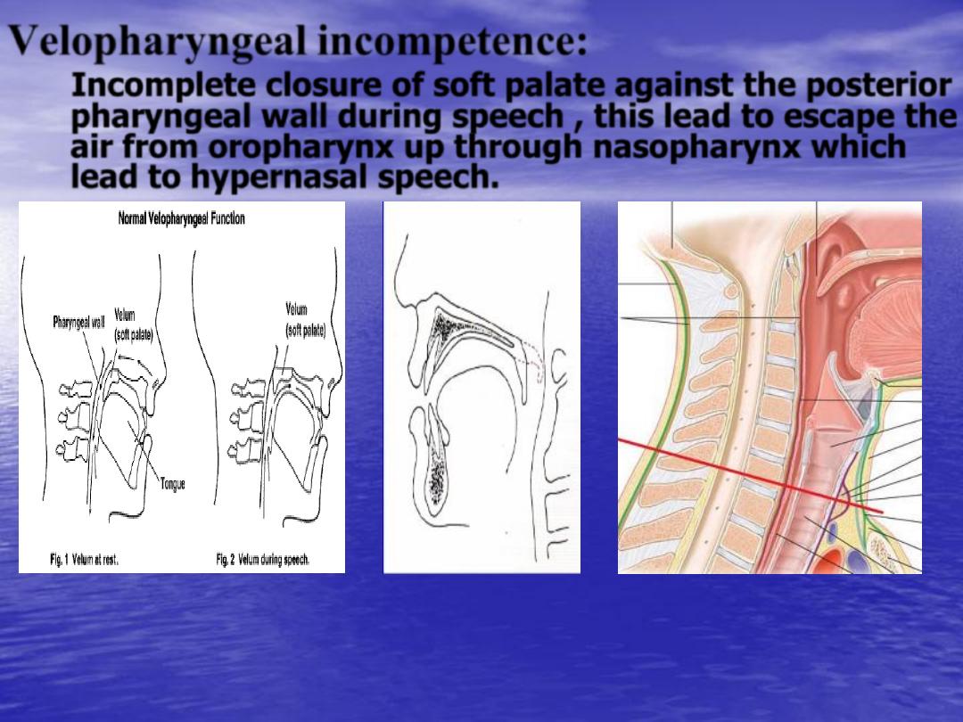
Velopharyngeal incompetence:
Incomplete closure of soft palate against the posterior
pharyngeal wall during speech , this lead to escape the
air from oropharynx up through nasopharynx which
lead to hypernasal speech.
•
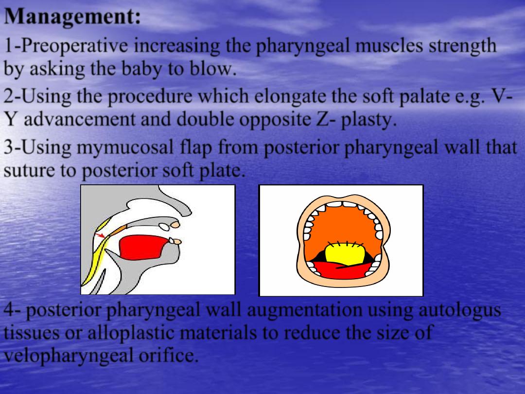
Management:
•
1-
Preoperative increasing the pharyngeal muscles strength
by asking the baby to blow.
•
2-
Using the procedure which elongate the soft palate e.g. V-
Y advancement and double opposite Z- plasty.
•
3-
Using mymucosal flap from posterior pharyngeal wall that
suture to posterior soft plate.
•
4-
posterior pharyngeal wall augmentation using autologus
tissues or alloplastic materials to reduce the size of
velopharyngeal orifice.

Thank you