Respiratory SystemDr.Ezdehar Nassif Ali

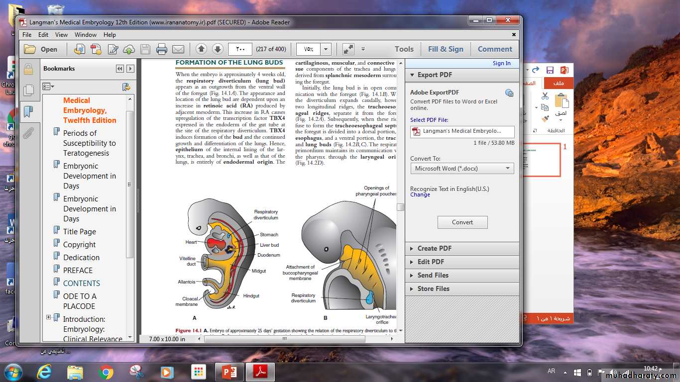
When the embryo is approximately 4 weeks old, the respiratory diverticulum (lung bud) appears as an outgrowth from the ventral wall of the foregut . The appearance and location of the lung bud are dependent upon an increase in retinoic acid (RA) produced by adjacent mesoderm.
The epithelium of the larynx, trachea, bronchi, and alveoli originates in the endoderm. The cartilaginous, muscular, and connective tissue components arise in the mesoderm.
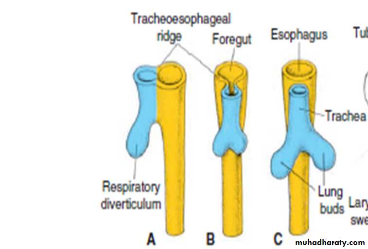
Initially, the lung bud is in open communication with the foregut .
When the diverticulum expands caudally, however, two longitudinal ridges, the tracheoesophageal ridges, separate it from the foregut. Subsequently, when these ridges fuse to form the tracheoesophageal septum, the foregut is divided into a dorsal portion, the esophagus, and a ventral portion, the trachea and lung buds.
LARYNX
The internal lining of the larynx originates from the endoderm ,but the cartilages and muscles originate from mesenchyme of the fourth and sixth pharyngeal arches. As a result of rapid proliferation of this mesenchyme, the laryngeal orifice changes in appearance from a sagittal slit to a T-shaped opening. The laryngeal ventricles are bounded by folds of tissue that differentiate into the false and true vocal cord. All laryngeal muscles are innervated by branches of the tenth cranial nerve ,the vagus nerve.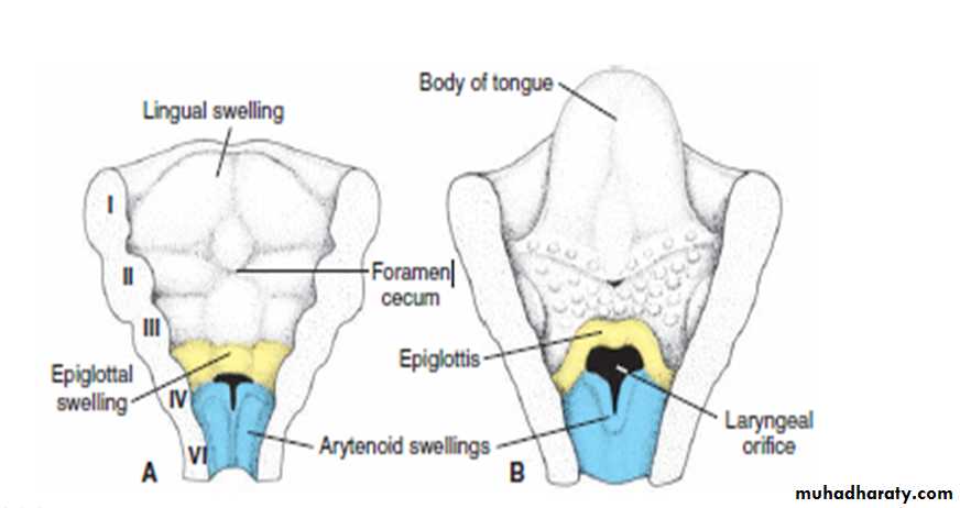
Trachea , Bronchi, and Lungs
At the beginning of the fifth week ,each of these lung buds enlarges to form right and left main bronchi .The right then forms three secondary bronchi, and the left ,two secondary bronchi.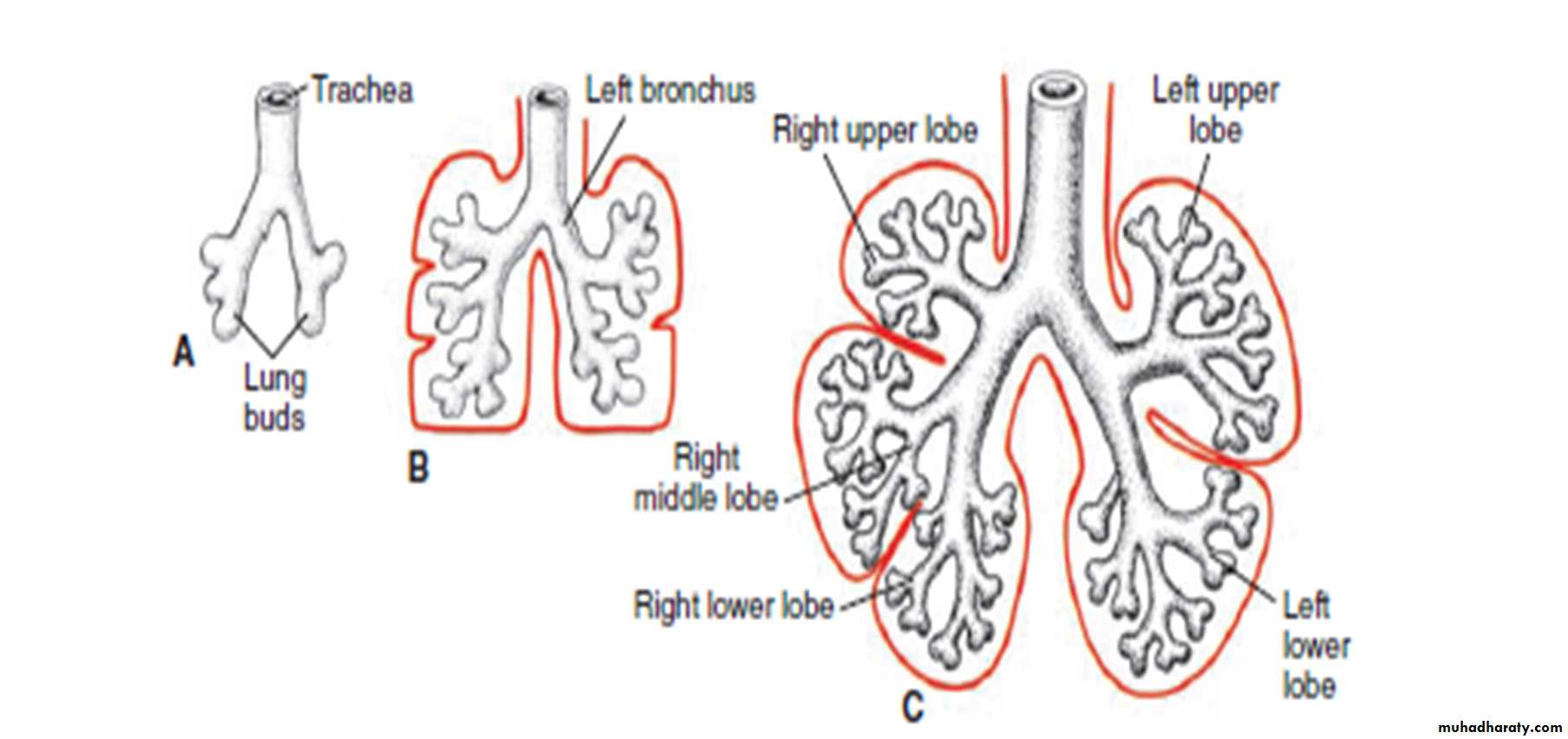
The mesoderm, which covers the outside of the lung develops into the visceral pleura. The somatic mesoderm layer, covering the body wall from the inside, becomes the parietal pleura . The space between the parietal and visceral pleura is the pleural cavity.
pleural cavity.
visceral pleuraparietal pleura
During further development, secondary bronchi divide repeatedly, forming 10 tertiary (segmental) bronchi in the right lung and 8 in the left, creating the broncho pulmonary segments of the adult lung.
By the end of the sixth month, approximately 17 generations of subdivisions have formed. Before the bronchial tree reaches its final shape , however, an additional six divisions form during postnatal life. All of these new subdivisions are occurring and the bronchial tree is developing ,the lungs assume a more caudal position , so that by the time of birth, the bifurcation of the trachea is opposite the fourth thoracic vertebra.
Maturation of the lungs
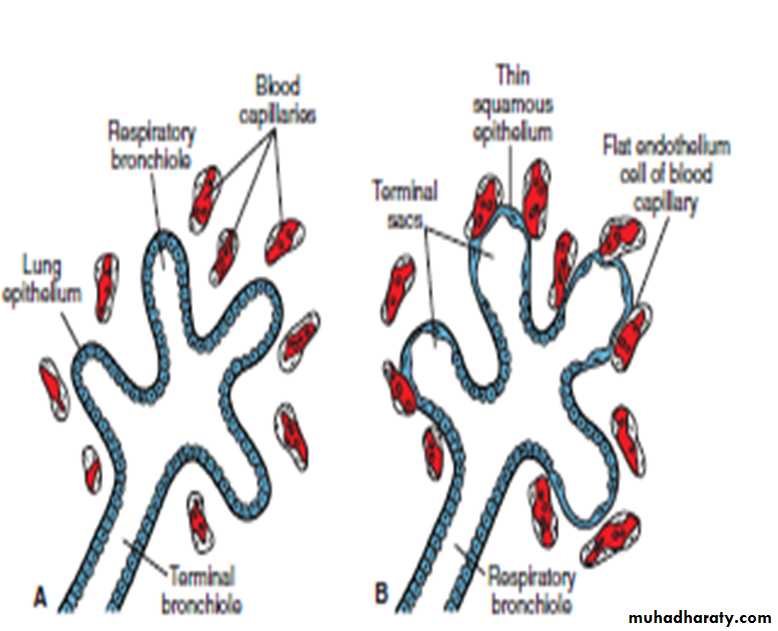
Up to the seventh prenatal month, the bronchioles divide continuously into more and smaller canals (canalicular phase) and the vascular supply increases steadily .
Terminal bronchioles divide to form respiratory bronchioles and each of these divides into three to six alveolar ducts .
The ducts end in terminal sacs (primitive alveoli) that are surrounded by flat alveolar cells in close contact with neighboring capillaries.
By the end of the seventh month, sufficient numbers of mature alveolar sacs and capillaries are present to guarantee adequate gas exchange, and the premature infant is able to survive.
During the last 2 months of prenatal life and for several years thereafter, the number of terminal sacs increases steadily. In addition, cells lining the sacs, known as type I alveolar epithelial cells, capillaries protrude into the alveolar sacs become thinner, so that surrounding capillaries protrude into the alveolar sacs.
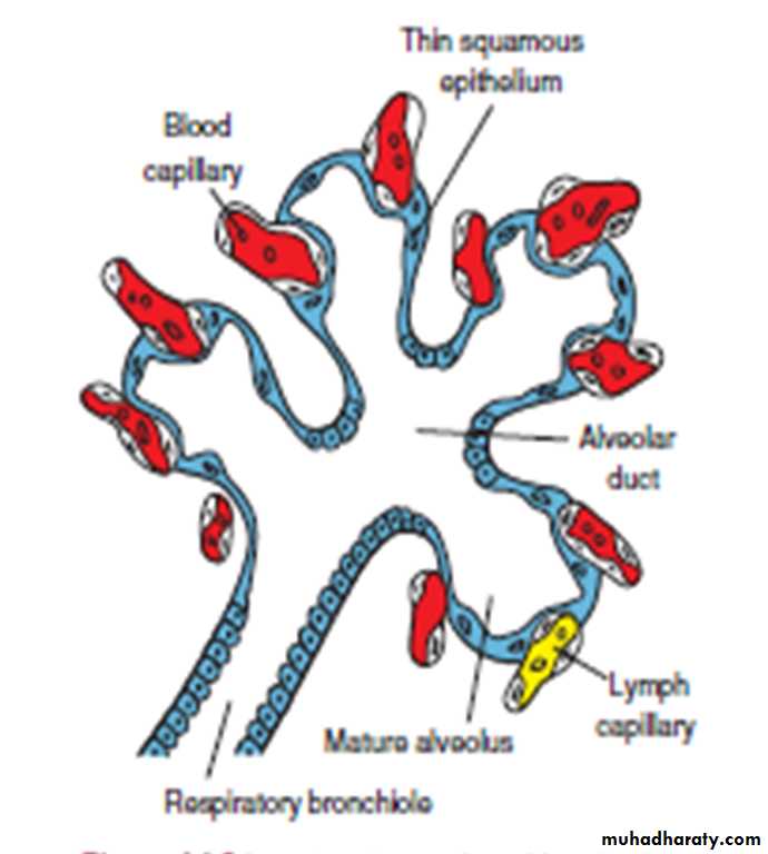
This intimate contact between epithelial and endothelial cells makes up the blood–air barrier. Mature alveoli are not present before birth.
In addition to endothelial cells and flat alveolar epithelial cells, another cell type develops at the end of the sixth month.
These cells, type II alveolar epithelial cells, produce surfactant, a phospholipid-rich fluid capable of lowering surface tension at the air–alveolar interface.
.
Fetal breathing movements begin before birth and cause aspiration of amniotic fluid. These movements are important for stimulating lung development and conditioning respiratory muscles.
Respiratory movements after birth bring air into the lungs, which expand and full the plural cavity . Although the alveoli increase somewhat in size, growth of the lungs after birth is due primarily to an increase in the number of respiratory bronchioles and alveoli.
It is estimated that only one –sixth of the adult number of alveoli are present at birth .
The remaining alveoli are formed during the first 10 years of postnatal life through the continuous formation of new primitive alveoli.
Absent or insufficient surfactant in the premature baby causes respiratory distress syndrome (RDS) because of collapse of the primitive alveoli.