The Meninges
The MeningesThree protective membranes that surround the brain, cerebellum and the spinal cord.
• The Dura mater (tough)
• The Arachnoid mater (delicate)
• The Pia mater (thin but firmly attached)
Dura Mater
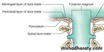
The Dura Mater:fibrous layer that is divided into two layers (except@ venous sinuses).
• Endosteal layer: (periosteum)
• Do not extend beyond the skull.
• Fuse with periosteum of the skull outside.
• Fuse with sutural ligaments.
• Meningeal layer: (dura mater proper)
• Extend beyond the skull @ foramen magnum.
• Fuse with epineurium of cranial nerves.
• Sends inward septa to form cranial partitions.
Dura Partitions
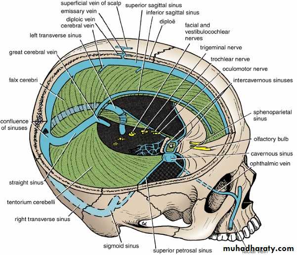
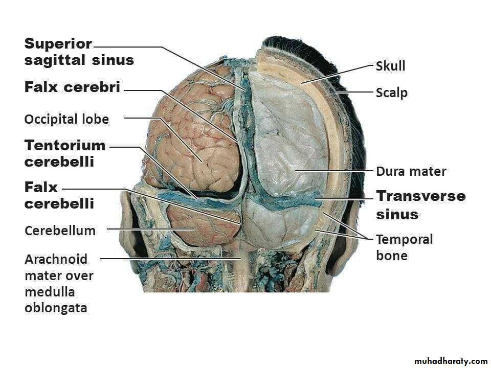
• Falx Cerebri• Tentorium Cerebelli
• Falx Cerebelli
• The diaphragma sellae
Dura Partitions – Falx Cerebri
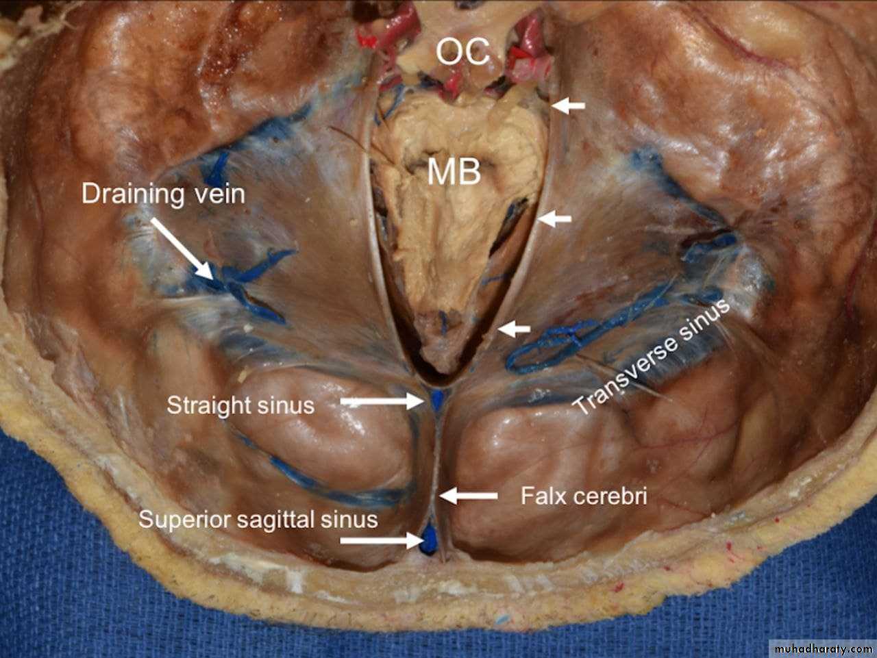
• Falx Cerebri:Sickle-shaped fold of dura mater.
Its ant. end is attached to the internal frontal crest and the crista galli. Its post. end blends with the upper surface of the tentorium cerebelli.
The superior sagittal sinus runs in its upper fixed margin
The inferior sagittal sinus in its lower free margin
The straight sinus runs along its attachment to the tentorium cerebelli.
Dura Partitions – Tentorium Cerebelli
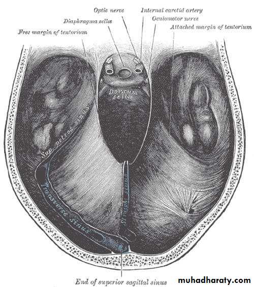
• Tentorium Cerebellicrescent-shaped fold of dura mater.
anteriorly, the tentorial notch for the midbrain.
its outer border is attached to the posterior clinoid processes, the superior borders of the petrous bones, and the margins of the grooves for the transverse sinuses on the occipital bone.
It inner free border crosses the attached border, and is attached to the anterior clinoid process.
The tentorium cerebelli, the second-largest dural reflection, is a crescent-shaped dura fold that extends over the posterior cranial fossa, separating the occipital and temporal cerebral hemisphere from the cerebellum and infratentorial brainstem .
distinguishing landmark and divides the cranial cavity into the supratentorial and infratentorial spaces .
has a free and fixed margin.
The fixed margins of the tentorium cerebelli are attached to the superior borders of the petrous part of the temporal bone, known as the posterior clinoid process via the anterior and posterior petroclinoid folds.and along the transverse sinuses grooves on the occipital bone posteriorly.
The free margin is located at the anterior edge and forms a U-shape termed the tentorial notch or the incisura tentoria.
Dura Partitions – Falx Cerebelli
• Falx Cerebelli:is a small, sickle-shaped fold of dura mater.
Its fixed posterior margin is attached to the internal occipital crest and contain the occipital sinus.
Its free border anteriorly separate the two cerebellar hemispheres.
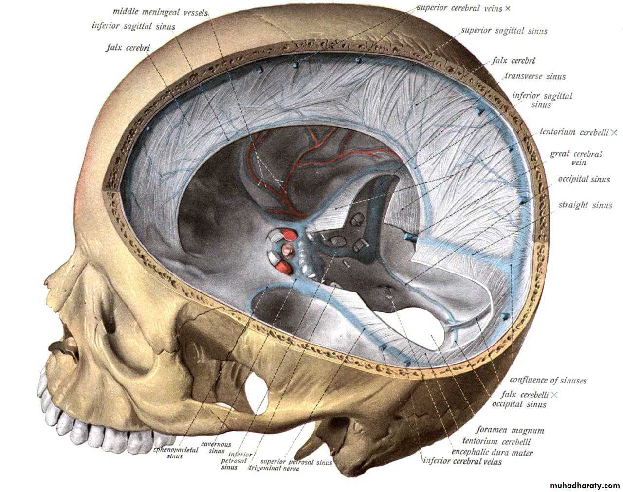
Dura Partitions - The diaphragma sellae
• The diaphragma sellaeis a small circular fold of dura mater
it forms the roof for the sella turcica.
It has a small opening in its center for stalk of the pituitary gland.
Innervation of the Dura
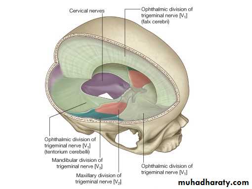
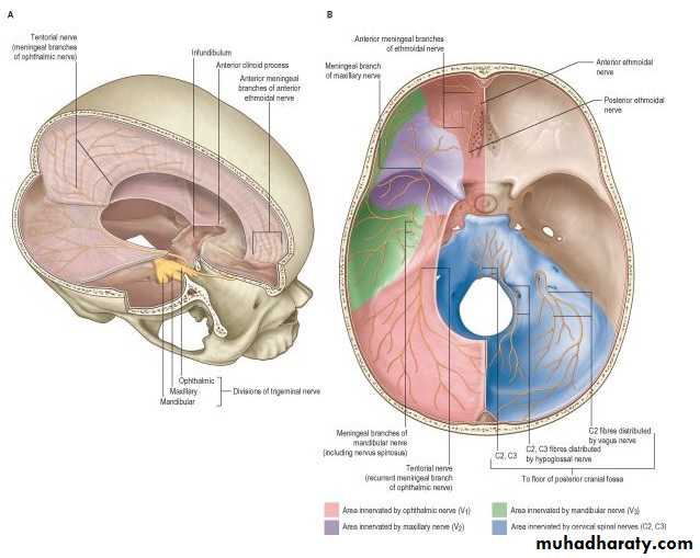
• Dura is sensitive to ?? --► headache• Dura of Posterior cranial fossa by branches from cervical spinal nerves (C1, C2 & C3)
• Dura of ACF, MCF, Falx cerebri & Tentorium Cerebelli by Trigeminal nerve:
1- Anterior meningeal nerves2- Tentorial nerve
3- Meningeal branches of maxillary and Mandibular divisions of Trigeminal nerve
Dural Nerve Supply
Branches of the trigeminal, vagus, and first three cervical nerves and branchesfrom the sympathetic system pass to the dura. Numerous sensory endings are in the dura.
The dura is sensitive to stretching, which produces the sensation of headache.
Stimulation of the sensory endings of the trigeminal nerve above the level of the tentorium cerebelli produces referred pain to an area of skin on the same side of the the head.
Stimulation of the dural endings below the level of the tentorium produces referred pain to the back of the neck and back of the scalp along the distribution of the greater occipital nerve
Dura Nerve Supply
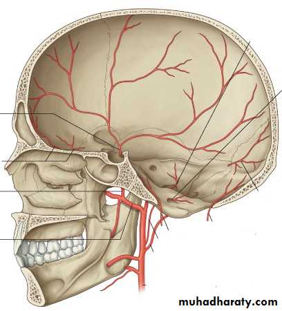
Arterial Supply of Dura
Anterior meningeal artery (branches of ethmoidal arteries which are branches of maxillary artery (ECA).Middle meningeal artery and accessary meningeal artery: branches of Maxillary artery (ECA)
Posterior meningeal artery (terminal branch of ascending pharyngeal artery (ECA) & other meningeal branches from:
1- Ascending pharyngeal artery
2- Occipital artery
3- Vertebral artery
Dural Arterial Supply
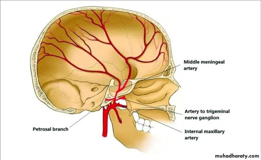
Numerous arteries supply the dura mater from the internal carotid, maxillary,ascending pharyngeal, occipital, and vertebral arteries. From a clinical standpoint,
the most important is the middle meningeal artery, which is commonly damaged in
head injuries.
MMA arises from the maxillary artery in the infratemporal fossa. It enters the cranial cavity and runs forward and laterally in a groove on the upper surface of the squamous part of the temporal bone.
To enter the cranial cavity, it passes through the foramen spinosum to lie between the meningeal and endosteal layers of dura. The anterior (frontal) branch deeply grooves or tunnels the anteroinferior angle of the parietal bone. The posterior (parietal) branch curves backward and supplies the posterior part of the dura mater
Venous Drainage
Venous drainage of dura usually follow the arterial arrangement of meningeal arteries:1- anterior Meningeal veins
2- middle Meningeal veins
3- Posterior Meningeal veins
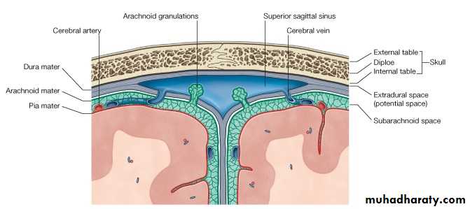
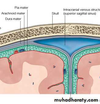
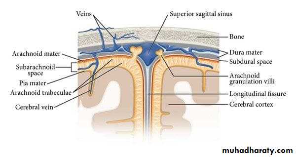
Arachnoid mater
• Delicate avascular layer (lies against but not firmly attached to dura mater). It sends numerous trabeculae toward the pia mater.
• Subarachnoid space?? CSF?
• Arachnoid cisternae.
• All cerebral arteries and veins lie in subarachnoid space.
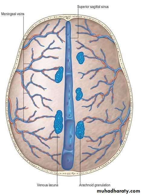
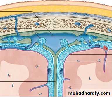
• Arachnoid villi: numerous projections to the venous sinus.
• Arachnoid granulations: aggregations of these villi at which CSF diffuses to venous sinuses.
• Arachnoid mater fuses with epineurium of nerves at the exit foramina except for optic nerve.
Arachnoid granulations, also known as Pacchionian granulations, are projections of the arachnoid membrane (villi) into the dural sinuses that allow CSF to pass from the subarachnoid space into the venous system
Pia mater
thin vascular layer that is adherent to the brain surface.It extends with cerebral sulci and cover gyri and fuses with epineurium of cranial nerves at their exit foramina.
Venous Sinuses
• Blood-filled spaces situated between the layers of the dura mater.• The sinuses have no valves.
• They receive tributaries from the brain, the diplo of the skull, the orbit, and the internal ear.
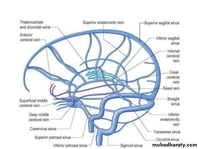
• The superior sagittal sinus - receives the superior cerebral veins.
• The inferior sagittal sinus - joins the great cerebral vein to form the straight sinus. It receives cerebral veins from the medial surface of the cerebral hemisphere.
• The straight sinus - it drains into the left transverse sinus.
• The left transverse sinus is a continuation of the straight sinus & end on each side by becoming the sigmoid sinus.
• The right transverse sinus is as a continuation of the superior sagittal sinus.
• The sigmoid sinuses - leave through jugular foramen --►internal jugular vein.
• The occipital sinus - communicates with the vertebral veins through the foramen.
• The cavernous sinus Anteriorly, the sinus receives the inferior ophthalmic vein and the central vein of the retina & drains posteriorly into the transverse sinus through the superior petrosal sinus.
• Intercavernous sinuses connect the two cavernous sinuses through the sella turcica.
The Venous Blood Sinuses
The venous sinuses of the cranial cavity are blood-filled spaces situated between the layers of the dura mater; they are lined by endothelium.Their walls are thick and composed of fibrous tissue; they have no muscular tissue.
The sinuses have no valves.
They receive tributaries from the brain, the diploë of the skull, the orbit, and the internal ear.
The superior sagittal sinus
lies in the upper fixed border of the falx cerebri.It runs backward and becomes continuous with the right transverse sinus.
The sinus communicates on each side with the venous lacunae. Numerous arachnoid villi and granulations project into the lacunae. The superior sagittal sinus receives the superior cerebral veins.
The inferior sagittal sinus
lies in the free lower margin of the falx cerebri.
It runs backward and joins the great cerebral vein to form the straight sinus.
It receives cerebral veins from the medial surface of the cerebral hemisphere
The straight sinus
lies at the junction of the falx cerebri with the tentorium cerebelli.Formed by the union of the inferior sagittal sinus with the great cerebral vein,
it drains into the left transverse sinus.
The right transverse sinus
begins as a continuation of the superior sagittal sinusthe left transverse sinus is usually a continuation of the straight sinus.
Each sinus lies in the lateral attached margin of the tentorium cerebelli, and they end on each side by becoming the sigmoid sinus.
The sigmoid sinuses
are a direct continuation of the transverse sinuses.Each sinus turns downward behind the mastoid antrum of the temporal bone and then leaves the skull through the jugular foramen to become the internal jugular vein.
The occipital sinus lies in the attached margin of the falx cerebelli. It communicates with the vertebral veins through the foramen magnum and the transverse sinuses.
Cavernous sinuses
Each cavernous sinus lies on the lateral side of the body of the sphenoid bone. Anteriorly, the sinus receives the inferior ophthalmic vein and the central vein of the retina. The sinus drains posteriorly into the transverse sinus through the superior petrosal sinus. Intercavernous sinuses connect the two cavernous sinuses through the sella turcica.
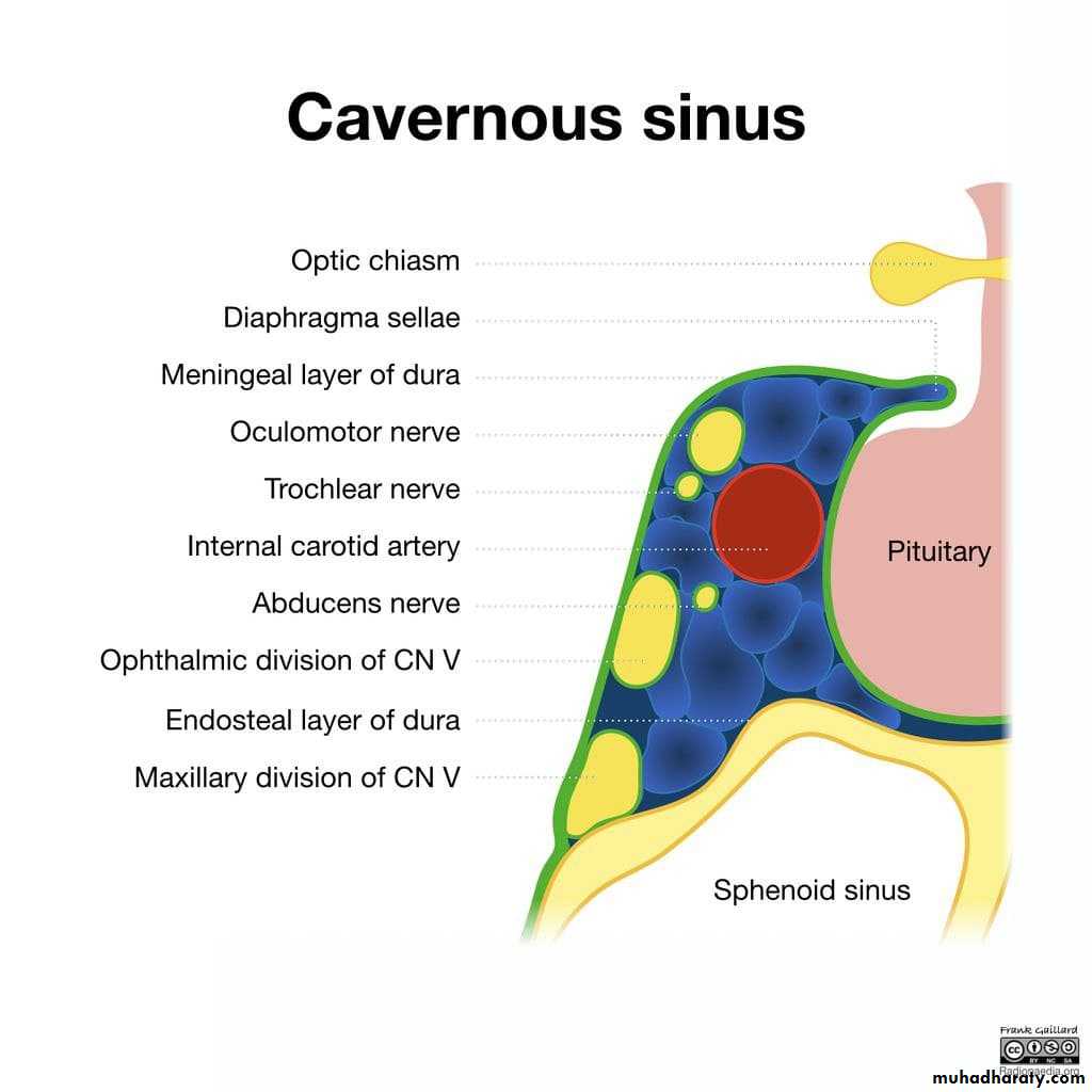
Content of cavernous sinus..Mnemonic O TOM CAT
O: oculomotor nerveT: trochlear nerve
O: ophthalmic branch of trigeminal nerveM: maxillary branch of trigeminal nerve
C: internal carotid arteryA: abducens nerve
T: trochlear nerve