Primary myocardial diseases:
1- Myocarditis:-It's an inflammatory processes of the myocardium that result in injury to the cardiac myocytes. It's either secondary to other heart diseases as ischemic injury, or primary myocarditis which's caused by several types of microorganisms as viruses, parasites and bacterial infections or may be caused by immune mediated reactions that cross react with myocardial cells as occur in rheumatic heart disease .
Morphology:-
The heart may be of normal size or dilated, the myocardium is flabby and pale with small areas of hemorrhage.Microscopically:- The microscopical changes depend on causative agents but in general it consists of inflammatory cells infiltrate as lymphocytes, mononuclear cells and even neutrophils, with degeneration and/or necrosis of myocytes.
Cardiomyopathies:- Or heart muscle disease, it is heart disease result from a primary abnormality in the myocardium.
It's divided into 3 major groups:
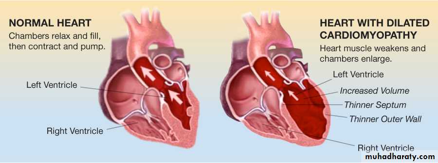
(I) Dilated cardiomyopathy:-
It's usually idiopathic but it may be secondary to other causes, it's characterized by progressive cardiac hypertrophy, dilation and contractile (systole) dysfunction.Dilated cardiomyopathy occur at any age but it usually common between ages of 20-60 years.
Morphology:-
The heart is enlarged by dilation and hypertrophy of all chambers. The dilation and poor contractile function cause stasis of blood in cardiac chambers and predispose to development of fragile mural thrombi and emboli.
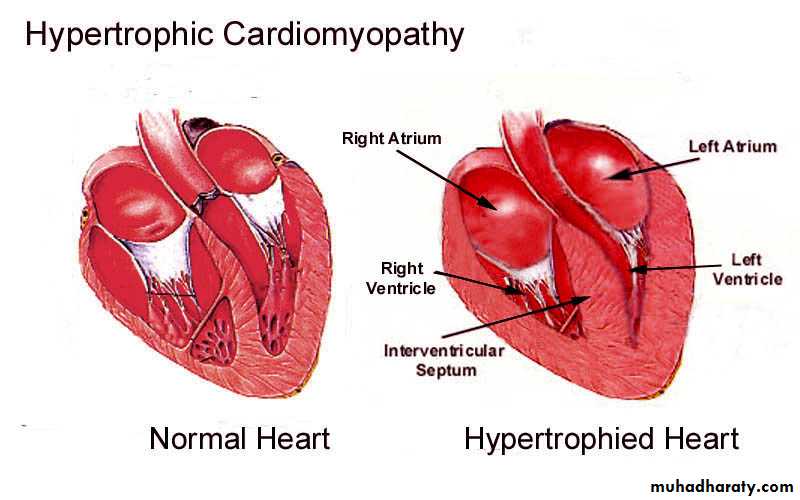
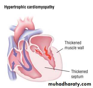
(II) Hypertrophic cardiomyopathy:-
It consists of a symmertric septal hypertrophy and idiopathic hypertrophic subaortic stenosis, it' s characterized by myocardial hypertrophy, abnormal diastolic filling and intermittent ventricular outflow obstruction.Morphology:-
The essential feature of hypertrophic cardiomyopathy is myocardial hypertrophy which is most pronounced in the left ventricle and inter-ventricular septum.The hypertrophy is usually conspicuous in sub-aortic region of the septum, so this asymmetric hypertrophy is often associated with ventricular outflow obstruction during systole, so it's called "idiopathic hypertrophic sub-aortic stenosis" .
Ventricular dilation is uncommon but left atrium may be dilated because of impaired diastolic filling of thickened rigid left ventricle.
Restrictive cardiomyopathy:-
It's a disorder characterized by a primary decrease in ventricular compliance, resulting in impaired ventricular filling during diastole, most common cause of this decrease of compliance due to endomyocardial fibrosis.
Hypertensive heart disease:- Hypertensive heart disease (HHD) is a consequence of the increased demands placed on the heart by hypertension, causing pressure overload and ventricular hypertrophy. Myocyte hypertrophy is an adaptive response to pressure overload; there are limits to myocardial adaptive capacity, however, and persistent hypertension eventually can culminate in dysfunction, cardiac dilation, CHF, and even sudden death. Although hypertensive heart disease most commonly affects the left side of the heart secondary to systemic hypertension, pulmonary hypertension also can cause right-sided hypertensive changes—so-called “cor pulmonale.”
Pericardial diseases:-
The most diseases of pericardium are:1- Inflammatory condition (pericarditis).
2- Pericardial effusion.
Pericardial effusion:-
It's accumulation of fluid in pericardial space is often asymptomatic if it accumulate slowly, but rapid developing effusions may cause tamponade the most cause of effusions are congestive heart failure, hypoalbuminemia, malignancy, and mediastinal lymphatic obstruction.Congenital heart diseases:-
It's most common types of congenital malformations and it's most common cause of heart disease in children.Its causes in 90% idiopathic, while 10% of it reveal either genetic factors or environmental factors such as congenital rubella infection.
The types of congenital heart diseases include:
1- Malformations causing a left-to-right shunt.
2- Malformations causing a right-to-left shunt (cyanotic congenital heart diseases).
3- Malformations causing obstruction.
(I) Left-to-right shunts:-
is most common types of cardiac malformations, this is usually a cyanotic in early stage but in later stages can cause cyanosis when produced significant pulmonary hypertension and reversal blood flow through the shunt occurs, that include:-
1- Atrial septal defects.
2- Ventricular septal defects.
3- Patent ductus arteriosus.
1- Atrial septal defects:-
Is caused most commonly by failure to close of foramen ovale after birth, the foramen ovale is a flap of tissue in septum between 2 atrium act as a one-way valve allowing blood to keep flowing from right to left during intrauterine life at the time of birth as pulmonary vascular resistance fall and systemic arterial pressure increases, so pressure in the left atrium rises above that in right atrium and must cause functional closure of the foramen ovale.Patency persist in about 25% of general population and this will cause shunt of blood from left to right atrium, this defect usually well tolerated if it's less than 1 cm in diameter but even larger lesions do not produce any symptom in childhood because the flow of blood is from left to right, but with time when pulmonary vascular resistance increase and pulmonary hypertension developed so reversal of shunt so the shunt become right to left and developed cyanosis.
Morphology:-
Manifested as right atrial and ventricular dilation, right ventricular hypertrophy and dilation of pulmonary artery, pulmonary hypertension developed.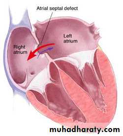
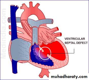
Ventricular septal defect:-
VSDs are the most common congenital heart defects and this like ASDs occur in isolation or in association with other cardiac malformations, the size and location of defect is variable ranging from minute to large defect and this may close spontaneously during infancy or childhood.Morphology:- In large defect associated with significant left to right shunt so right ventricle is hypertrophied and dilated, with pulmonary hypertension developed if this occur so reverse of shunt and cyanosis occur.
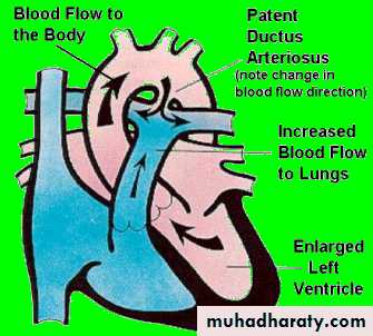
Patent ductus arteriosus:-
Ductus arteriosus: is an arterial channel that courses between the pulmonary artery and aorta. During intrauterine life, the DA permits blood to flow freely from the pulmonary artery to the aorta. Complete irreversible closure occurs within the 1st few months after birth, this closure may be delayed or failed to occur to give PDA condition.Morphology:-
The oxygenated blood flows from the left ventricle to the lungs and is returned to the left atrium, so form volume overload to cause dilation and hypertrophy of left atrium and ventricle.
The proximal pulmonary arteries are also dilated with development of pulmonary hypertension and cause right ventricular hypertrophy and dilation and right atrial dilation.
(II) Right-to-left shunts:-
Cardiac malformations associated with right-to-left shunts are distinguished by cyanosis at or near the time of birth, this occur because poorly oxygenated blood from the right side of the heart is introduced directly into the arterial circulation.2 important conditions cause cyanotic congenital heart disease:
1-Tetralogy of Fallot
2- Transposition of great vessels.
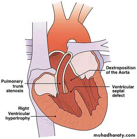
Tetralogy of Fallot:-
TOF is the most common cause of cyanotic congenital heart disease, the four components of TOF:1- VSD.
2- Dextraposed aortic root that overrides the VSD.
3- Right outflow obstruction.
4- Right ventricular hypertrophy.
Patient with TOF is at risk to develop infective endocarditis, systemic emboli and brain abscesses.
Morphology:- The heart is enlarged externally by right ventricular hypertrophy, the proximal aorta is larger than pulmonary trunk, so that because of stenotic pulmonary artery, this will lead to shunt unoxygenated blood from right ventricle through VSD to go to left ventricle and aorta.
Transposition of great arteries:-
There's abnormal truncal septation, the aorta arises from the right ventricle and the pulmonary artery from the left ventricle, so all unoxygenated blood with pump from right ventricle to systemic circulation by aorta and cause cyanosis but with presence of another defect as ASD, VSD and PDA, this allowsoxygenated blood to reach aorta.
Congenital obstructive lesions:-
As :1- valvular aortic stenosis.
2- Pulmonic stenosis.
3- Coarctation of aorta.
Cardiac tumors:-
Metastatic carcinoma:-It's more common than primary one, the most common sites of metastasis to the heart are: lungs, breast and malignant melanoma, the metastatic cells usually goes to pericardium to cause pericarditis and hemorrhagic pericardial effusions.
Primary neoplasms:-
Less common and the common primary neoplasm are:
1- Myxoma: is a benign tumor,
2- Cardiac rhabdomyoma: is a benign tumor.
3- Lipoma.