Normal embryology and anatomy
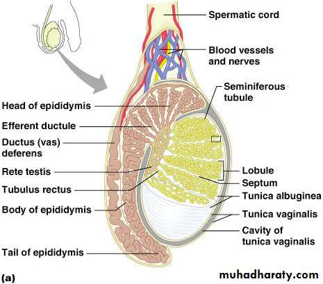
The normal adult testis is a paired organ that lies within the scrotum suspended by the spermatic cord. The average weight of each testis is 15 to 19 g,
the right usually being 10% heavier than the left.
The organ is covered by a capsule composed of three layers the outer serosa or tunica vaginalis (covered by a flattened layer of mesothelial cells), the tunica albuginea, and the inner tunica vasculosa The posterior portion of the capsule, called the mediastinum, contains blood and lymph vessels, nerves, and the mediastinal portion of the rete testis
The parenchyma is divided into approximately 250 lobules,
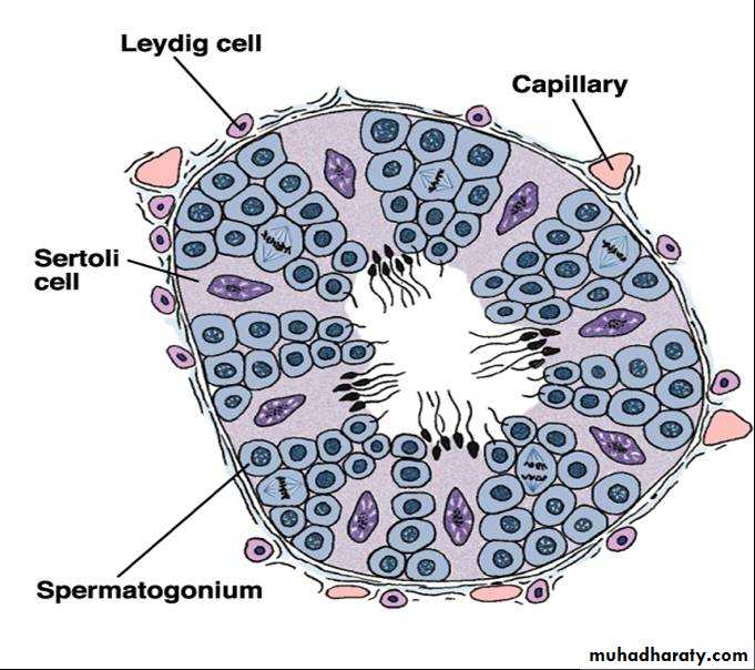
each lobule containing up to four seminiferous
Inside the Scrotum
Each testes is enclosed by the tunica vaginalis, a continuation of the peritoneum that lines the abdominopelvic cavity.A fibrous capsule covers each testis called the tunica albuginea.
Testicle
• The tunica albuginea gives rise to septa (partitions) that divide the testis into lobules (about 250)
• Each lobule contains 3 or 4 highly coiled seminiferous tubules
• These converge to become rete testis which transport sperm to the epididymis
3
Seminiferous Tubules
4
Seminiferous Tubules Histology
5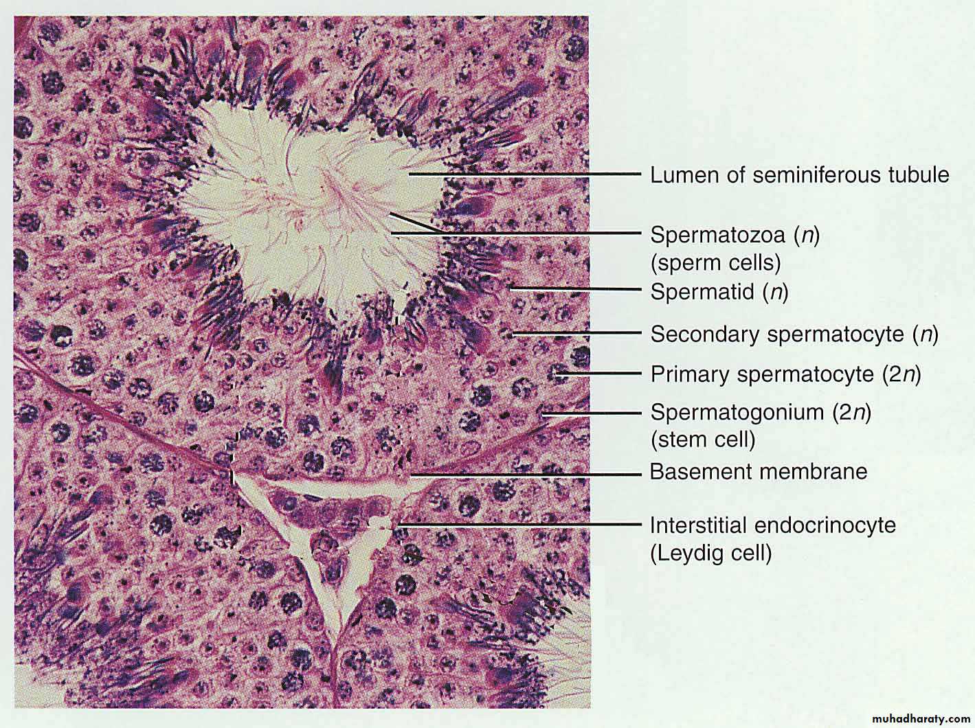
cryptorchidism
In one out of every ten males, the testis has not descended into the scrotum at the time of birth but has remained in the inguinal region or abdomenMost of these "retained" or "retractile" testes descend into the scrotum during the first year of life
. In only 1 in 100 individuals will a permanent retention of the testis out side the scrotum occur a condition known as cryptorchidism
The exact pathogenesis is unknown, but most evidence
favors a role for testosterone under the influence of the
hypothalamic-pituitary axis.
Cryptorchidism is unilateral in 80% of the case
Long-term consequences of cryptorchidism may include testicular malignancy and infertility/subfertility.
Grossly,
cryptorchid testes in adults are small and brown.The testicular tubules are atrophic, and their basement membrane is greatly thickened
In some instances, atypical germ cells are seen at the base ofthe tubules; these are indicative of intratubular germ cell Neoplasm
A cryptorchid testis is 30 to 50 times more likely to
develop a malignant neoplasm than is a normally placed organ.
Atrophy and infertility
Atrophy of the testis may result from a large variety of
causes: the already mentioned cryptorchidism;
the orchitis of mumps, especially when the infection occurs at or after puberty;
liver cirrhosis, as a result of increased circulating endogenous estrogens not metabolized by The diseased liver
administration of estrogens or gonadotropin-releasing hormone analog in the treatment of prostatic carcinoma
radiation exposure;
chemotherapy,
In advanced testicular atrophy of any of these etiologies, the tubules are small, with thick basement membranes and few or no germ cells
. The interstitial tissue shows varying degree of fibrosis, and there may be
an increased number of Leydig cells.
The causes of male infertility fall into one of three categories:
pretesticular,testicular,
and post-testicular.
The pretesticular causes are extragonadal endocrine disorders usually originating in the pituitary or adrenal gland.
The testicular causes are primary diseases of the
testes, and little treatment is available at the present time.
The post-testicular causes consist mainly of obstructions
of the ducts leading away from the testes.
The evaluation of the infertile male includes
• clinical history and examination
• semen analysis
• quantitation of leukocytes in semen,
• search for antisperm antibodies.
• testicular biopsy particularly useful in the individual with azoospermia and normal endocrine findings
The material should be handled with extreme care Zenker's and
Bouin's fixatives are used.Biopsy specimens from infertile men with total lack of
spermatozoa (azoospermia) usually show one of the followingconditions:
1. Germ cell aplasia (Sertoli cell-only syndrome) (29%),
In which the tubules are populated by only Sertoli cells,
thickening of the tubular basement membrane;
germ cells are completely absent
2. Spermatocytic arrest (26%), characterized by a halt of
the maturation sequence, usually at the stage of the
primary spermatocyte no spermatids or spermatozoa are present despite the presence ofabundant cells in division
3. Generalized fibrosis (18%).
4 .Normal spermatogenesis(27 %).
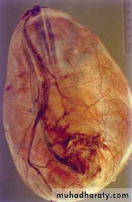
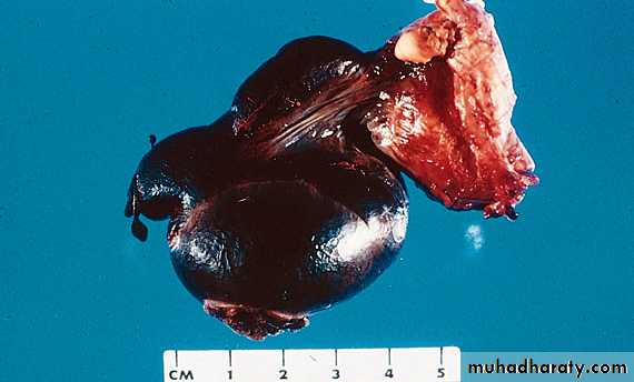
Torsion of the testis:- (rotation of the organ 360ْ around it longitudinal axis resulting in interruption of its blood supply) Sudden severe physical exercise or congenital anomalies that leads to increasing the mobility of the testis and epididymis such high attachment of tunica vaginalis on the spermatic cord, incomplete descened of the testis or absent of the scrotal ligments. These above causes may lead to complete torsion with sudden onset of severe scrotal pain followed by swelling and hemorrhagic infarction of the testicular germ cells within few hours. Recurrent incomplete torsion of the spermatic cord results in small fibrotic testis.
Vascular Related
TorsionVenous compression
Hemorrhagic infarct
Young men
At night
Very painful
Can be reduced
Inflammatory Lesions
Inflammatory lesions of the testis are more common in the epididymis than in the testis proper. Some of the more important inflammatory diseases of the testis are associated with venereal disease.Other causes of testicular inflammation include nonspecific epididymitis and orchitis, mumps, and tuberculosis.
Nonspecific epididymitis and orchitis usually begin as a primary urinary tract infection with secondary ascending infection of the testis through the vas deferens or lymphatics of the spermatic cord. The involved testis is typically swollen and tender and contains a predominantly neutrophilic inflammatory infiltrate.
Orchitis complicates mumps infection in roughly 20% of infected adult males but rarely occurs in children. The affected testis is edematous and congested and contains a predominantly lymphoplasmacytic inflammatory infiltrate. Severe cases may be associated with considerable loss of seminiferous epithelium with resultant tubular atrophy, fibrosis, and sterility.
Epididmitis
Disorders of the testicular tunica:-
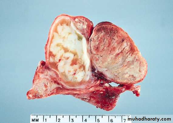
Hydrocele: - is the most common cause of the scrotal swelling and it refers to collection of serous fluid in the scrotal sac (tunica vaginalis). It is either congenital or acquired due to inflammatory disorders of the epididymis and the testis. Uncomplicated hydrocele usually presented with unilateral scrotal swelling.Hematocele:- refers to hemorrhage into a hydrocele
Hydrocele
Fluid filled scrotal cyst.Benign
Often with inguinal hernia
Transilluminates
Fluid will recollect if aspirated.
Can be large