Dr Hadi Al-Sagur
1

Content:
• Introduction
• Light properties
• Measurement of light and its units
• Applications of visible light in medicine
Curved Surfaces
Endoscope
Transillumination
Phototherapy
• Application of ultraviolet and infrared light in medicine
• Hazarads
• Laser in medicine
• Applications of microscopes in medicine
CH 14 Light in Medicine
2

1. Introduction
CH 14 Light in Medicine
In this chapter we discuss the medical applications of light in
diagnosis
and
therapy
and also
hazards
of light.
Definition:
Light or visible light is
electromagnetic radiation
within the
portion of the electromagnetic spectrum.
1. Travel at the speed of light
2. Have no electric charge
3
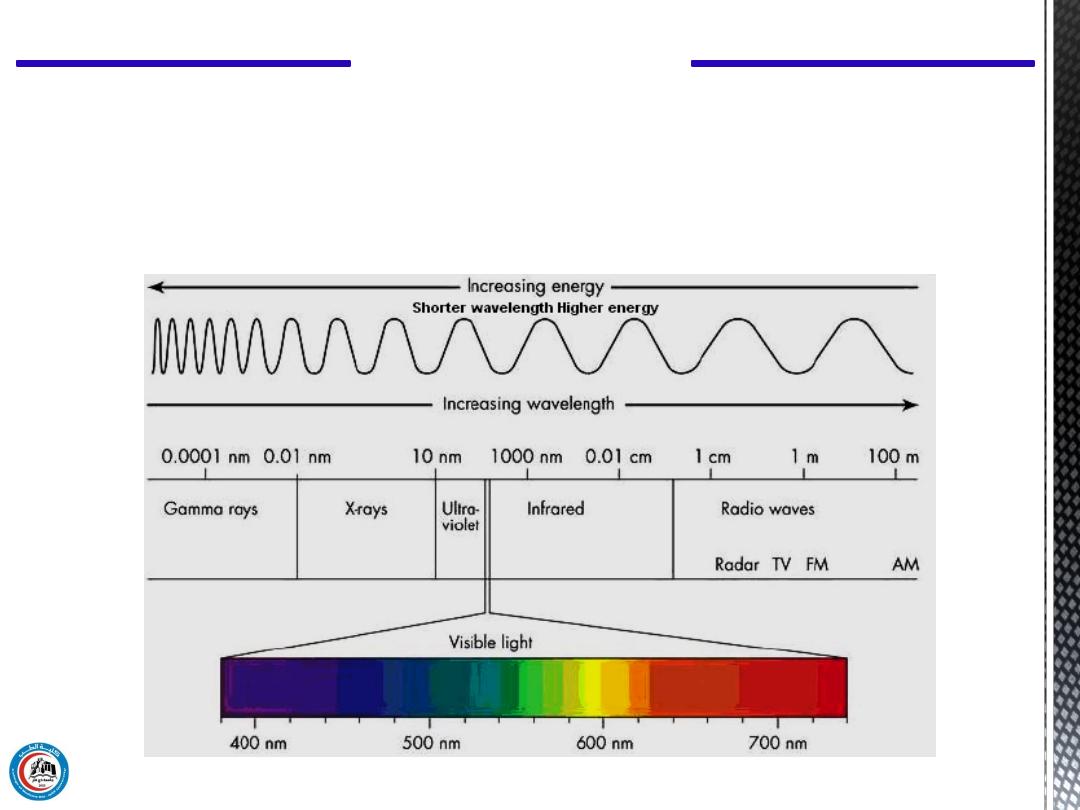
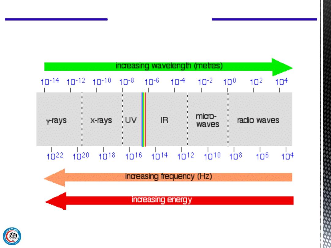
We will study light in three categories
(visible & ultraviolet & infrared)
CH 14 Light in Medicine
4
CH 14 Light in Medicine
5

CH 14 Light in Medicine
2. Light properties
Light has some interesting properties, many of
which are used in medicine:
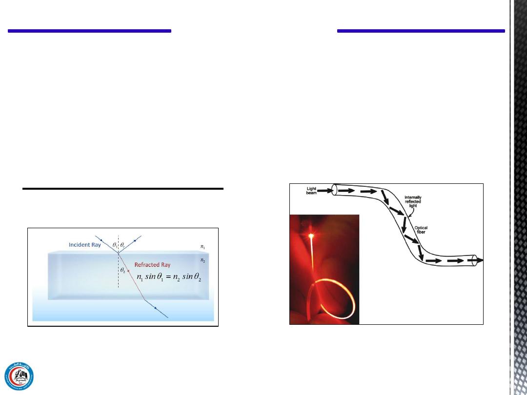
1. The speed of light changes when it goes from
material into another. The ratio of the speed of light
in a vacuum to its speed in a given material is
called the
index of refraction
. If a light beam
meets a new material at an angle other than
perpendicular, it bends, or is refracted.
6

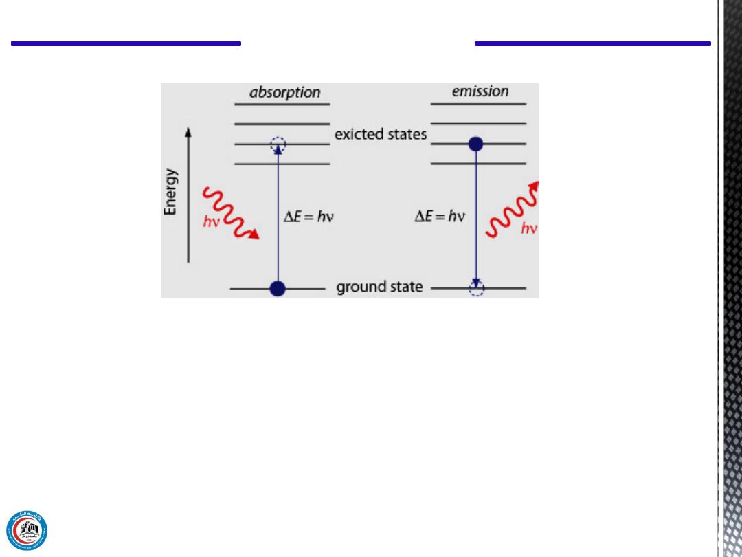
2. Light behaves both as a wave and as a particle. As a wave it
produces interference and diffraction. As a particle it can be
absorbed by a single molecule.
When a light photon is absorbed its energy is used in various
ways. It can cause a chemical change in the molecule that in
turn can cause an electrical change.
Photons are the particle form of light.
This is basically what happens when a light photon is absorbed
in one of the sensitive cells of the retina. The chemical change
in a particular point of the retina triggers an electrical signal to
the brain to inform it that a light photon has been absorbed at
that point.
CH 14 Light in Medicine
7
3. When light is absorbed, its energy generally appears as
heat. This property is the basic for the use in medicine of lR
light to heat tissues. Also, the heat produced by laser beams is
used to "weld" a detached retina to the back of the eyeball and
to coagulate small blood vessels in the retina.
CH 14 Light in Medicine
8

4. Sometimes when photon is absorbed, a lower energy light
photon is emitted. This property is known as fluorescence, it
is the basis of the fluorescent lightbulb. Certain materials
fluoresce in the presence of UV light, sometimes called
"black light," and give off visible light.
The amount of fluorescence and the color of the emitted light
depend on the wavelength of the UV light and on the
chemical composition of the material that is fluorescing.
CH 14 Light in Medicine
9
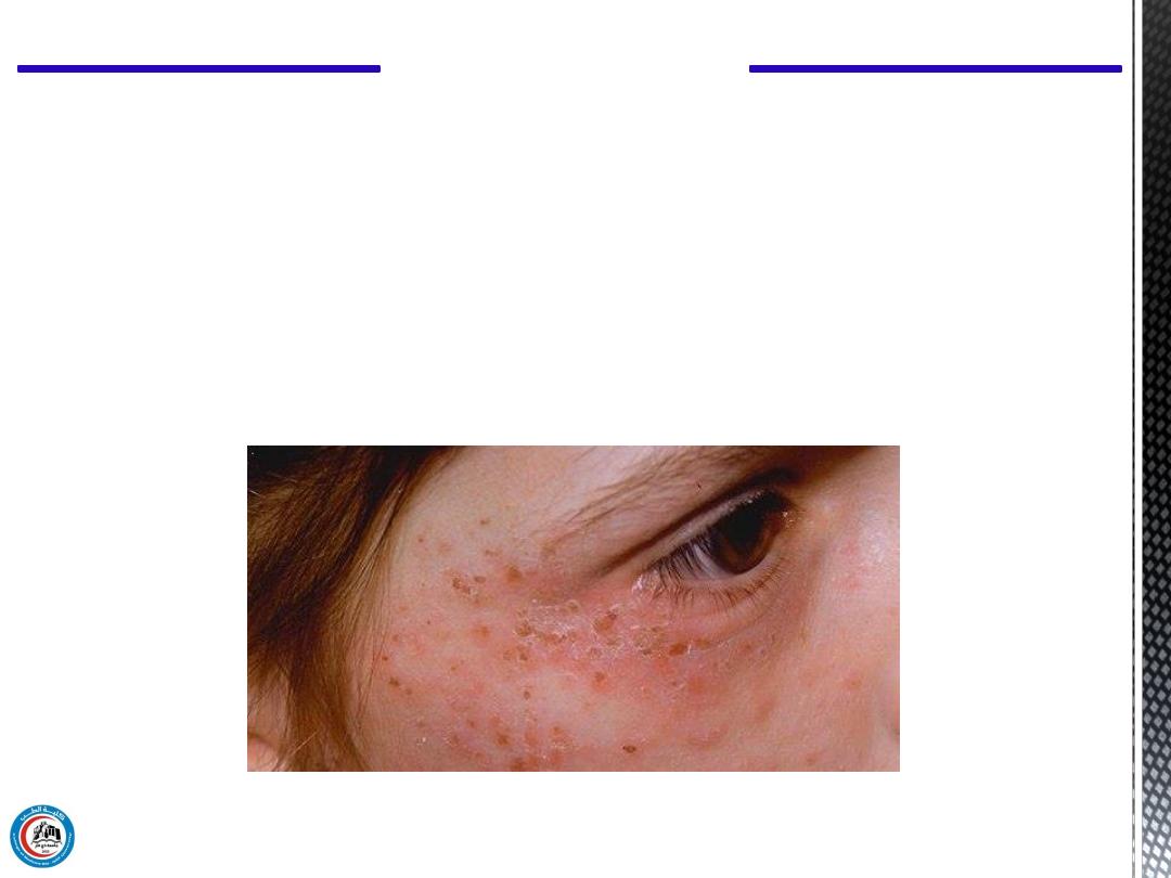
One way fluorescence is used in medicine is in the
detection
of porphyria
, a condition in which the teeth fluoresce red
when irradiated with UV light. Another important application
is in fluorescent microscopes.
CH 14 Light in Medicine
10
A skin rash in a person with porphyria
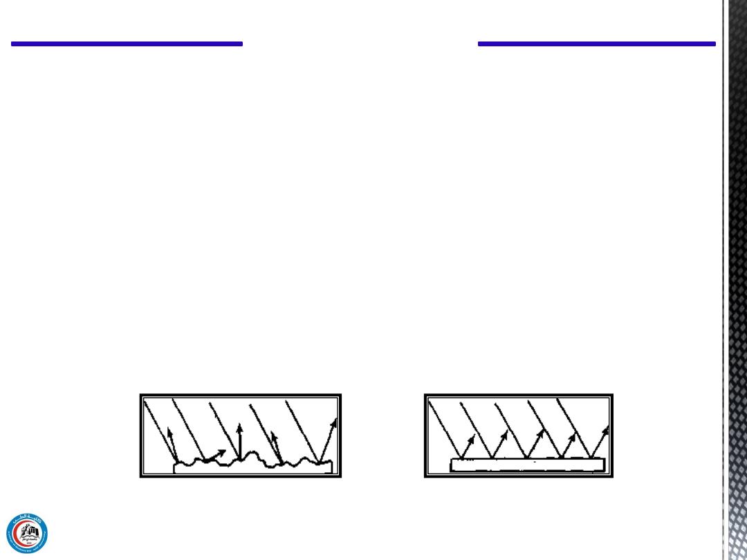
5. Light is reflected to some extent from all surfaces. There are
two types of reflection. Diffuse reflection occurs when rough
surfaces scatter the light in many directions.
Specular reflection is more useful types of reflection; it is
obtained from very smooth shiny surfaces such as mirrors where
the light is reflected at an angle that is equal to the angle at which
it strikes the surface. Mirrors are used in many medical
instruments.
CH 14 Light in Medicine
11
Specular reflection
Diffuse reflection

The three general categories of light-UV, visible, and IR
are defined in term of their wavelengths.
Wavelengths
of light used to be measured in
microns (1 μ = 10
-6
m) or in
angstroms (1 Å = 10
-10
m),
but at present the recommended unit is the nanometer
(1 nm = 10
-9
m).
Ultraviolet light has wavelengths from about 100 to 400
nm; visible light extends from about
400 to 700 nm
; and
IR light extents from about 700 to over 10
4
nm.
CH 14 Light in Medicine
3. Measurement of light and its units
12
CH 14 Light in Medicine
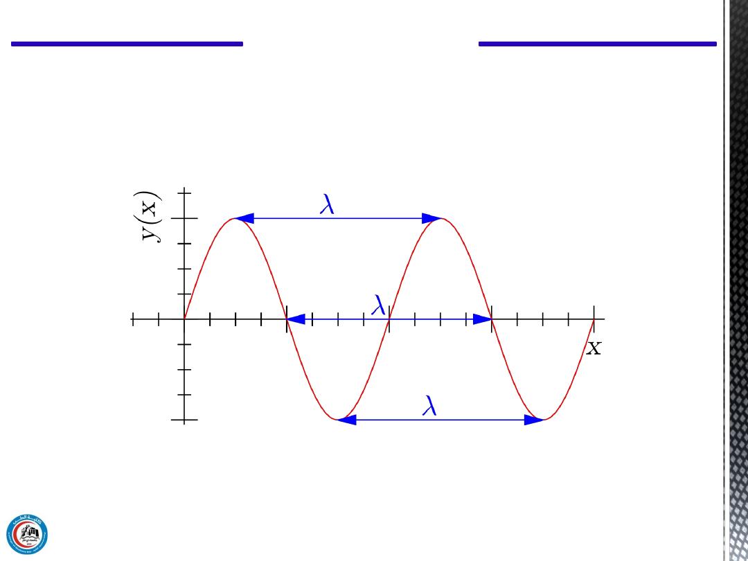
The wavelength λ represents the distance between two points with the
same phase, such as between crests (on top), or troughs (on bottom).
Sine wave
13

CH 14 Light in Medicine
Prefix/Symbol
Meaning
Multiplier
giga (G)
One billion
10
9
1,000,000,000
mega (M)
One million
10
6
1,000,000
kilo (k)
One thousand
10
3
1,000
hector (h)
One hundred
10
2
100
deca (da)
Ten
10
10
1
deci (d)
One-tenth
10
-1
0.1
centi (c)
One-hundredth
10
-2
0.01
milli (m)
One-thousandth
10
-3
0.001
micro (μ)
One-millionth
10
-6
0.000001
nano (n)
One-billionth
10
-9
0.000000001
14
a) Curved Surfaces
Curved lenses (concave, convex & cylindrical) lenses
Curved mirrors which are used in:
a. ophthalmoscope for locking into the eye.
b. otoscope for locking into the ear.
CH 14 Light in Medicine
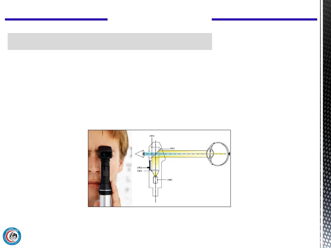
4. Applications of visible light in medicine
15
Figure shows retinoscope. An integrated lamp or LED light source (4)
shines light through a collimating lens (3) onto a partially reflective
mirror (2), which directs the light to the eye.
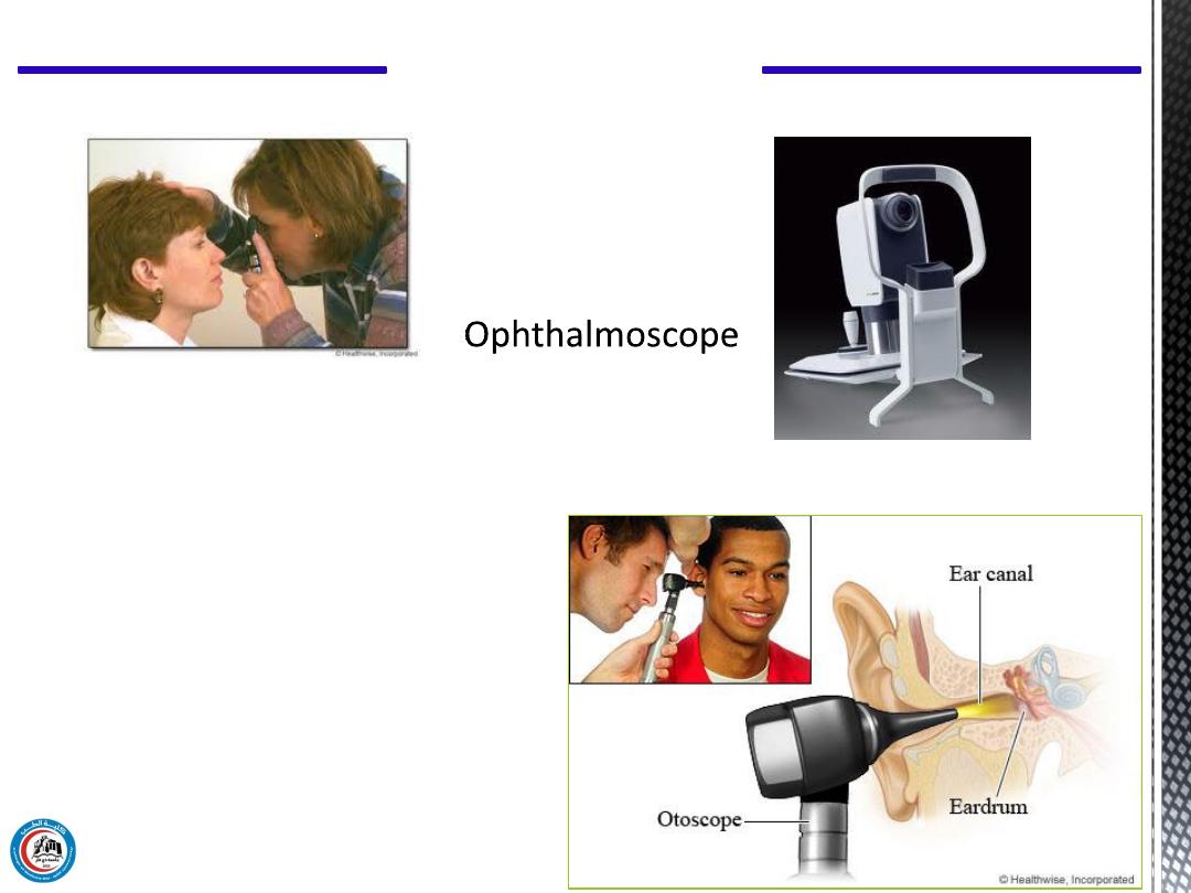
Otoscope
CH 14 Light in Medicine
16
b) Endoscopes
are used for viewing internal body cavities as:
a. cyctoscope for examination of bladder.
b. proctoscope for examination of rectum.
c. bronchoscope for examination of air passages into the lung.
CH 14 Light in Medicine
17
Optical Principles of the Endoscope
The mathematical expression that describes the refraction phenomena is
known as
Snell’s law
,
n
1
sin θ
1
=n
2
sin θ
2
CH 14 Light in Medicine
18
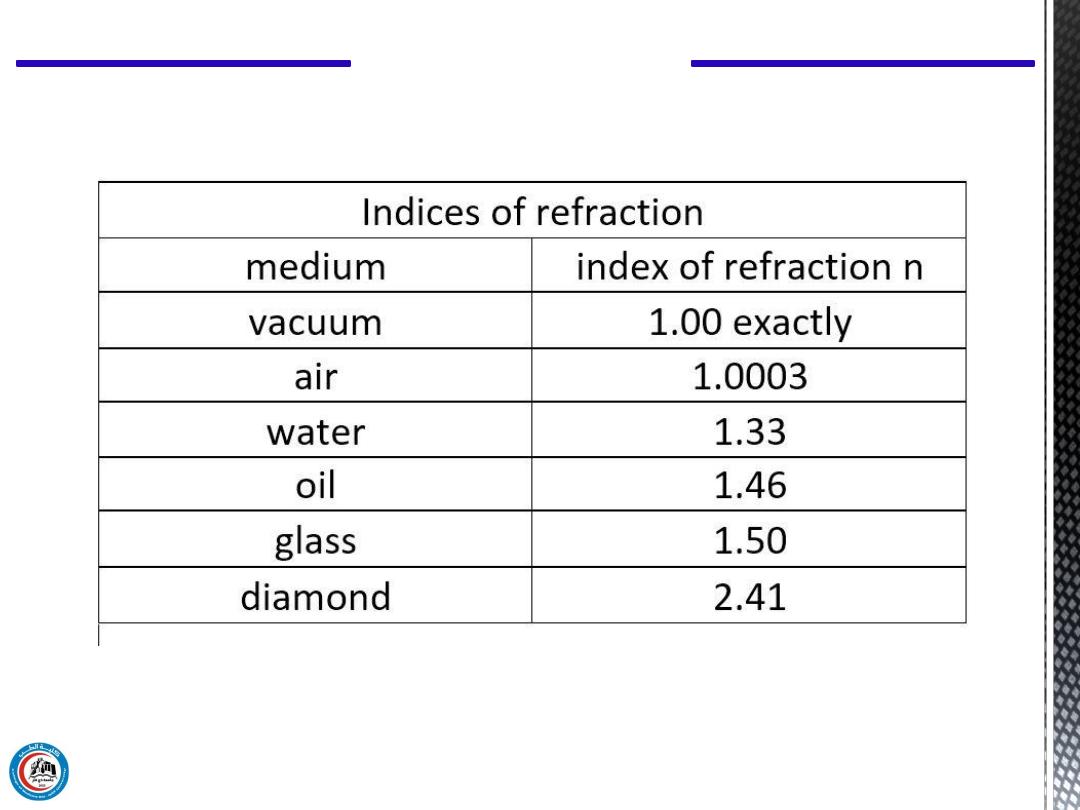
Refractive index of some materials:
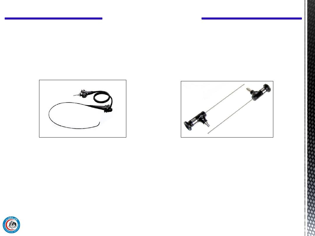
The development of fiberoptic techniques permitted the
construction of flexible endoscopes. Flexible endoscopes can
be used to obtain information from regions of the body that
cannot be examined with rigid endoscopes, such as the small
intestine and much of the large intestine.
Some flexible endoscopes are over a meter in length.
The image obtained with a flexible endoscope is not as good as that
obtained with a rigid endoscope, but often the only alternative to a
flexible endoscopic examination is exploratory surgery.
CH 14 Light in Medicine
19
Flexible endoscopes
Rigid endoscopes
To treat a variety of gastrointestinal problems
To visualize the surface of organs, their vessels, or
pathological changes
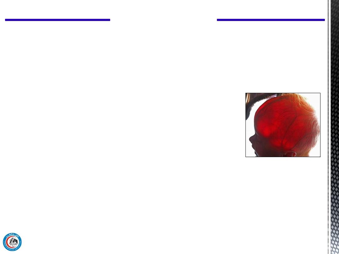
c) Transillumination
It is the transmission of light through the tissue of the body.
It used clinically in the detection of:
a. hydrocephalus (water head) in infant.
b. pneumothorax (collapsed lungs) in infant.
c. the sinuses
d. the gums
e. the breast
f. the testes.
CH 14 Light in Medicine
20

d) Phototherapy
Premature infant recover from jaundice when they exposed to
visible light.
A premature infant is a baby born before 37 completed weeks of gestation.
CH 14 Light in Medicine
21

The wavelengths adjacent to the visible spectrum also have
important uses in medicine. Ultraviolet photons have energies
greater than visible photons, while IR photons have lower energies,
because of their higher energies, UV photons are more useful than
IR photons.
Ultraviolet light with wavelengths below about 290 nm is
germicidal
-that is, it can kill germs and it is sometimes used to
sterilize medical instruments
CH 14 Light in Medicine
5. Applications of ultraviolet and infrared light in medicine
22

Ultraviolet light also produces more reactions in the
skin than visible light. Some of these reactions are
beneficial, and some are harmful. One of the major
beneficial effects of UV light from the sun is the
conversion of molecular products in the skin into
vitamin D
CH 14 Light in Medicine
23

A
blacklight
(or often black light), also referred to as a UV-A
light, Wood's lamp, or ultraviolet light, is a lamp that emits
long-wave (UV-A) ultraviolet light and very little visible light.
Robert Williams Wood
in 1903 using "Wood's glass", it was
in 1925
A
Wood's lamp
is a diagnostic tool used in dermatology by
which ultraviolet light is shone (at a wavelength of
approximately 365 nanometers)
Wood's glass is an optical filter glass invented in 1903 by
American physicist
Wood
(1868–1955), which allows
ultraviolet and infrared light to pass through, while blocking
most visible light
CH 14 Light in Medicine
24
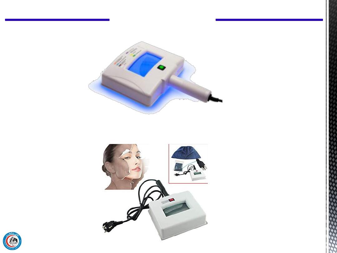
Wood lamp Ultraviolet light lamp for skin diagnose and analysis
Wood's lamp
CH 14 Light in Medicine
25

SKIN CONDITION DISPLAYED COLOR:
• Thick epidermis White fluorescence
• Necrosis cells White spot
• Healthy skin Blue and white
• Water deficiency (thin skin) Purple
• Water deficiency Light purple
• Water abundance Bright fluorescence
• Dark fleck Brown
• Oiled part and pimple Yellow or pink
Wood's lamp
CH 14 Light in Medicine
26

Ultraviolet light from the sun affects the melanin in the skin to
cause tanning. However, UV can produce sunburn as well as tan
the skin. The wavelengths that produce sunburn are around 300
nm.
Solar UV light is also the major cause of skin cancer in humans.
The high incidence of skin cancer among people, who have been
exposed to the sun a great deal, such as fishermen and
agricultural workers, may be related to the fact that the UV
wavelengths that produce sunburn are also very well absorbed
by the DNA in the cells.
CH 14 Light in Medicine
6. Hazards
27

IR light
Two types of IR photography are used in medicine: reflective IR
photography and emissive IR photography. The latter, which uses
the long IR heat waves emitted by the body that give an
indication of the body temperature, is usually called
thermograph.
Reflective IR photography, which uses wavelengths of 700 to
900 nm to show the patterns of veins just below the skin.
Some of these veins are visible to the eye, but many more can be
seen on a near-IR photograph of the skin.
CH 14 Light in Medicine
28

Infrared can also be used to photograph the pupil of the eye
without stimulating the reflex that changes its size.
CH 14 Light in Medicine
29

A laser is a unique light source, that emits a narrow beam of light
of a single wavelength (monochromatic light) in which each
wave is in phase with the others near it (coherent light). Laser is
an acronym for Light Amplification by Stimulated Emission of
Radiation.
While the basic theory for lasers was proposed by Albert
Einstein in 1917, the first successful laser was not made until
1960, when T. H. Maiman produced a laser beam from a ruby
crystal. Since 1960 scientists have made many types of lasers
using gases and liquids as well as solids as the laser materials.
CH 14 Light in Medicine
7. Laser in medicine
30

In a laser, energy that has been stored in the laser material
(e.g., ruby) is released as a narrow beam of light-either
as a steady beam continuous wave (CW) or an intense
pulse. The beam remains narrow over long distances and
can be thought of as an ideal "spot" light. A laser beam
can be focused to be a spot only a few microns in
diameter. When all of the energy of the laser is
concentrated in such a small area, the power density
becomes very large. The total energy of a typical laser
pulse used in medicine, which is measured in millijoules
(mJ), can be delivered in less, than a microsecond, and
the resultant instantaneous power may be in megawatts
Two types for laser pumping is
1.
CW
2.
PW
CH 14 Light in Medicine
31

Useful
1.
Since in medicine lasers are used primarily to deliver
energy to tissue, the laser wavelength used should be
strongly absorbed by tissue.
2.
The curve varies for different individuals, but the short
wavelengths (400 to 600 nm) are always absorbed better
than the long wavelengths (~700 nm).
3.
The laser is routinely used in clinical medicine only in
ophthalmology.
4.
Its effectiveness in treating certain types of cancer and its
usefulness as a "bloodless knife" for surgery are under
active investigation.
5.
Lasers are also being used in medical research for special
three-dimensional imaging called holography.
CH 14 Light in Medicine
32
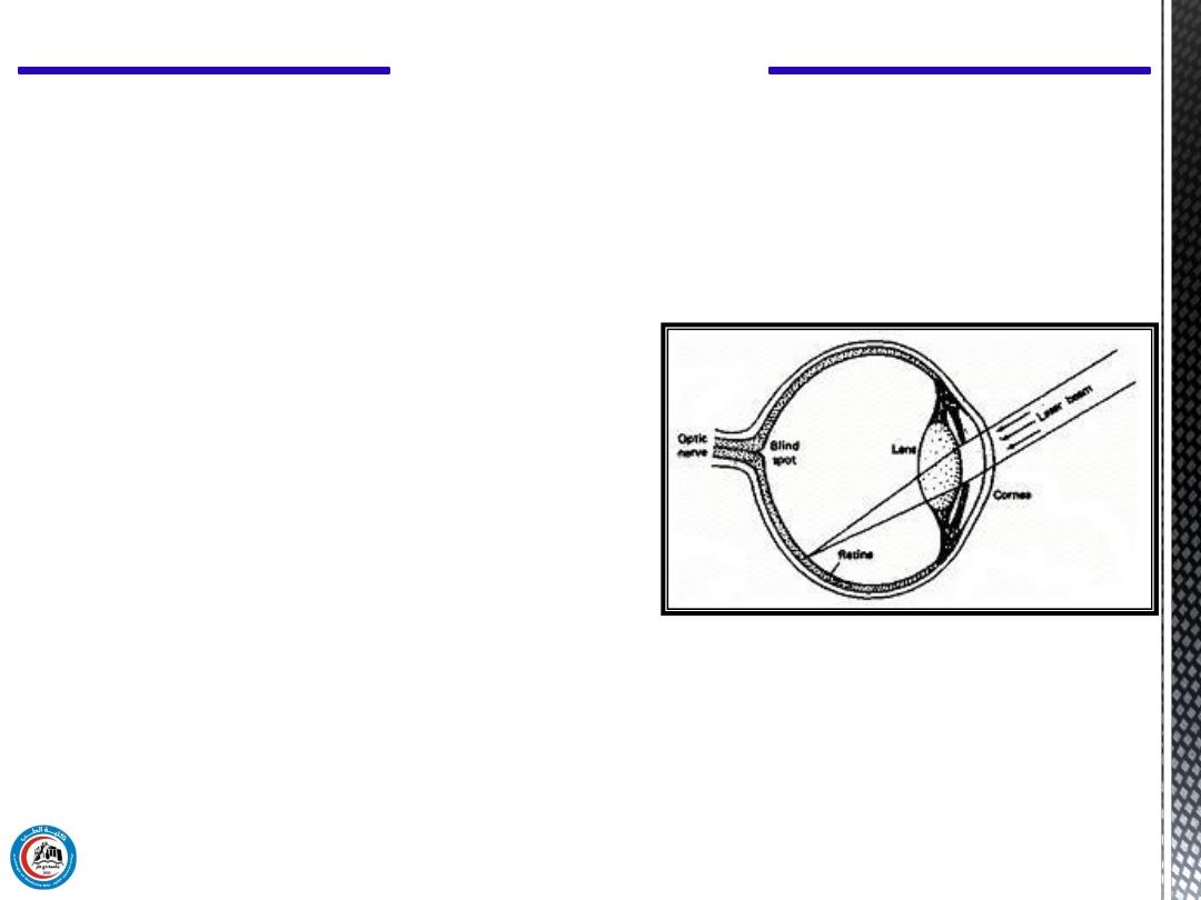
6.
In
ophthalmology lasers are
primarily used for
photocoagulation of the retina that is, heating a blood vessel to
the point where the blood coagulates and blocks the vessel.
The amount of laser energy needed for photocoagulation
depends on the spot size used. In general, the proper dose is
determined visually by the ophthalmologist at the time of the
treatment.
CH 14 Light in Medicine
33
The minimum amount of laser energy that will do
observable damage to the retina is called the minimal
reactive dose (MRD).
CH 14 Light in Medicine
34

There have been few breakthroughs in science that have had as
great an impact as the invention of the microscope by
Leeuwenhoek (1670). The use of the microscope in the
pathology laboratory is as common as the use of the thermometer
in the clinic.
The standard light microscope usually can be set at any of several
magnifications by changing the power of the eyepiece or of the
objective lens. The highest magnification that can be obtained is
limited by the wavelength of visible light. Since the wavelength
of visible light range from 400 to 700 nm, the smallest object that
can be resolved is about 1 μm in diameter. Since most cells are 5
to 50 μm in diameter, this type of microscope is adequate for
resolving all but subcellular objects.
CH 14 Light in Medicine
8. Applications of microscopes in medicine
35

1.
If you put a thin slice of tissue under a microscope you will
not see much because most cells are transparent to all
wavelengths of visible light-red blood cells are an exception.
2.
In order to distinguish different cells it is usually necessary to
stain them with a chemical that strongly absorbs certain
visible wavelengths.
3.
It is sometimes advantageous to use UV light or x-rays in
microscopy. Since our eyes cannot see wavelength shorter
than those of visible light, it is necessary to convert the image
produced by UV light or x-ray beams into images that use
visible light.
CH 14 Light in Medicine
36

Thank you
37

Questions
38