Dr Hadi Al-Sagur
1
Practical Medical Physics
College of Medicine
Physiology and Medical Physics Department
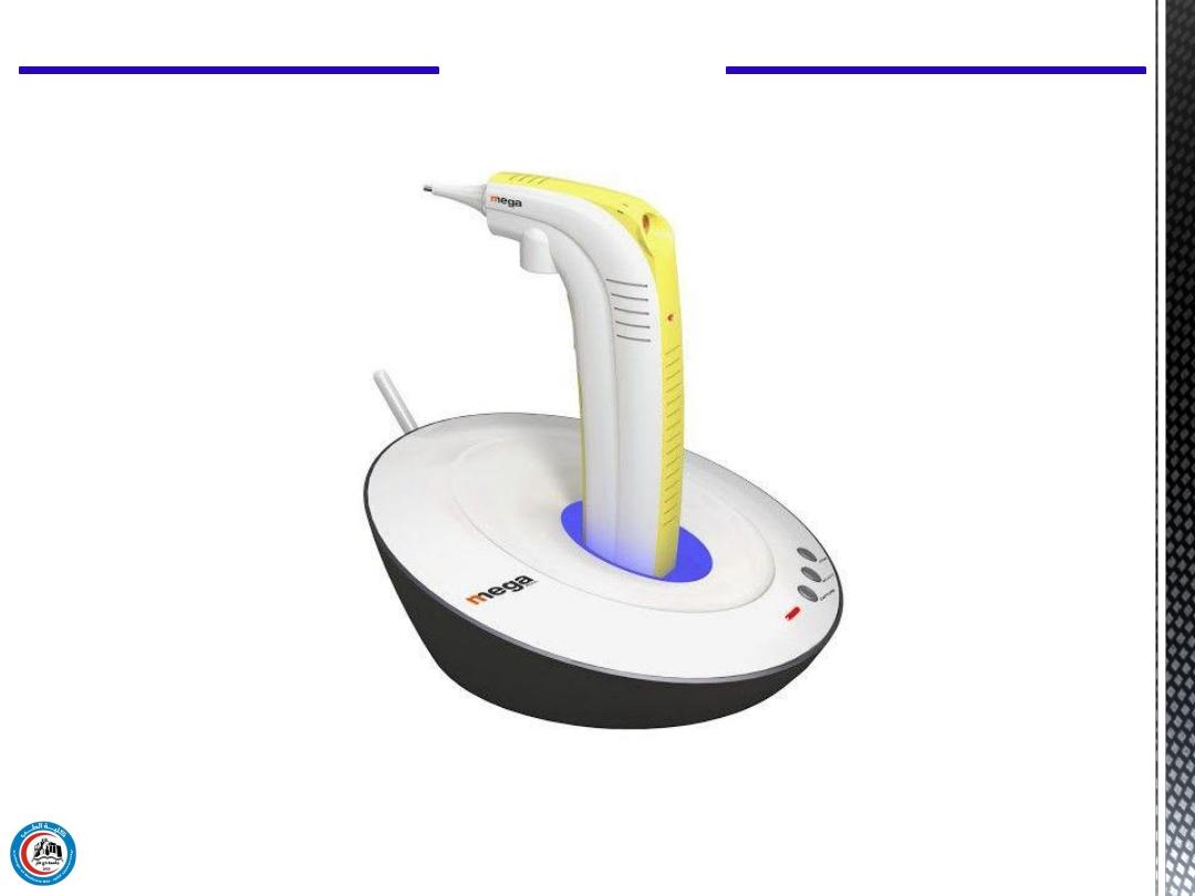
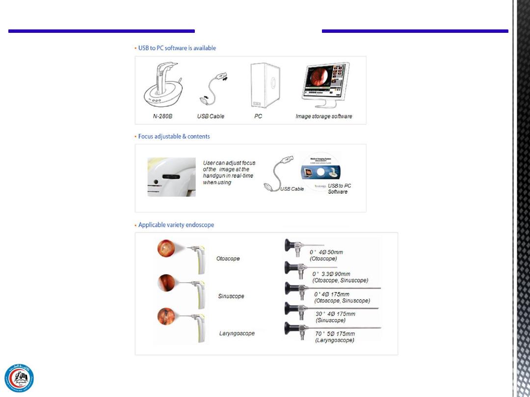
Figure 1: NET-280B Series Wireless Visual System for ENT
Practical Medical Physics
2

In this experiment we have a visual system used for correct
diagnosis and procedures in the field of otorhinolaryngology.
Practical Medical Physics
3
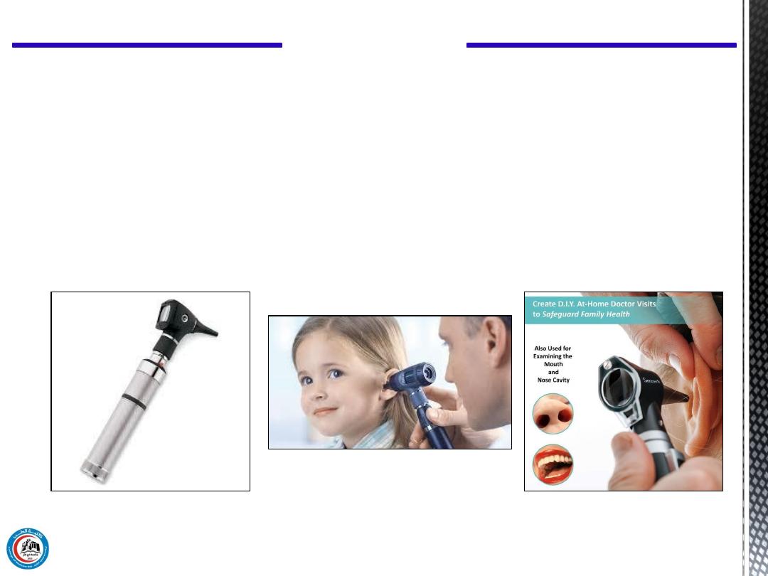
Otoscope or auriscope:
is a medical device which is used to look into
the ears.
Introduction
This is performed in order to examine the ear canal that leads
from the outer ear (pinna) to the eardrum.

Practical Medical Physics
4
Otorhinolaryngology:
The word otorhinolaryngology or its shorter form,
otolaryngology
, is
derived from the Greek root words: otos (ear), rhino (nose), laryngo
(windpipe) and logos (science).Ear, Nose, Throat (ENT).
Also it is a surgical subspecialty within medicine that deals with the
surgical and medical management of conditions of the head and neck.
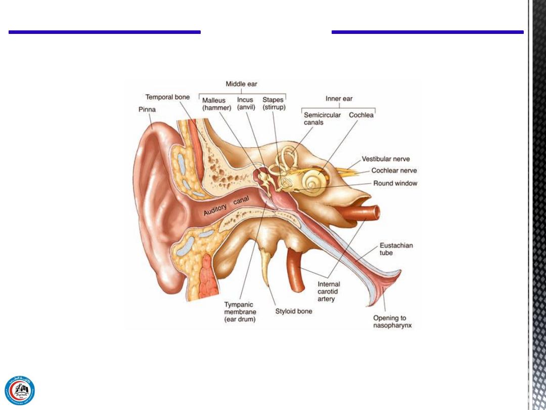
Figure 2: Cross-section of the ear
Practical Medical Physics
5
Practical Medical Physics
6
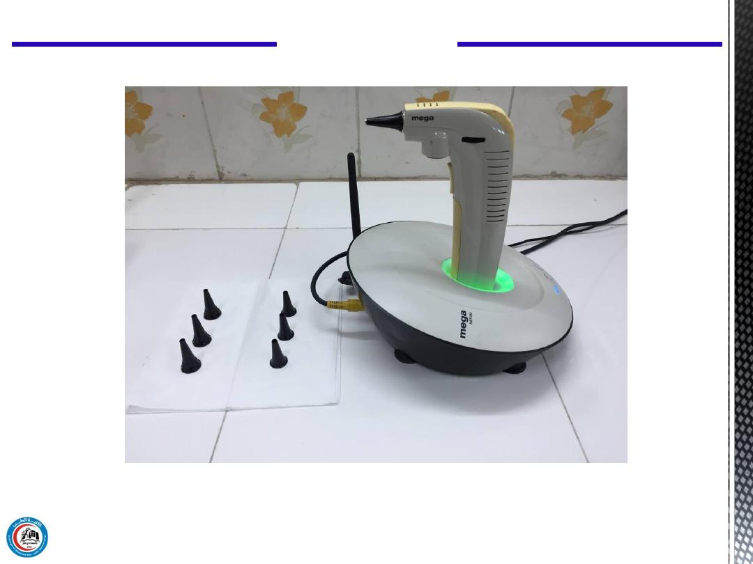
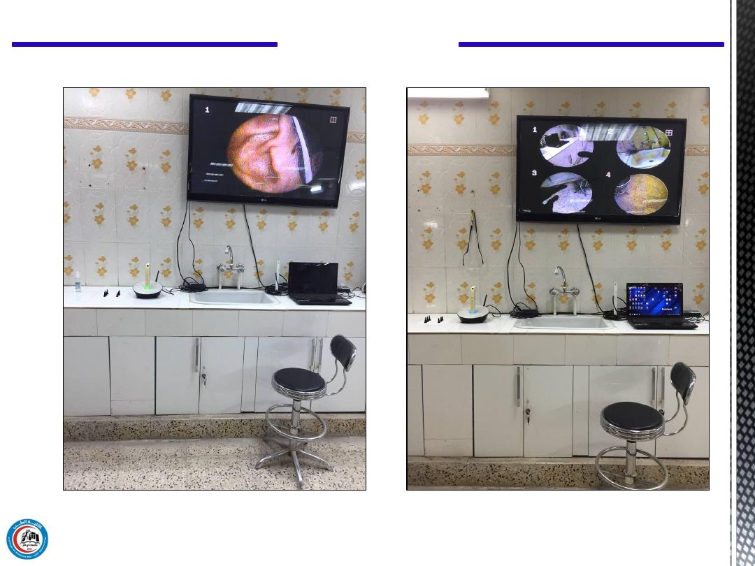
Figure 3: NET-280B Series Wireless Visual System in our lab
Practical Medical Physics
7
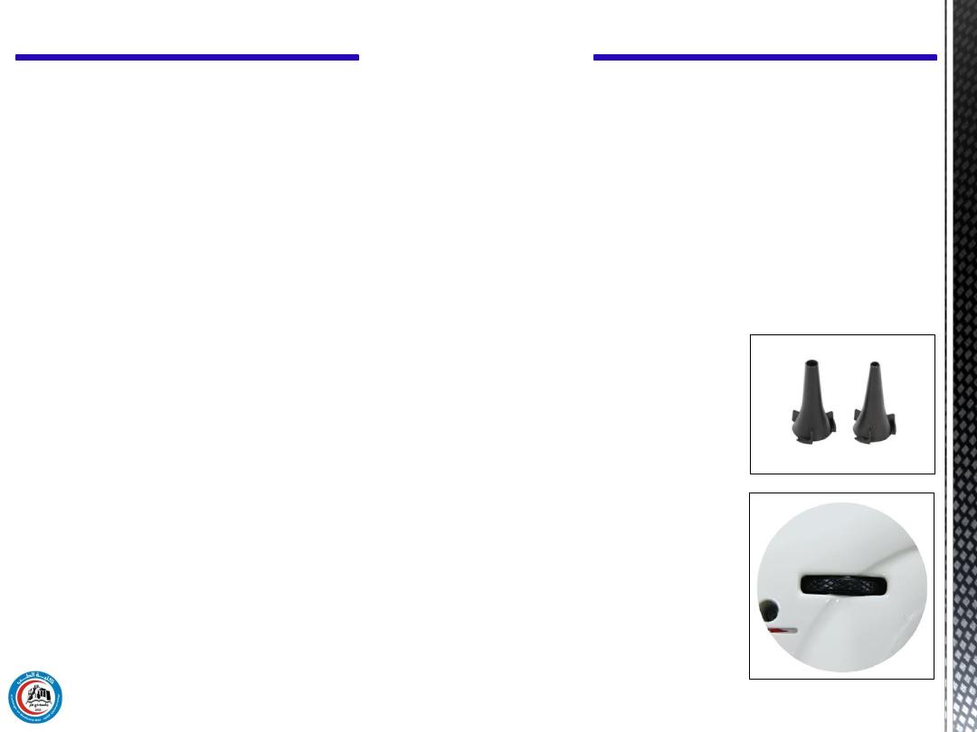
Standard Accessories
The standard accessories of our otoscope are:
- Wireless handgun camera
- Wireless camera cradle
- Freezing system (built-in)
- Telescope(Built-in)
- AC Power adaptor
- Protection tip
- LAN Cable
- PC S/W CD
Focus Adjustable:
This is used for enhance the captured picture.
Protection tip
: ENT Otoscope disposable specula tips.
Practical Medical Physics
8
Figure 4: Setting and adjusting of the otoscope
Practical Medical Physics
9
Figure 5: Practical test in our lab
Practical Medical Physics
10
Examination of the tympanic membrane and middle ear by
otoscopic examination can help providers diagnose a wide variety
of conditions, including:
Clinical Significance (Usage)
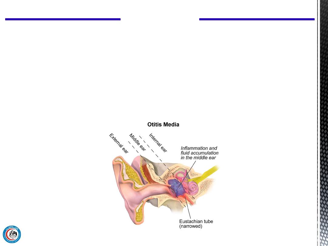
It is a painful type of ear infection. It occurs when the area behind
the eardrum, the middle ear becomes inflamed and infected.
1. Acute otitis media
Figure 6: cross-section of the ear shows the acute otitis media
Practical Medical Physics
11
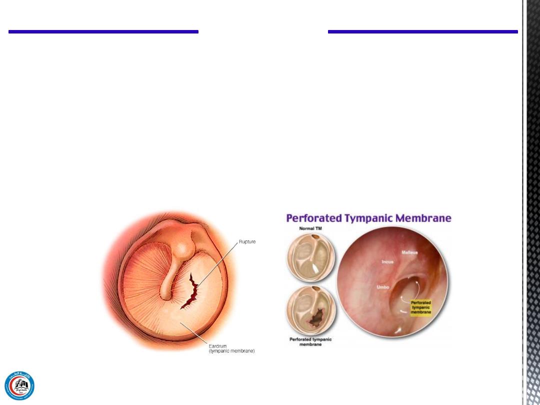
2. Traumatic perforation (ruptured) of the tympanic membrane
This ruptured causes sudden severe pain sometimes followed
by bleeding from the ear, hearing loss, and tinnitus.
Figure 7: Ruptured of the tympanic membrane
Practical Medical Physics
12
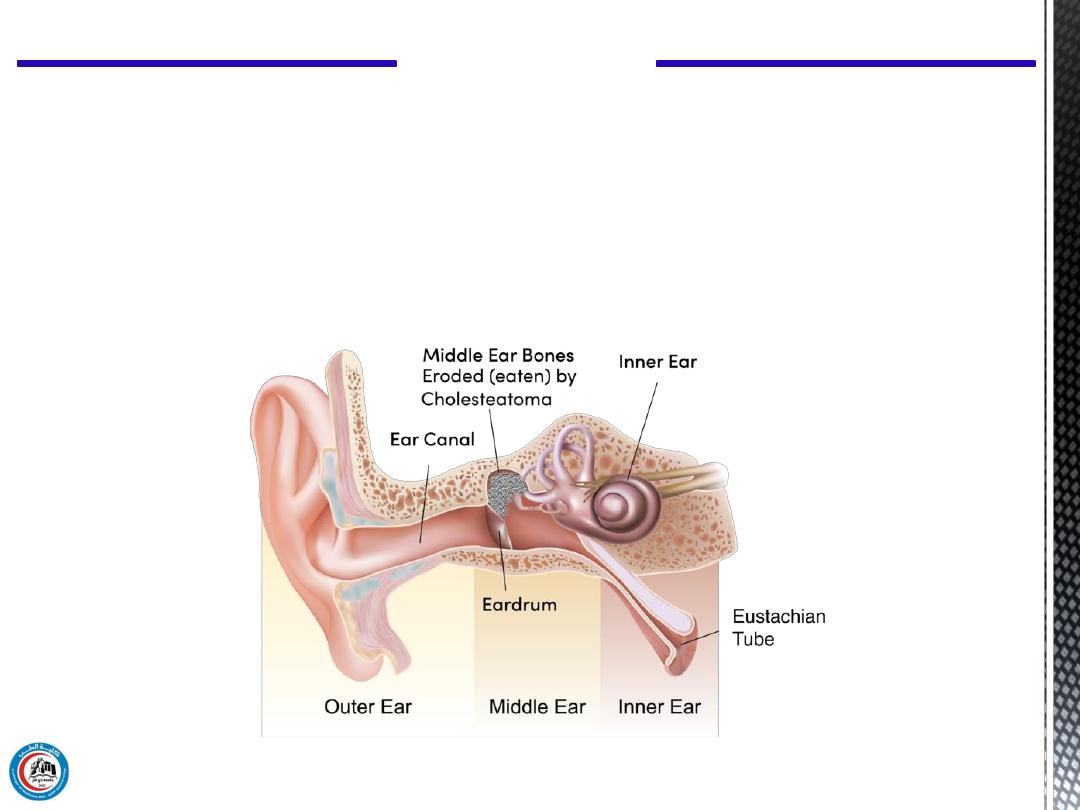
3. Cholesteatoma
It is an abnormal growth of skin within the middle ear. It
begins in the middle ear and may spread to the mastoid bone
behind the ear.
Figure 8: Cholesteatoma grows around and destroys middle ear structures
Practical Medical Physics
13
Other generations of otolaryngology instruments:
Figure 9: Otoscope
1. Otoscope
Practical Medical Physics
14
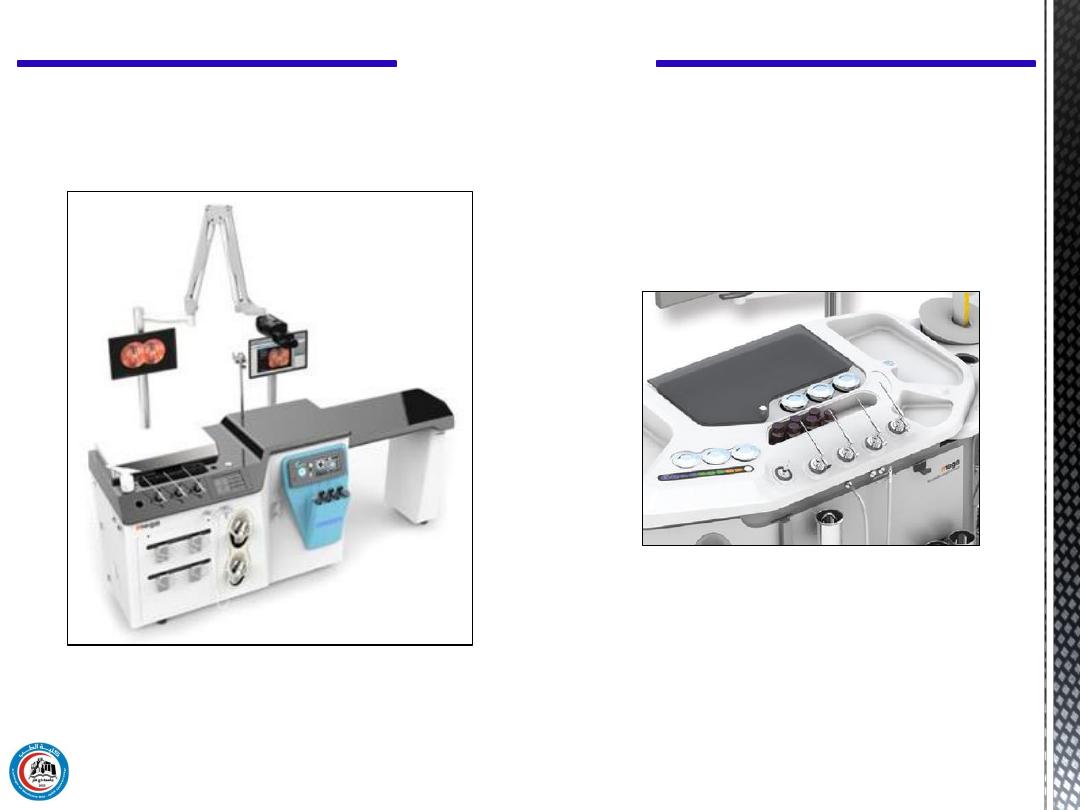
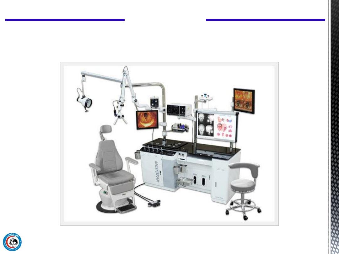
Workstation
Figure 10: NET-280B Series Wireless Visual System for ENT
2. NET-280B Series Wireless Visual System for ENT
Practical Medical Physics
15
Figure 11: ENT Total Treatment Unit
3. ENT Total Treatment Unit

Thank you
16

Questions
17