
-
Physics of Diagnostic X-Rays
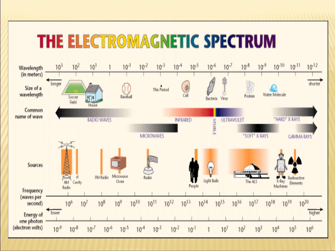
The X-ray photon is a member of the electromagnetic
family that includes light of all types, radio waves,
radar and television signals, and gamma rays.
The field of radiology has three major branches:
Diagnostic Radiology.
Radiation Therapy.
Nuclear Medicine.
Chapter 16

The characteristics of X-Rays:-
1. had very great penetrating power.
2. could ionize a gas.
3. traveled in a straight line from the source
and were not deflected in passing through
electric and magnetic .
4. X-Rays are neutral do not have any change.
5. X-Rays are transverse wave similar to light
wave.
6. X-Rays wave are diffracted by crystal and
have very short wave length
.

In the fall of 1895, W. C. Roentgen, a physicist at
the
University of Wurzburg in Germany,
was studying cathode rays in his
laboratory.
He was using a fairly high voltage across a
tube covered with black paper that had
been evacuated to a low pressure.
When he "excited" the tube with high
voltage, he noticed that some crystals
on a nearby bench glowed
and that the rays causing this fluorescence
could pass through solid matter
.

Production of X-ray beams
The maim component of modern X-ray unit are:
1-A filament (
Cathode
)
–as a source of electrons.
2-A target (
anode
)
–which the electrons strike to
produce x-ray.
3-
High positive voltage-to
accelerate the
negative electrons
travel from the cathode to the anode.
4- An
evacuated space-in
which to speed up the
electrons.
5- Water or oil to cool the target
.
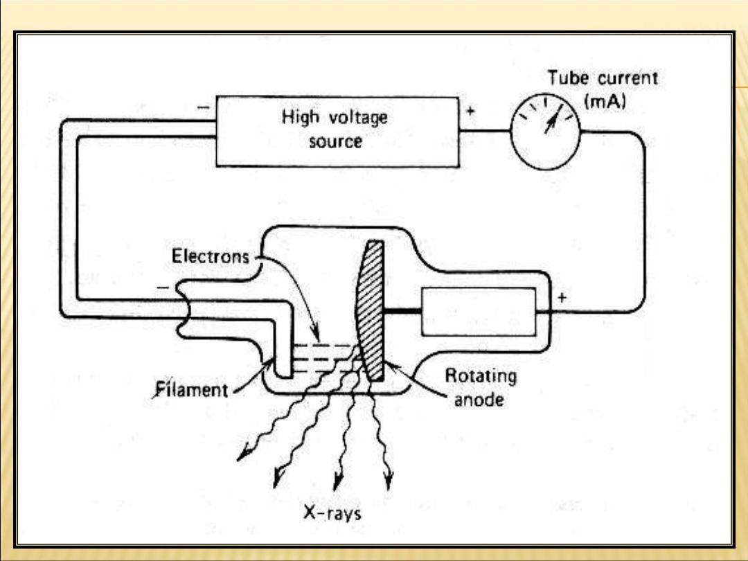
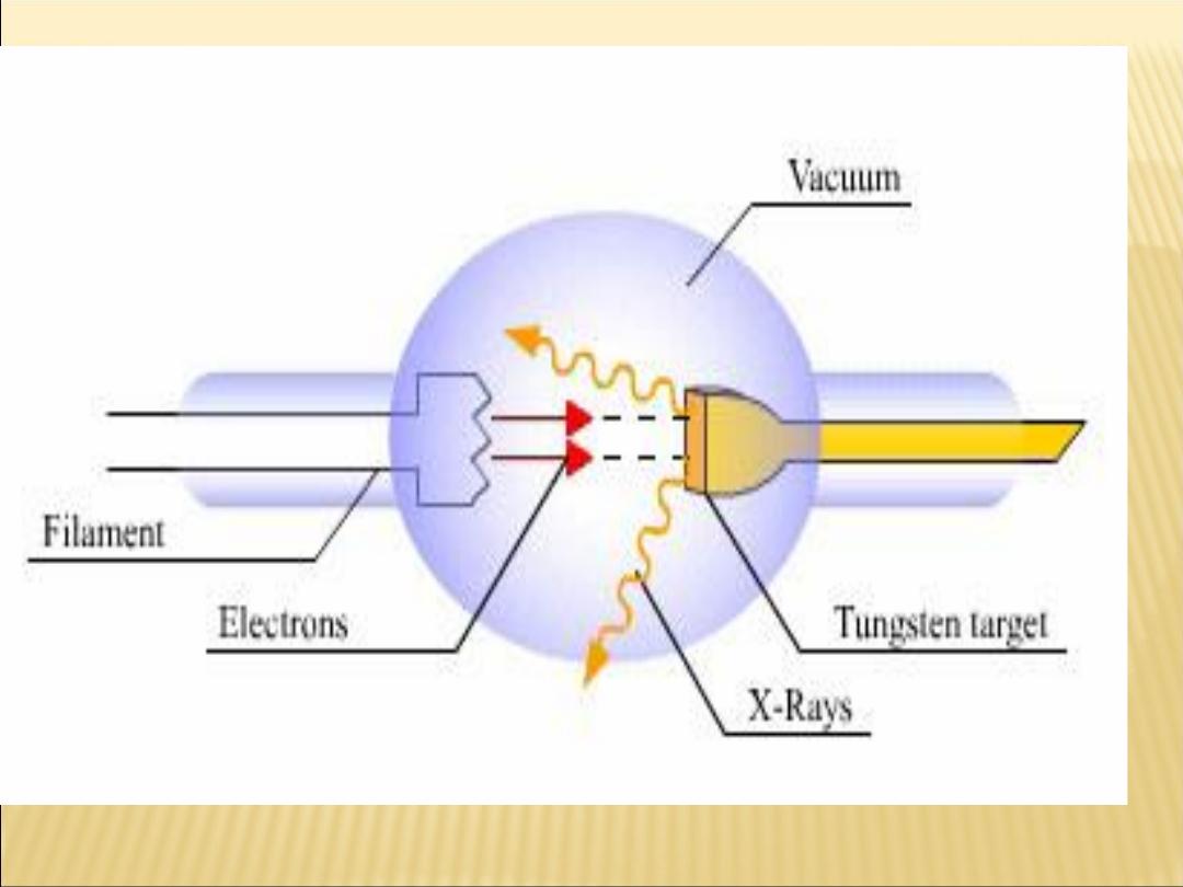

A high-speed electron
(
the electron librated from the
cathode will be accelerated toward the
anode),
can convert some or all of its
energy into an X-ray photon when it
strikes an atom
.

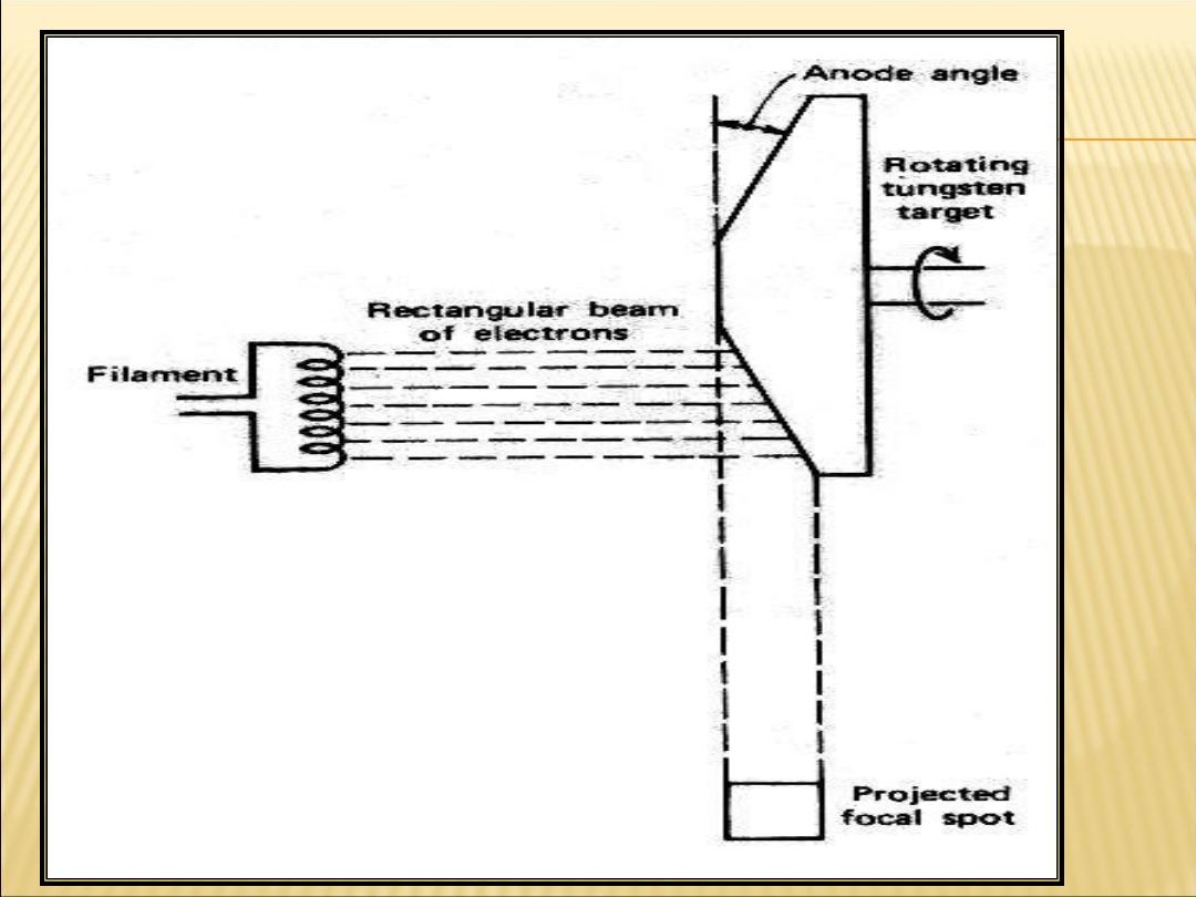
The target of X-ray unit, it should have
A-
high atomic number
(tungsten Z=74)
B-
high melting point
(for tungsten
≈ 340 C.
C-Rotated at 3600 rpm
at minimum and the heat
is spread over a large area as the anode rotates.
D-increase the area
on the target struck by
Electrons to avoid overheating without increasing
the blurring of the X-ray image. This technique,
Called the
line-focus principle
..

General notes
1-In a modern X-ray tube the
number
of electrons
accelerated toward the anode depends on the
temperature of the filament.
2-The maximum
energy
of the X-ray photons
produced is determined by the accelerating
voltage-kilovolt peak (KVP
3-The energy of most electrons striking the
target (99.3%) is dissipated in the form of
heat.The remaining few energy (0.7%) produce
useful X-ray.
Type of X-rays
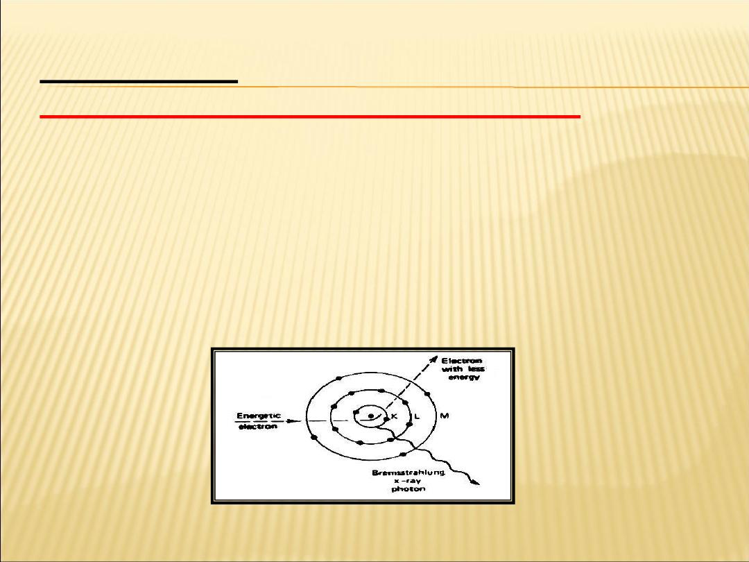
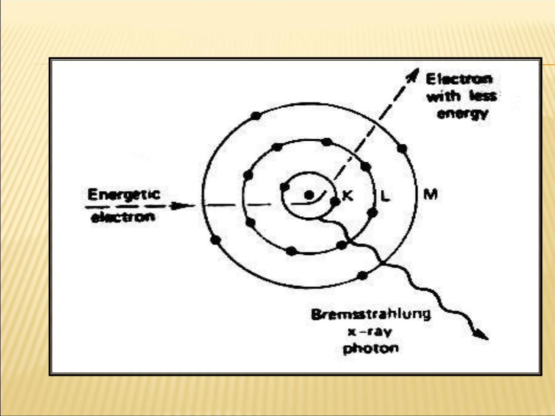
1-Bremsstrahlung (continuous) X-rays:
This type of X-rays is produce by the sudden
decelerating of high energy electron.
When the electrons gets close enough to the
nucleus of a target atom to be diverted from its path
and emits an X-ray photon that has some of its
energy.
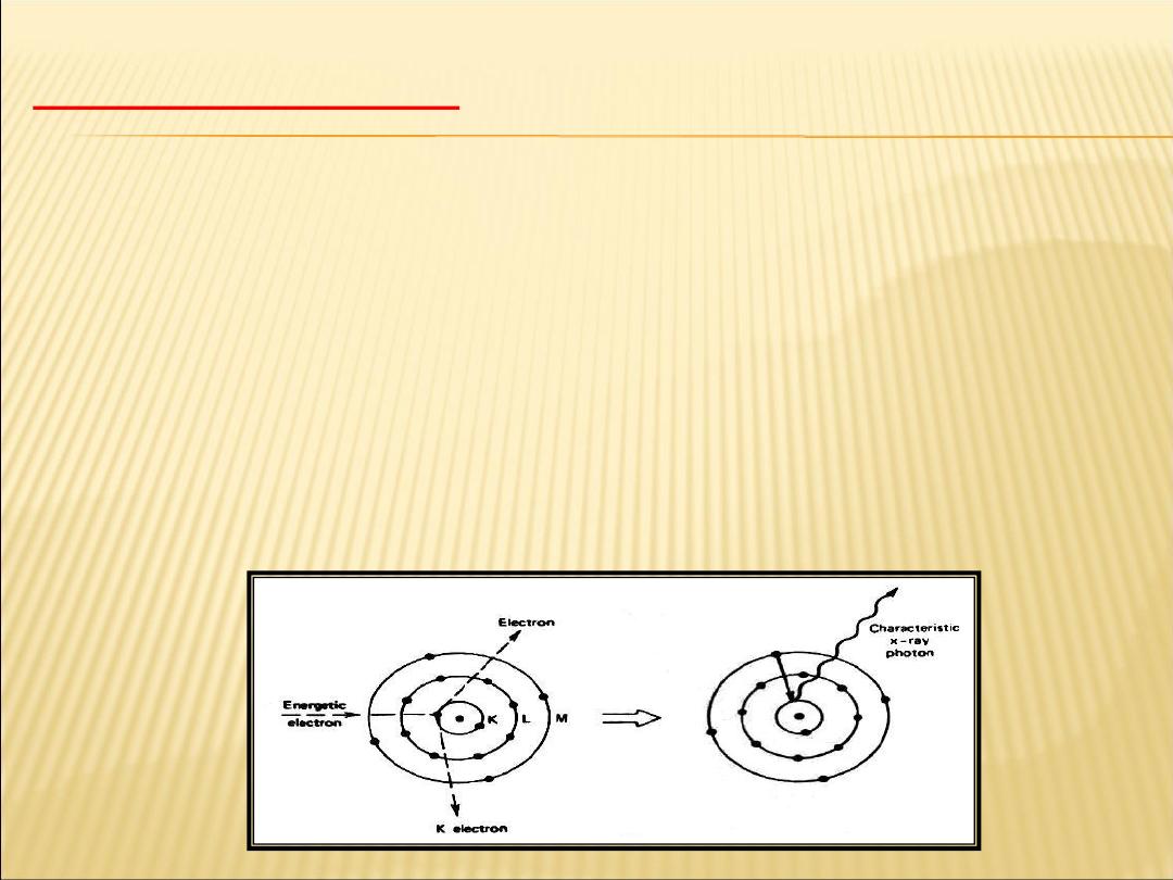
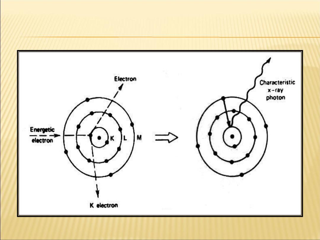
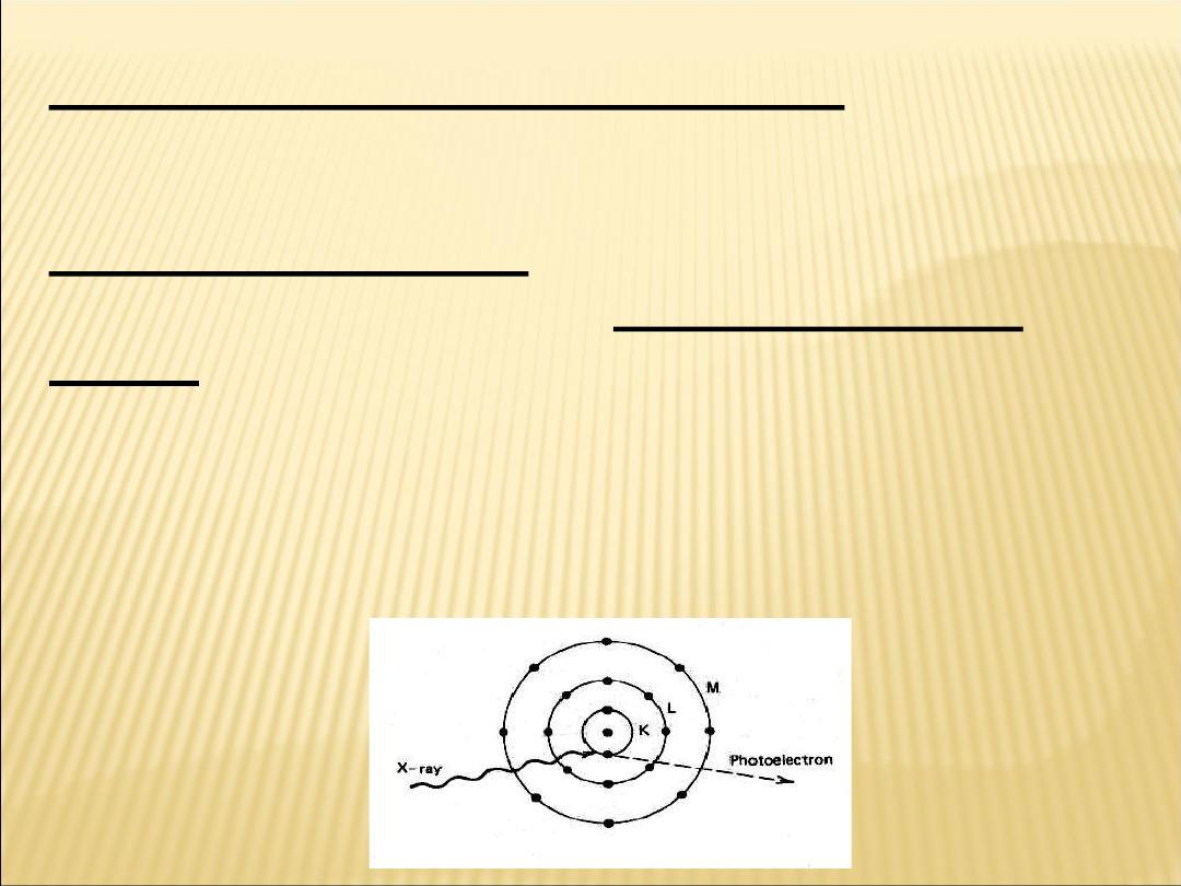
2-Characteristic X-rays
Sometimes a fast electron strikes a K electron in a target atom and knocks it
out of its orbit and free of the atom. The vacancy in the K
shell is filled almost immediately when an electron from
an outer shell of the atom falls into it,
and in the process, a characteristic K X-ray photon is emitted.
An X-ray photon emitted when an electron falls from the
L level to the K level is called a
Kα characteristic X-ray,
and that emitted when an electron falls from the M shell to the K
shell is called a
Kβ X-ray.
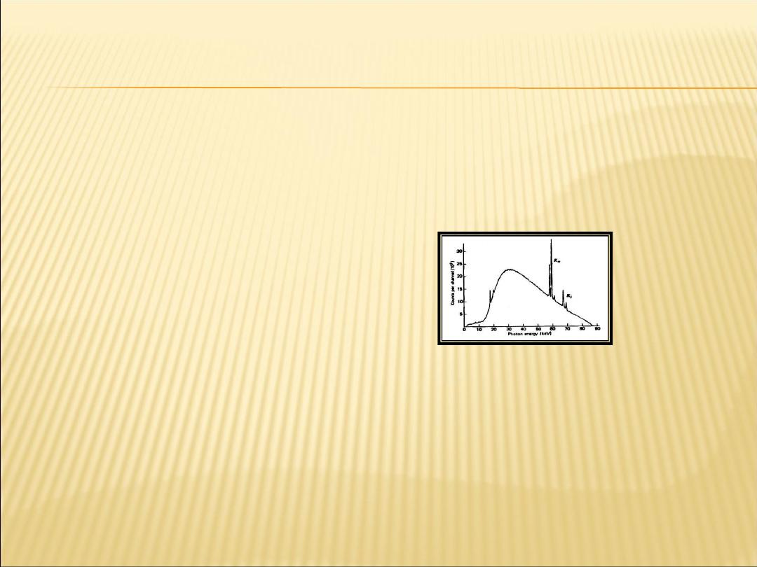
The spectrum of X-rays
produced by a
modern X-ray
generator. The broad
smooth curve is due
to the
bremsstrahlung, and
the spikes represent
the characteristic X-
rays.
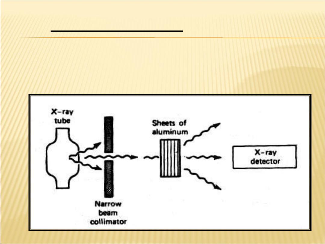
The attenuation of an X-ray beam is its
reduction due to
the absorption and scattering of some of the photons
out of the beam
Attenuation of X-rays:

−
= e
I
I
o
Io=The unattenuated beam intensity
I = attenuated beam intensity
e=2.718,
x is the thickness of the attenuator,
μ is the linear attenuation coefficient of the attenuator.
The linear attenuation coefficient is dependent on the energy
of the X-ray photons; as the beam becomes harder, it decreases.

693
.
0
=
HVL
−
= e
I
I
o
693
.
0
=
HVL
Half-value layer (HVL) :
Is the thickness of a given material that will reduce the beam
intensity by one-half (50%).
The half-value layer is related to the linear attenuation coefficient by: -
Ln (I/
Iо)=-μ(HVL)
Ln(50/100)=-
μ(HVL)

The mass attenuation coefficient
μm
is used to remove the effect of density when comparing
attenuation in several materials.
The mass attenuation coefficient of a material is equal to
the linear attenuation coefficient
μ
divided by
the density
ρ
of the materials.
I
=
Ioe
–
(μ/ρ)(ρx)
=
Ioe
–
μ
m
(ρx)
Calculate the percentage of X-ray beam absorbed by a
bone of thickness 3cm and U/
ρ of the bone to that X-ray
energy is 0.2cm
2
/gm, and
ρ of the bone is 1.9gm/cm
3
.
3
.
0
718
.
2
14
.
1
14
.
1
)
3
9
.
1
)(
2
.
0
(
)
)(
/
(
)
)(
/
(
=
=
=
=
=
=
−
−
−
−
−
e
e
I
I
e
I
I
e
I
I
x
o
x
m
o
x
m
o

693
.
0
=
HVL
−
= e
I
I
o
693
.
0
=
HVL
Half-value layer (HVL): Is the thickness of a given
material that will reduce the beam intensity by one-half
(50%).
The half-value layer is related to the linear attenuation coefficient by:
-
Ln (I/
Iо)=-μ(HVL)
Ln(50/100)=-
μ(HVL)
Ln1/2=-
μ(HVL)
Ln=0.693-----------------
●
Interaction of X-ray with materials :-
X-rays are interaction with the matter when it pass
through the matter.
1- Photoelectric effect
The incoming X-ray photon transfers all of its
energy to an electron which will use it to Overcome
the binding energy and get away from the Nucleus.
(then escapes from the atom) Photoelectric effect
is more common in elements with
high Z than in those with low Z.(It is
dominant when hf<50Kev.)
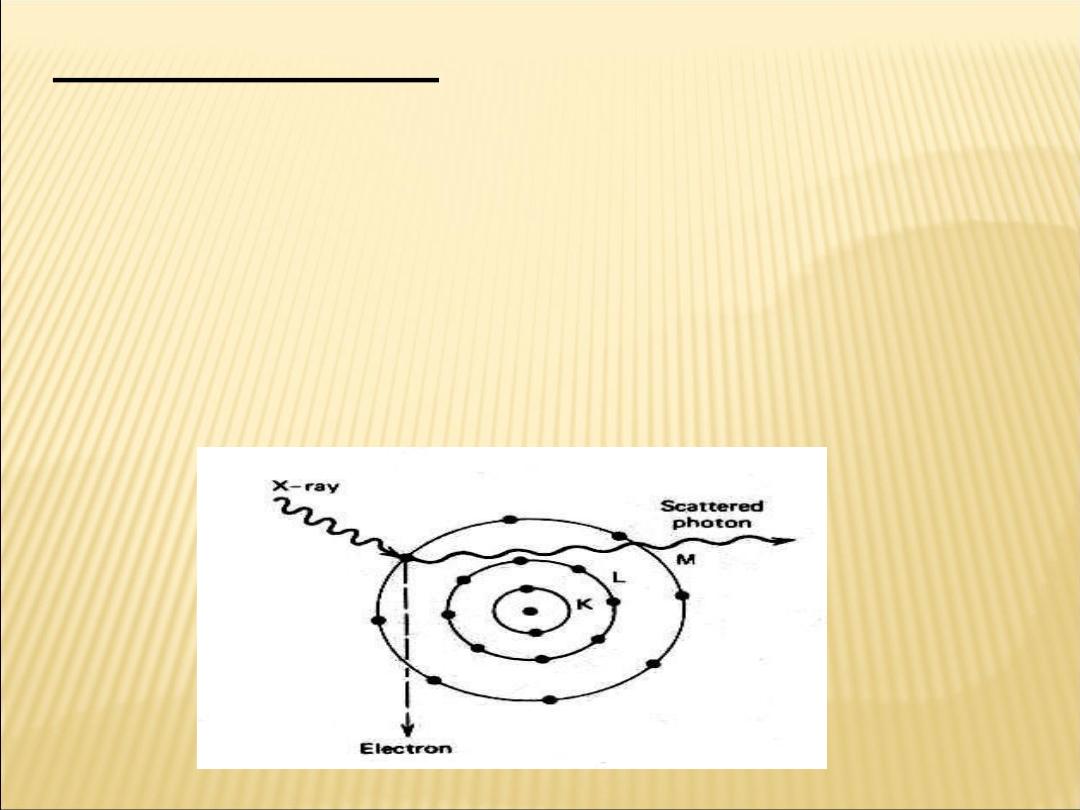
2-Compton effect :-
that an x-rays photon can collide with loosely
bound outer electron. much like a billiard ball
collides with another billiard ball.
At the collision the electron receives part of the
energy and the reminder is given to a
Compton(scattering) photon which then travels in
direction different from the original x-ray.
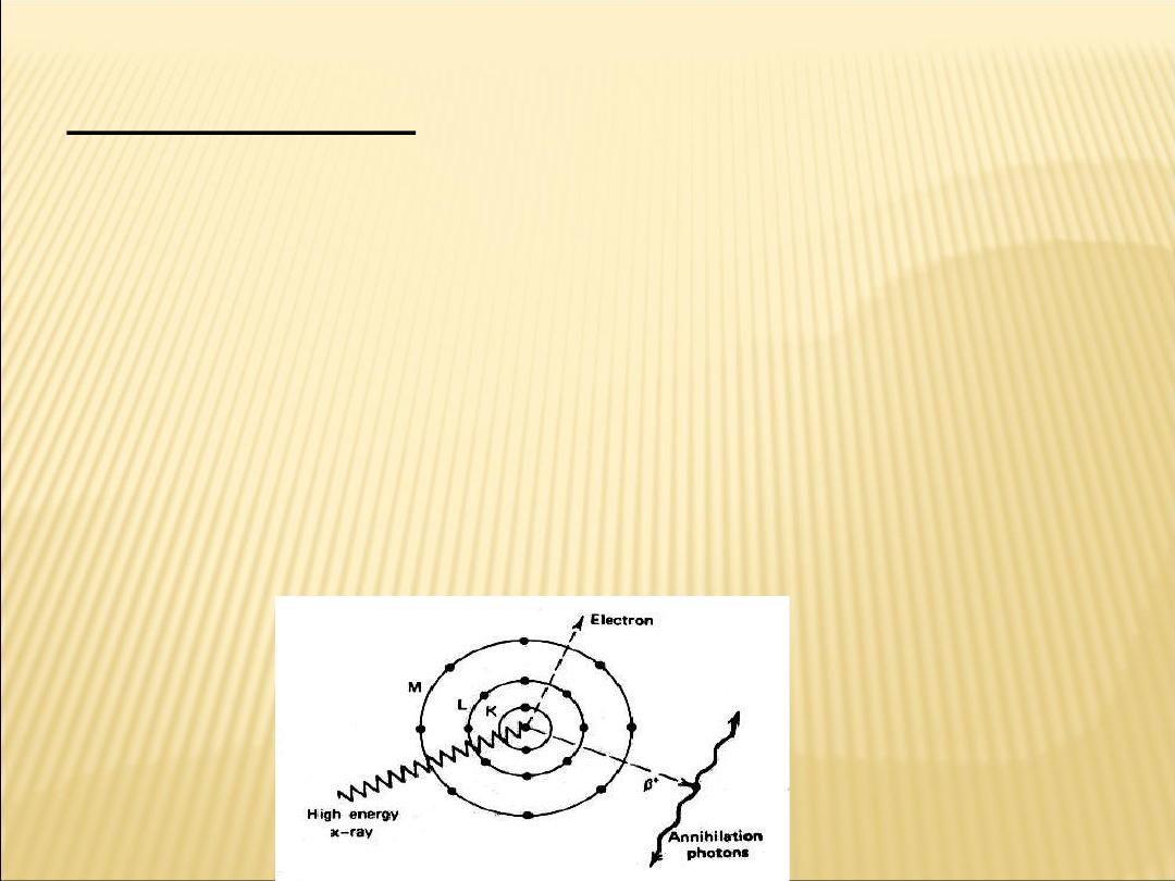
3-Pair production:-
when a very energetic photon enters the intense electric field
of nucleus , it may be convert into two particles as electron
and positron B+ or positive electron
After , it has spent it's kinetic energy ionization , it does a
death dance with an electron. Both then vanish and their
mass energy usually appears as two photons of 511 kev
each called annihilation radiation.

Contrasting
Radiologist often inject high Z material into different
part of the body which are called (contrasting media)
inject high Z materials, or contrast media,
into different parts of the body.
1-Iodinin injected into the bloodstream to show the
arteries
2-An oily mist containing iodine is sometimes sprayed
into the lungs to make the airways visible.
3-Barium compounds orally to see parts of the upper
gastrointestinal tract (upper GI)
4-barium enemas to view the other end of the digestive
system (lower GI).
In a double-contrast study, barium and air are used
separately to show the same organ.
5- Air is used to replace some of fluid ventricles of the
brain ,which a pneumocephalogram is taken
.

The problems involved in obtaining good X-ray shadows are:
1-blurring :It can be reduced by using a small focal spot.
2-positioning: The patient as close to the film as possible to
reduce the penumbra.
3-increasing the distance between the X-ray tube and the film
as much as possible).
4- Reducing the amount of scattered radiation striking the
film as much as possible.
5-Avoid motion during the exposure, since motion causes
blurring.
6- Holding breath when having a chest X-rays ,to reduce
motion which in turn reduces blurring .However, it is not
possible to hold your heart motion, and X-rays of the heart
are somewhat blur. This blurring can be reduced by making
the exposure as short as possible
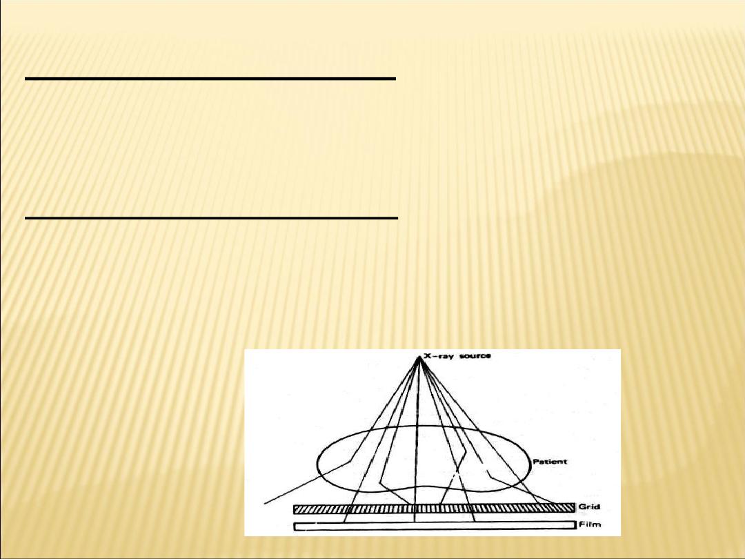
To reducing the scattered :
The most significant way of reducing the amount of
scattered radiation striking the film is by using a grid
consisting of a series of lead and plastic strips.
The strips are aligned so that
1-unscattered X-rays will go through the plastic
strips and strike the film
2-the scattered radiation will strike the lead strips
and be absorbed.

X-ray film:
It is sensitive photographic plat, which is easily effected by X-
ray.
This is because of containing silver halogens(Agcl,AgI,AgBr).
Density : The brightness of a specific point on a film is
determined by the density of the film at the point.
A density of 0 means that the film is completely clear and all
light 100% penetrate.